You’re cruising through a derm Q-bank when a “pearly papule” shows up and you think, “Basal cell carcinoma—easy.” But the highest-yield learning isn’t just picking the right label; it’s knowing why every other answer is wrong and what detail would have made it right. That’s the difference between pattern recognition and exam-ready clinical reasoning.
Tag: Dermatology > Skin Disorders
The Clinical Vignette (Q-bank style)
A 67-year-old man presents with a slowly enlarging lesion on the left nasal ala. He has a history of chronic sun exposure and says the spot “sometimes bleeds when I wash my face.” Exam shows a 4-mm pearly, flesh-colored papule with telangiectasias and a rolled border with central ulceration. No lymphadenopathy.
Question: What is the most likely diagnosis?
Correct Answer: Basal Cell Carcinoma (BCC)
Why BCC is the best fit
This vignette is basically the BCC highlight reel:
- Older age + chronic sun exposure
- Head/neck location (especially nose, periorbital, ears)
- Pearly papule/nodule
- Rolled (“raised”) borders
- Surface telangiectasias
- Ulceration/bleeding (“rodent ulcer”)
High-yield pathology + mechanism
- Arises from basal cells of epidermis/hair follicles.
- Classically associated with PTCH (patched) tumor suppressor loss → constitutive Hedgehog signaling.
- Think: Gorlin syndrome (basal cell nevus syndrome) = multiple BCCs, odontogenic keratocysts, etc.
Histology buzzwords (Step 1 favorites)
- Basaloid nests of tumor cells
- Peripheral palisading of nuclei
- Retraction artifact (clefting between tumor islands and stroma)
Behavior: locally destructive, rarely metastatic
- Metastasis is rare, but BCC can cause significant local tissue destruction, especially on the face.
Management (Step 2–relevant)
Depends on size/location/risk:
- Excision for many lesions
- Mohs micrographic surgery for:
- Face (“H-zone”: nose, eyelids, lips, ears)
- Recurrent tumors
- Ill-defined borders or high-risk histology
- Topicals for superficial BCC (selected cases): imiquimod, 5-FU
- Vismodegib (Hedgehog pathway inhibitor) for advanced/unresectable BCC
Why Every Other Answer Choice Matters (Systematic Distractor Breakdown)
Below are common “BCC look-alikes” and what clue would pivot you away from BCC.
1) Squamous Cell Carcinoma (SCC)
Why it’s tempting: also linked to sun exposure and can occur on the face.
Why it’s wrong here:
- SCC classically appears as an erythematous, scaly plaque or ulcerated nodule—more “rough” than “pearly.”
- BCC’s pearly papule + telangiectasias + rolled border is more specific.
What would make SCC correct:
- Hyperkeratotic, crusted lesion on sun-exposed skin (or lip/ear)
- May arise from actinic keratosis
- Higher metastasis risk than BCC (still not huge, but real)
Key histology:
- Keratin pearls and intercellular bridges
- Often driven by p53 mutations from UV damage
Classic clinical pearl:
- Lower lip SCC (vs upper lip BCC)
2) Melanoma
Why it’s tempting: any changing lesion raises melanoma anxiety.
Why it’s wrong here:
- The lesion described is flesh-colored/pearly, not pigmented and irregular.
- No ABCDE warning signs given.
What would make melanoma correct:
- ABCDE:
- Asymmetry
- Border irregularity
- Color variation
- Diameter mm (rule of thumb)
- Evolving (change in size/shape/color or symptoms)
- History of blistering sunburns, numerous nevi, family history
High-yield pathology:
- BRAF V600E (common in superficial spreading melanoma)
- Worst prognostic factor: Breslow depth (tumor thickness)
Exam tip:
- Melanoma can be amelanotic, but then you’ll usually get other cues (rapid growth, atypical vascularity, high suspicion prompting biopsy).
3) Actinic Keratosis (AK)
Why it’s tempting: sun-exposed elderly patient.
Why it’s wrong here:
- AK is typically a rough, sandpaper-like, scaly lesion—not pearly or ulcerated.
What would make AK correct:
- Multiple gritty macules/papules on bald scalp, face, dorsal hands
- Considered premalignant (can progress to SCC)
Management:
- Cryotherapy for isolated lesions
- Field therapy: topical 5-FU, imiquimod, photodynamic therapy
Path clue:
- Atypia confined to basal layer (early dysplasia)
4) Seborrheic Keratosis
Why it’s tempting: common in older adults and can be pigmented.
Why it’s wrong here:
- Seborrheic keratoses look “stuck on,” waxy, often verrucous—not pearly, not ulcerated with rolled borders.
- Usually on trunk/face but without the classic BCC telangiectasia/ulcer story.
What would make it correct:
- Multiple waxy brown plaques with a stuck-on appearance
Histology:
- Horn cysts
High-yield association:
- Leser-Trélat sign: sudden eruption of many seborrheic keratoses → possible internal malignancy (classically GI adenocarcinoma). Not common, but testable.
5) Keratoacanthoma
Why it’s tempting: sun-exposed older patient; can be dome-shaped.
Why it’s wrong here:
- Keratoacanthoma grows rapidly and typically has a central keratin plug/crater.
- BCC is slow-growing and pearly.
What would make it correct:
- “Rapidly enlarging” crateriform nodule with keratinous center on sun-exposed skin
High-yield nuance:
- Often treated like SCC because it can be hard to distinguish clinically/histologically and has malignant potential.
6) Pyogenic Granuloma (Lobular Capillary Hemangioma)
Why it’s tempting: it bleeds easily.
Why it’s wrong here:
- Pyogenic granuloma is a friable, bright red, pedunculated papule—often after minor trauma or in pregnancy.
- The vignette lesion is pearly with telangiectasias and rolled border.
What would make it correct:
- Rapid onset, very vascular red papule that bleeds with minimal contact (often fingers, face, oral mucosa)
One Table to Lock It In
| Condition | Classic appearance | Key clue | Cancer behavior |
|---|---|---|---|
| Basal cell carcinoma | Pearly papule, telangiectasias, rolled border, may ulcerate | Head/neck, “bleeds easily,” slow growth | Locally invasive, rarely metastasizes |
| SCC | Scaly, erythematous plaque/nodule; may ulcerate | Actinic keratosis precursor, lower lip/ear | Can metastasize |
| Melanoma | Pigmented lesion with ABCDE changes | “Ugly duckling,” evolving lesion | High metastatic potential |
| Actinic keratosis | Rough “sandpaper” papule | Premalignant, sun-exposed | Precursor to SCC |
| Seborrheic keratosis | “Stuck-on,” waxy plaque | Horn cysts | Benign |
| Pyogenic granuloma | Friable red papule | Pregnancy/trauma, rapid growth | Benign vascular proliferation |
USMLE-Style “Next Best Step” Pearls
If the stem asks what to do next
- Suspicious non-melanoma skin cancer on the face: biopsy (shave/punch depending on lesion and clinical context), then treat.
- High-risk facial BCC: Mohs surgery is a common “best next step” when offered.
If they test metastasis vs local destruction
- BCC: minimal metastasis risk, but can invade deeply (cartilage, bone).
- SCC: more metastatic potential than BCC.
Rapid-Fire Takeaways
- BCC = pearly + telangiectasias + rolled border + ulceration on sun-exposed head/neck.
- Histology: palisading nuclei + retraction artifact.
- Pathway: PTCH/Hedgehog (think: vismodegib).
- Don’t get baited by “sun exposure” alone—texture and border usually separate BCC (pearly/rolled) from SCC/AK (scaly/rough).
Visual explanations from the StepGenie QBank
Infographics StepGenie drew to explain questions in its own QBank, matched to this article's topic. Each caption is the takeaway of the question it explains.
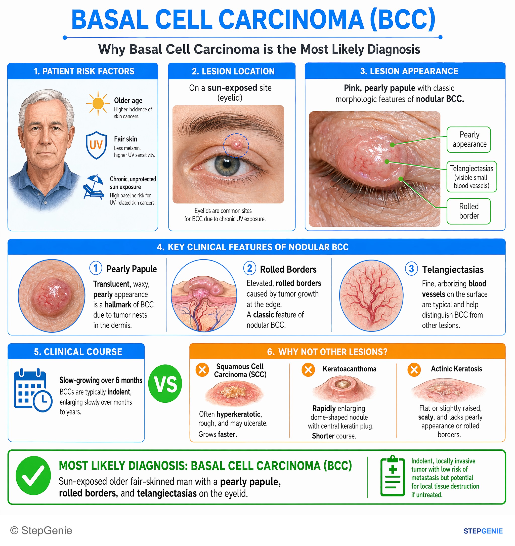
Basal cell carcinoma: sun-exposed older fair-skinned man with pearly papule, rolled borders, and telangiectasias on eyelid.
USMLE Step 2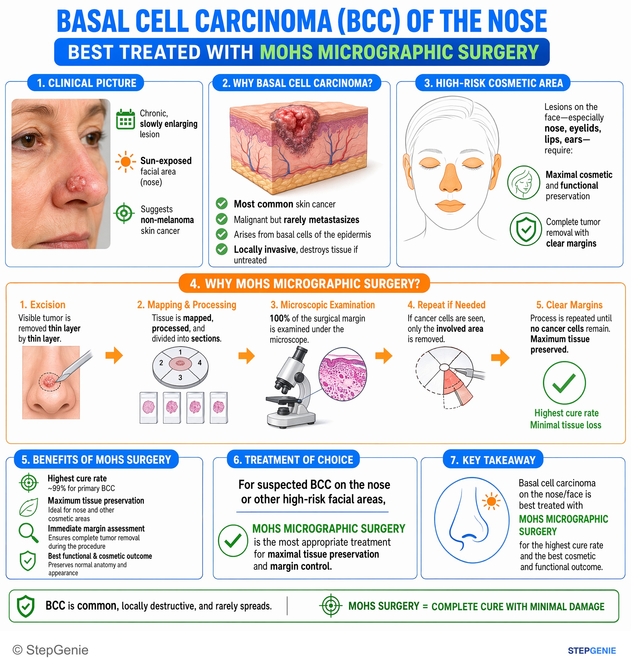
Basal cell carcinoma on a high-risk cosmetic area (nose/face) is best treated with Mohs micrographic surgery for maximal tissue…
USMLE Step 2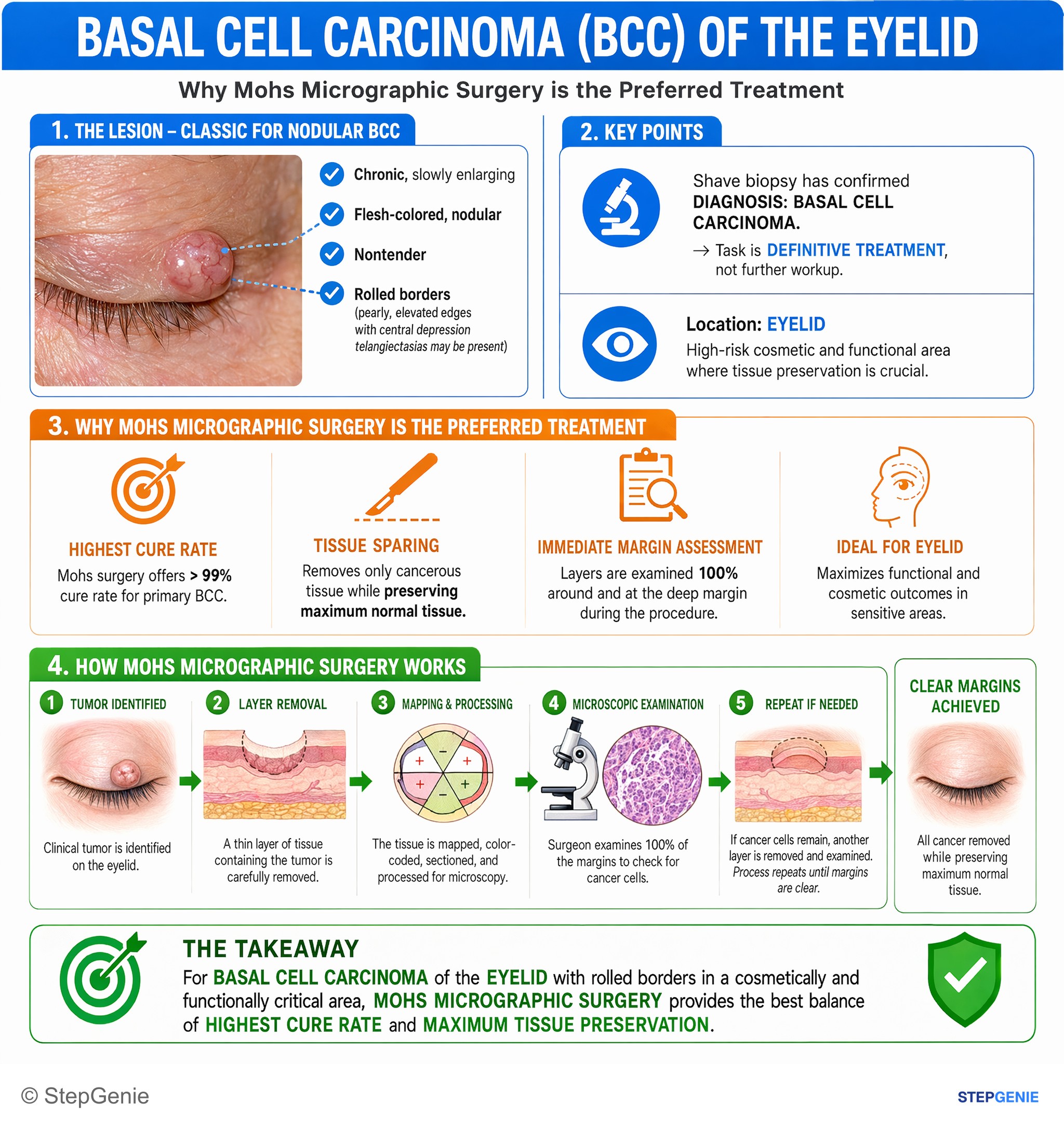
Basal cell carcinoma of the eyelid with rolled borders requires tissue-sparing, margin-controlled excision using Mohs micrographic surgery.
USMLE Step 2Frequently Asked Questions
What clinical features make basal cell carcinoma the most likely diagnosis in a pearly nasal lesion?
Basal cell carcinoma is most likely when an older patient with chronic sun exposure has a head or neck lesion that is a pearly, flesh-colored papule or nodule with telangiectasias, a rolled border, and central ulceration or bleeding. A slowly enlarging lesion on the nose that sometimes bleeds is a classic presentation, especially because BCC commonly occurs on sun-exposed facial areas and is locally destructive.
How do you tell basal cell carcinoma from squamous cell carcinoma on an exam question?
Basal cell carcinoma is suggested by a pearly papule with telangiectasias and rolled borders, whereas squamous cell carcinoma more often appears as an erythematous, scaly plaque or a rough, hyperkeratotic ulcerated nodule. SCC can also arise from actinic keratosis and has a real metastatic risk. Histologically, SCC shows keratin pearls and intercellular bridges, while BCC shows basaloid nests with peripheral palisading and retraction artifact.
What mutation and pathway are classically associated with basal cell carcinoma?
Basal cell carcinoma is classically associated with loss of the PTCH tumor suppressor, which causes constitutive Hedgehog signaling. This mechanism explains why Hedgehog pathway inhibitors such as vismodegib are used for advanced or unresectable disease. A related high-yield association is Gorlin syndrome, also called basal cell nevus syndrome, which features multiple basal cell carcinomas and odontogenic keratocysts.
What histology findings point to basal cell carcinoma?
The classic histology of basal cell carcinoma shows basaloid nests of tumor cells with peripheral palisading of nuclei and retraction artifact, meaning clefting between tumor islands and the surrounding stroma. These are the key buzzwords that distinguish BCC on pathology questions. The tumor arises from basal cells of the epidermis or hair follicles, matching its characteristic clinical appearance on sun-exposed skin.
What is the treatment for basal cell carcinoma, and when is Mohs surgery preferred?
Treatment for basal cell carcinoma depends on the lesion’s size, location, and risk, but many lesions are treated with surgical excision. Mohs micrographic surgery is preferred for tumors on the face, especially the H-zone such as the nose, eyelids, lips, and ears, as well as for recurrent tumors or lesions with ill-defined borders or high-risk histology. Superficial cases may be treated with topical imiquimod or 5-FU.