Aplastic anemia is failure of the hematopoietic stem cells, usually from an immune attack, that produces pancytopenia with a low reticulocyte count and a hypocellular, fat-replaced bone marrow without fibrosis or malignant cells. Work it up in order: confirm pancytopenia, show that reticulocytes are low, exclude look-alikes such as B12 or folate deficiency, hypoplastic myelodysplastic syndrome (MDS), leukemia and PNH, then confirm with a bone marrow biopsy and grade the severity. Severe disease in a patient about 40 or younger with a matched sibling donor is treated with hematopoietic stem cell transplant (HSCT); otherwise first-line therapy is horse antithymocyte globulin (ATG) plus cyclosporine, now combined with eltrombopag.
Step-by-step flowchart: pancytopenia to treatment
- Suspect it from the consequences of low counts. Anemia causes fatigue and dyspnea, neutropenia causes fever and infections, and thrombocytopenia causes petechiae and mucosal bleeding. The liver, spleen and lymph nodes are not enlarged; if they are, look for another diagnosis. (The exception is splenomegaly from transfusion-related iron overload.)
- Confirm pancytopenia. A CBC with differential shows cytopenias in two or more cell lines.
- Check the reticulocyte count. Low reticulocytes mean the marrow is not producing. High reticulocytes mean destruction or bleeding, which is not aplastic anemia.
- Exclude the mimics before the marrow. Look at the smear and send B12 and folate levels: macro-ovalocytes and hypersegmented neutrophils mean megaloblastic anemia, which can also cause pancytopenia with low reticulocytes. Blasts suggest leukemia. Hemolysis or thrombosis at unusual sites suggests PNH, so send flow cytometry for CD55- and CD59-deficient cells.
- Bone marrow aspirate and biopsy with cytogenetics. Aplastic anemia shows a hypocellular marrow replaced by fat, with no fibrosis, no malignant infiltration and usually normal cytogenetics. Dysplasia or a clonal cytogenetic abnormality in a hypocellular marrow points to hypoplastic MDS instead.
- Grade the severity. Severe aplastic anemia is marrow cellularity below 25% plus at least two of: absolute neutrophil count (ANC) below 500/µL, absolute reticulocyte count below 60,000/µL, platelets below 20,000/µL. Very severe disease meets these criteria with an ANC below 200/µL. Reticulocyte cutoffs vary between references: Merck and recent trials such as RACE use an absolute count below 60,000/µL, while StatPearls lists below 40,000/µL (or below 1%) in its diagnostic criteria.
- Look for a cause. Most acquired cases (roughly half to two-thirds) are idiopathic. Identifiable causes are listed below, and inherited marrow failure, especially Fanconi anemia, should be considered in children and young adults.
- Treat. Start supportive care (red cell and platelet transfusions, antimicrobial prophylaxis, prompt treatment of infections) and stop any suspect drug. Then choose definitive therapy:
- HSCT from a matched sibling donor for severe or very severe disease in patients about 40 or younger. It is curative.
- Immunosuppressive therapy for patients who are older or lack a matched sibling donor: horse ATG + cyclosporine + eltrombopag.
- Follow up for relapse and clonal evolution. Aplastic anemia can evolve into PNH, MDS or acute myeloid leukemia, so patients are screened for a PNH clone every year.
Quick flow: pancytopenia → low reticulocytes → smear, B12 and folate, PNH flow cytometry → marrow biopsy: hypocellular and fatty, no fibrosis or blasts → grade severity → young with a sibling donor: HSCT; otherwise horse ATG + cyclosporine + eltrombopag.
Causes to know
- Idiopathic, immune-mediated: T cells and their cytokines drive stem cell apoptosis. About half of patients with acquired aplastic anemia have a detectable PNH clone.
- Drugs and radiation: antineoplastic drugs and ionizing radiation damage stem cells directly; other implicated drugs include antibiotics, antiseizure drugs, NSAIDs, gold salts, penicillamine, acetazolamide and quinacrine. Chloramphenicol causes two types of marrow toxicity: a more common, dose-related, reversible suppression, and a less common idiosyncratic aplastic anemia that starts later and is more often fatal.
- Chemicals: benzene and other industrial solvents, and inorganic arsenic.
- Viruses: seronegative hepatitis (negative for the known hepatitis viruses) accounts for about 5–10% of cases; EBV and CMV are also implicated.
- Pregnancy: usually resolves after delivery but can relapse.
- Inherited: Fanconi anemia is the most common inherited cause; dyskeratosis congenita (a telomere disorder) and Shwachman-Diamond syndrome also occur. Telomere defects are found in 5–10% of adult-onset cases.
Fanconi anemia in one line: usually autosomal recessive DNA repair failure (rare X-linked and dominant forms), with short stature, café au lait macules, thumb and radial ray anomalies, bone marrow failure in the first decade, a roughly 35% risk of MDS or AML by age 40, and head and neck squamous cell carcinoma. Diagnose it with increased chromosome breakage after diepoxybutane (DEB) or mitomycin C.
Pancytopenia: aplastic anemia vs look-alikes
| Condition | Clue on the blood count, smear or exam | Marrow or confirmatory test |
|---|---|---|
| Aplastic anemia | Pancytopenia, low reticulocytes, no organomegaly, no blasts | Hypocellular, fatty marrow; no fibrosis or infiltrate; cytogenetics usually normal |
| Megaloblastic anemia (B12, folate) | Macro-ovalocytes, hypersegmented neutrophils; reticulocytes low | Megaloblasts; low B12 or folate level |
| Hypoplastic MDS | Dysplastic cells | Hypocellular marrow with dysplasia and/or a clonal cytogenetic abnormality |
| Acute leukemia | Blasts | Marrow infiltrated by blasts |
| PNH | Hemolysis, thrombosis at unusual sites | Flow cytometry: CD55- and CD59-deficient cells |
| Pure red cell aplasia | Only red cells low; white cells and platelets normal | Absent or rare red cell precursors; parvovirus B19, thymoma, Diamond-Blackfan anemia |
| Fanconi anemia | Child with short stature, thumb or radial anomalies, café au lait macules | Chromosome breakage with DEB or mitomycin C |
PNH and aplastic anemia overlap so often that it is worth reviewing the PNH overview, and hypoplastic MDS is covered in this myelodysplastic syndromes question breakdown.
Treatment details that show up on exams
- Horse ATG beats rabbit ATG. In a randomized NIH trial, 6-month hematologic response was 68% with horse ATG vs 37% with rabbit ATG, and 3-year survival was 96% vs 76%.
- Adding eltrombopag helps. In the randomized RACE trial, adding eltrombopag to horse ATG plus cyclosporine improved the rate, speed and depth of hematologic response in previously untreated severe aplastic anemia, without added toxicity.
- Boards convention vs current practice: older review books answer "ATG + cyclosporine"; current references, and eltrombopag's FDA label, add eltrombopag to standard immunosuppression as first-line therapy for severe disease when transplant is not chosen.
- Transplant candidates: HSCT from a matched sibling is favored for patients about 40 or younger with severe disease. If immunosuppression fails or the disease relapses, HSCT is preferred for eligible patients, and a haploidentical donor is an alternative when no matched sibling exists.
Exam traps
- High reticulocyte count: not aplastic anemia; think hemolysis or bleeding.
- Splenomegaly or lymphadenopathy: look for another diagnosis.
- B12 or folate deficiency can cause pancytopenia with low reticulocytes; hypersegmented neutrophils give it away.
- Parvovirus B19 infects red cell precursors through the P antigen and causes a transient aplastic crisis in chronic hemolytic anemias (sickle cell disease, hereditary spherocytosis). Only the red cell line falls; review it in this parvovirus B19 question breakdown.
- Child with absent thumbs and pancytopenia: Fanconi anemia; order a chromosome breakage test.
- After immunosuppressive therapy: relapse and clonal evolution (PNH, MDS, AML) remain long-term risks.
Worked example
Vignette. A 19-year-old man has 3 weeks of fatigue, gum bleeding and fever. Two months ago he had acute hepatitis with negative serologies for the hepatitis viruses. He has petechiae but no hepatosplenomegaly or lymphadenopathy. Hemoglobin is 7.2 g/dL, ANC 300/µL, platelets 12,000/µL and absolute reticulocytes 15,000/µL; the smear shows no blasts, and B12 and folate are normal. Bone marrow biopsy shows 10% cellularity with fat replacement and no fibrosis or abnormal cells. His brother is an HLA-identical match. What is the best definitive treatment?
Answer: Allogeneic hematopoietic stem cell transplant from his matched sibling.
Reasoning: This is hepatitis-associated severe aplastic anemia: cellularity below 25% with ANC below 500/µL, platelets below 20,000/µL and reticulocytes below 60,000/µL (not very severe, because the ANC is above 200/µL). A young patient with a matched sibling donor should be transplanted. Without a donor, or in an older patient, horse ATG plus cyclosporine and eltrombopag would be the answer.
Key takeaways
- Pancytopenia + low reticulocytes + hypocellular fatty marrow = aplastic anemia.
- No splenomegaly or lymphadenopathy; their presence suggests another diagnosis.
- Rule out megaloblastic anemia, hypoplastic MDS, leukemia and PNH before labeling it aplastic anemia.
- Severe: cellularity below 25% plus two of ANC below 500/µL, reticulocytes below 60,000/µL and platelets below 20,000/µL; very severe if ANC is below 200/µL.
- Most cases are idiopathic and immune-mediated; know chloramphenicol, benzene, radiation, seronegative hepatitis and Fanconi anemia.
- Young with a matched sibling: HSCT. Otherwise: horse ATG + cyclosporine + eltrombopag.
Sources
Visual explanations from the StepGenie QBank
Infographics StepGenie drew to explain questions in its own QBank, matched to this article's topic. Each caption is the takeaway of the question it explains.
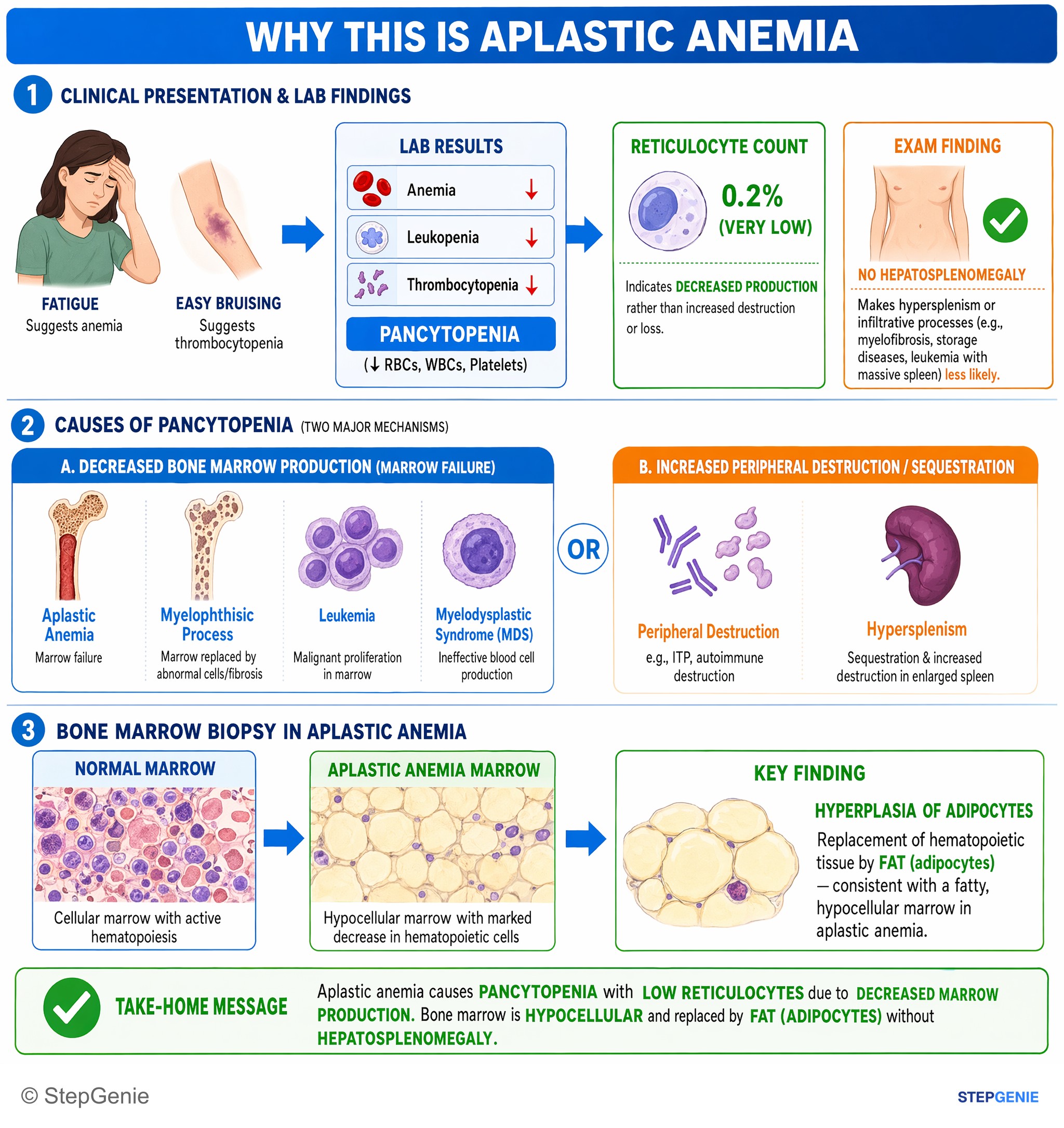
Aplastic anemia causes pancytopenia with low reticulocytes and hypocellular marrow replaced by fat (adipocytes) without splenomegaly.
USMLE Step 1Frequently Asked Questions
What are the criteria for severe aplastic anemia?
Severe aplastic anemia requires bone marrow cellularity below 25% plus at least two of the following: an absolute neutrophil count below 500/µL, an absolute reticulocyte count below 60,000/µL (some references use a lower reticulocyte cutoff) and a platelet count below 20,000/µL. Very severe aplastic anemia meets the same criteria with a neutrophil count below 200/µL. Severity, age and donor availability together guide treatment.
What is the most common cause of aplastic anemia?
Most acquired cases are idiopathic and are thought to result from T cells attacking hematopoietic stem cells. Identifiable causes include drugs such as chloramphenicol and antineoplastic agents, radiation, benzene, seronegative hepatitis and pregnancy. Fanconi anemia is the most common inherited cause.
What is the treatment for aplastic anemia?
All patients need supportive care with transfusions and prevention and prompt treatment of infections. For severe disease, hematopoietic stem cell transplant from a matched sibling donor is preferred in patients about 40 or younger, and it is curative. Patients who are older or lack a sibling donor receive immunosuppressive therapy with horse antithymocyte globulin, cyclosporine and eltrombopag.
Does aplastic anemia cause splenomegaly?
No. The liver, spleen and lymph nodes are not enlarged in aplastic anemia, and their enlargement should prompt a search for another cause of pancytopenia. The exception is splenomegaly caused by iron overload from repeated transfusions.
What is the difference between aplastic anemia and pure red cell aplasia?
Aplastic anemia affects all cell lines, causing pancytopenia with a hypocellular, fatty marrow. Pure red cell aplasia affects only red cell production, so there is anemia with very low reticulocytes but normal white cell and platelet counts. Causes of pure red cell aplasia include parvovirus B19, thymoma and Diamond-Blackfan anemia.