The rate-limiting enzyme of the urea cycle is carbamoyl phosphate synthetase I (CPS I), which uses ammonia, bicarbonate and 2 ATP to make carbamoyl phosphate in liver mitochondria and works only when its obligate activator, N-acetylglutamate, is bound. The rate-limiting enzyme of ketogenesis is mitochondrial HMG-CoA synthase (HMGCS2); its look-alike, HMG-CoA reductase, is the rate-limiting enzyme of cholesterol synthesis and the target of statins. The master table below covers every enzyme on the standard exam list with its activators, inhibitors, hormonal control and cell location, followed by a DKA question that explains every answer choice.
Rate-limiting enzymes: the master table
↑ = the hormone raises the enzyme's activity or amount; ↓ = it lowers it; — = no classic exam answer. These are the classic exam answers; textbooks add other control points (see below).
| Pathway | Rate-limiting enzyme | Activated by | Inhibited by | Hormones | Location |
|---|---|---|---|---|---|
| Glycolysis | Phosphofructokinase-1 (PFK-1) | AMP, fructose-2,6-bisphosphate (F2,6BP) | ATP, citrate, low pH | Insulin ↑ (raises F2,6BP); glucagon ↓ (lowers it in liver) | Cytosol |
| Gluconeogenesis | Fructose-1,6-bisphosphatase (FBPase-1) | Citrate; acetyl-CoA activates pyruvate carboxylase upstream | AMP, F2,6BP | Glucagon ↑; insulin ↓ | Cytosol (liver, kidney) |
| TCA cycle | Isocitrate dehydrogenase (NAD⁺-linked) | ADP, Ca²⁺, citrate | ATP, NADH | — | Mitochondrial matrix |
| Glycogen synthesis | Glycogen synthase | Glucose-6-phosphate; dephosphorylation | Phosphorylation (PKA, GSK-3) | Insulin ↑; glucagon (liver), epinephrine ↓ | Cytosol (liver, muscle) |
| Glycogen breakdown | Glycogen phosphorylase | AMP (muscle); phosphorylation | ATP, glucose-6-phosphate; glucose (liver) | Glucagon (liver), epinephrine ↑; insulin ↓ | Cytosol (liver, muscle) |
| Pentose phosphate pathway (HMP shunt) | Glucose-6-phosphate dehydrogenase (G6PD) | NADP⁺ | NADPH | — | Cytosol |
| De novo pyrimidine synthesis | Carbamoyl phosphate synthetase II (CPS II) | PRPP | UTP, UMP | — | Cytosol |
| De novo purine synthesis | Glutamine-PRPP amidotransferase | PRPP (substrate and allosteric activator) | AMP, GMP, IMP | — | Cytosol |
| Urea cycle | Carbamoyl phosphate synthetase I (CPS I) | N-acetylglutamate (required); arginine raises it | — | — | Liver mitochondria |
| Fatty acid synthesis | Acetyl-CoA carboxylase (ACC; biotin) | Citrate | Palmitoyl-CoA; phosphorylation by AMPK, PKA | Insulin ↑; glucagon, epinephrine ↓ | Cytosol |
| Fatty acid oxidation | Carnitine palmitoyltransferase I (CPT I) | Falling malonyl-CoA | Malonyl-CoA | Glucagon ↑, insulin ↓ (via malonyl-CoA) | Outer mitochondrial membrane |
| Ketogenesis | Mitochondrial HMG-CoA synthase (HMGCS2) | Acetyl-CoA supply from β-oxidation | — | Glucagon ↑; insulin ↓ (main brake) | Liver mitochondria |
| Cholesterol synthesis | HMG-CoA reductase | — | Cholesterol (shuts off SREBP-2, speeds enzyme breakdown); phosphorylation; statins | Insulin, thyroid hormone, estrogen ↑; glucagon, glucocorticoids ↓ | ER membrane |
| Bile acid synthesis | Cholesterol 7α-hydroxylase (CYP7A1) | Bile acid loss (e.g., cholestyramine) | Bile acids (feedback) | — | Hepatocyte ER |
| Heme synthesis | ALA synthase (needs vitamin B₆) | CYP450-inducing drugs, fasting (liver ALAS1) | Heme, glucose (liver ALAS1) | — | Mitochondria |
| Catecholamine synthesis | Tyrosine hydroxylase (needs BH₄, Fe²⁺) | Phosphorylation | Catecholamines, especially dopamine; metyrosine | — | Cytosol of adrenal medulla and catecholamine neurons |
| Serotonin synthesis | Tryptophan hydroxylase (needs BH₄) | — | Telotristat (drug) | — | Gut enterochromaffin cells (TPH1); serotonergic neurons |
The question
A 28-year-old man with type 1 diabetes comes to the emergency department with 1 day of abdominal pain, vomiting and rapid, deep breathing. He ran out of insulin 2 days ago, and his breath smells fruity. Laboratory studies show:
- Glucose: 410 mg/dL
- Sodium 133 mEq/L, chloride 97 mEq/L, bicarbonate 10 mEq/L (anion gap 26 mEq/L)
- Arterial pH 7.26, PaCO₂ 23 mm Hg
- Serum β-hydroxybutyrate: 6.0 mmol/L (markedly elevated)
His liver is producing large amounts of ketone bodies. Which enzyme catalyzes the rate-limiting step of the pathway that makes them?
A. Carnitine palmitoyltransferase I (CPT I)
B. HMG-CoA synthase
C. HMG-CoA reductase
D. Acetoacetyl-CoA thiolase
E. Acetyl-CoA carboxylase
F. Succinyl-CoA:3-oxoacid CoA transferase (SCOT)
Answer: B. HMG-CoA synthase (the mitochondrial isoform)
The labs meet every standard DKA criterion, even at the strictest cut-off in each range: pH below 7.30, bicarbonate below 15–18 mEq/L, an anion gap above 10–12, ketones in serum or urine and glucose above 200–250 mg/dL. A PaCO₂ of 23 mm Hg is the expected respiratory compensation (Winter's formula: 1.5 × 10 + 8 = 23 ± 2).
With no insulin and high glucagon, unrestrained lipolysis sends free fatty acids to the liver. Low insulin also means less acetyl-CoA carboxylase activity, so malonyl-CoA falls and stops blocking CPT I, and fatty acids pour into mitochondria for β-oxidation. Oxaloacetate is being pulled out of the TCA cycle for gluconeogenesis, so the flood of acetyl-CoA cannot be oxidized and is diverted into ketogenesis inside liver mitochondria:
- Thiolase: 2 acetyl-CoA ⇌ acetoacetyl-CoA (reversible)
- HMG-CoA synthase (HMGCS2), the committed, regulated step: acetoacetyl-CoA + acetyl-CoA → HMG-CoA
- HMG-CoA lyase: HMG-CoA → acetoacetate + acetyl-CoA
- β-Hydroxybutyrate dehydrogenase: acetoacetate + NADH → β-hydroxybutyrate; in DKA, β-hydroxybutyrate is usually the predominant ketoacid
- Decarboxylation: acetoacetate → acetone (spontaneous and enzymatic), the fruity breath
HMGCS2 catalyzes the first irreversible step of ketogenesis and is expressed far more in the liver than in any other tissue. Insulin is the main brake on the pathway; glucagon and the other counterregulatory hormones push it.
The dipstick trap: urine ketone strips (nitroprusside) detect acetoacetate, not β-hydroxybutyrate, so they understate ketosis; measure serum β-hydroxybutyrate. During treatment, β-hydroxybutyrate is converted back to acetoacetate, so ketone clearance on the strip can lag behind real improvement. For the whole pathway, see ketogenesis for Step 1; for the clinical comparison, see DKA vs HHS.
Why the other choices are wrong
A. CPT I: the gate into β-oxidation. CPT I, on the outer mitochondrial membrane, moves long-chain fatty acids into mitochondria and is inhibited by malonyl-CoA. In DKA it is wide open and supplies the acetyl-CoA, but it is the regulated step of fatty acid oxidation, not of ketone synthesis. Its board angle is deficiency: CPT1A deficiency causes hypoketotic hypoglycemia after fasting or illness, because fat cannot get into mitochondria to make ketones. See the carnitine shuttle.
C. HMG-CoA reductase: same substrate, different pathway. It turns HMG-CoA into mevalonate on the endoplasmic reticulum membrane, the rate-limiting step of cholesterol synthesis and the statin target. It uses HMG-CoA from a separate cytosolic synthase, and insulin, which this patient lacks, raises it while glucagon lowers it, so it is not what drives his ketone output. Drill it with the HMG-CoA reductase question.
D. Thiolase: first, but reversible. Thiolase joins two acetyl-CoA molecules into acetoacetyl-CoA, and the reaction runs both ways. Being first does not make a step rate-limiting; the first irreversible, committed step is HMG-CoA synthase.
E. Acetyl-CoA carboxylase: the fed-state enzyme. This biotin-dependent cytosolic enzyme makes malonyl-CoA, the rate-limiting step of fatty acid synthesis. Citrate and insulin turn it on; palmitoyl-CoA and phosphorylation by AMPK or glucagon-activated PKA turn it off. In DKA its activity is low, which is exactly why malonyl-CoA falls and CPT I opens.
F. SCOT: ketone use, not ketone synthesis. Succinyl-CoA:3-oxoacid CoA transferase (often called thiophorase) lets tissues outside the liver, including the brain when glucose is scarce, activate acetoacetate and burn it. The liver does not express SCOT, so it cannot oxidize the ketones it makes and exports them instead.
HMG-CoA synthase vs HMG-CoA reductase
| Mitochondrial HMG-CoA synthase (HMGCS2) | HMG-CoA reductase | |
|---|---|---|
| Rate-limiting for | Ketogenesis | Cholesterol synthesis |
| Location | Mitochondria, mainly liver | ER membrane |
| Reaction | Acetoacetyl-CoA + acetyl-CoA → HMG-CoA | HMG-CoA → mevalonate |
| Turned up by | Low insulin, high glucagon (fasting, DKA) | Insulin, thyroid hormone, estrogen |
| Turned down by | Insulin | Cholesterol, glucagon, glucocorticoids, statins |
| Clinical hook | HMGCS2 deficiency: hypoketotic hypoglycemia, encephalopathy, hepatomegaly | Statins: competitive inhibitors; hepatocytes respond with more LDL receptors |
The catch: the cytosol has its own HMG-CoA synthase that makes HMG-CoA for cholesterol, and there the controlled step is the next enzyme, HMG-CoA reductase. Only the mitochondrial isozyme controls ketogenesis. Cholesterol restrains the reductase three ways: it shuts off SREBP-2-driven transcription, lowers translation of the mRNA and speeds sterol-triggered degradation of the enzyme.
Urea cycle: CPS I and N-acetylglutamate
CPS I catalyzes the first and rate-limiting step of the urea cycle: NH₄⁺ + HCO₃⁻ + 2 ATP → carbamoyl phosphate, in the mitochondrial matrix of hepatocytes. It needs N-acetylglutamate (NAG) as an allosteric activator. NAG synthase makes NAG from acetyl-CoA and glutamate in the mitochondrial matrix, and arginine stimulates NAG synthase. Ornithine transcarbamylase (OTC), the second step, is also mitochondrial; citrulline then moves to the cytosol, where the rest of the cycle makes urea.
| Proximal defect | Inheritance | Plasma citrulline | Urine orotic acid | Specific treatment |
|---|---|---|---|---|
| NAG synthase deficiency | Autosomal recessive | Low | Normal or low | Carglumic acid, a NAG analog that activates CPS I |
| CPS I deficiency | Autosomal recessive | Low | Normal or low | Carglumic acid helps some patients |
| OTC deficiency (most common urea cycle disorder) | X-linked | Low | Markedly high | Arginine supplementation |
All three share protein restriction and nitrogen scavengers. Acute hyperammonemia is treated with IV sodium phenylacetate and sodium benzoate and, when severe, dialysis (high-dose continuous kidney replacement therapy is now preferred), and many patients eventually need liver transplantation.
Why orotic acid splits them: when OTC is missing, carbamoyl phosphate that cannot enter the urea cycle spills into the cytosolic pyrimidine pathway and becomes orotic acid. With a CPS I or NAGS defect, no excess carbamoyl phosphate is made, so orotic acid stays normal or low.
| CPS I | CPS II | |
|---|---|---|
| Pathway | Urea cycle | De novo pyrimidine synthesis |
| Location | Liver mitochondria | Cytosol (part of the multifunctional CAD protein) |
| Nitrogen source | Ammonia | Glutamine |
| Regulation | Requires N-acetylglutamate | Activated by PRPP; inhibited by UTP |
The pyrimidine side is covered in de novo vs salvage nucleotide synthesis.
What "rate-limiting" really means
The exam framing: each pathway has one named rate-limiting enzyme, almost always the committed step: the first essentially irreversible reaction unique to the pathway, where allosteric effectors, phosphorylation and hormones act. Berg's Biochemistry agrees that enzymes catalyzing essentially irreversible reactions are potential control sites and that the enzyme catalyzing the committed step is, in general, the most important control element; in glycolysis that is PFK-1, which commits fructose 6-phosphate to fructose 1,6-bisphosphate.
What biochemistry texts add: control is shared. Metabolic control analysis gives each enzyme a flux control coefficient (the fractional change in pathway flux per fractional change in that enzyme's activity), and the coefficients of all the enzymes in a pathway sum to 1 (the summation theorem). A truly rate-limiting enzyme would have a coefficient of 1 and every other enzyme 0, which rarely holds, so one textbook calls the term "somewhat misleading". Raising PFK 3.5-fold in fermenting yeast, for example, did not change ethanol production.
Where exam tables and textbooks part ways:
| Pathway | Exam answer | Other control points textbooks stress |
|---|---|---|
| Glycolysis | PFK-1 | Hexokinase (inhibited by its product, glucose-6-phosphate; liver glucokinase is not); liver pyruvate kinase, switched off by glucagon-driven phosphorylation |
| Gluconeogenesis | FBPase-1 | Pyruvate carboxylase (activated by acetyl-CoA) and PEPCK (controlled mainly through gene expression: cAMP raises it, insulin lowers it); some references call PEPCK the rate-limiting enzyme |
| TCA cycle | Isocitrate dehydrogenase | Citrate synthase and α-ketoglutarate dehydrogenase, tuned by the NADH/NAD⁺ and ATP/ADP ratios; Ca²⁺ activates the dehydrogenases |
| Ketogenesis | HMGCS2 | Fatty acid supply from lipolysis and entry through CPT I; all rise when insulin falls |
Exam rule: give the enzyme from the master table. If a stem asks for the committed step, pick the first irreversible step unique to the pathway; if it asks why flux changed, look upstream at substrate supply and hormones. Worked questions: glycolysis regulation and gluconeogenesis key enzymes.
Drugs and diseases tied to rate-limiting enzymes
| Enzyme | Drug that acts on it | Clinical link |
|---|---|---|
| HMG-CoA reductase | Statins (competitive inhibitors) | Lower LDL: hepatocytes make more LDL receptors |
| Cholesterol 7α-hydroxylase | Bile acid sequestrants (cholestyramine) up-regulate it | Pulls cholesterol into bile acid synthesis |
| ALA synthase 1 (liver) | Hemin, IV dextrose and givosiran turn it down | Acute porphyrias: attacks when ALAS1 is driven up |
| ALA synthase 2 (erythroid) | Pyridoxine (B₆) helps some patients | Sideroblastic anemia (loss of function); X-linked protoporphyria (gain of function) |
| CPS I | Carglumic acid activates it | NAG synthase and CPS I deficiency: neonatal hyperammonemia |
| Tyrosine hydroxylase | Metyrosine | Pheochromocytoma (drug use); TH deficiency: infantile dopa-responsive dystonia |
| Tryptophan hydroxylase | Telotristat ethyl | Carcinoid syndrome diarrhea (drug use) |
| CPT I, HMGCS2 | — | Deficiency of either: hypoketotic hypoglycemia |
| G6PD | — | G6PD deficiency (X-linked): drugs, foods or infection trigger hemolysis |
| Glycogen phosphorylase | — | McArdle disease (muscle, GSD V); Hers disease (liver, GSD VI) |
- Metyrosine (Demser and generics) cuts catecholamine synthesis by about 35–80% in pheochromocytoma; it is used before surgery, when surgery is not possible, and long term for malignant disease. Sedation is the most common adverse effect.
- Telotristat ethyl (Xermelo, approved 2017; label revised 2026) is added to a somatostatin analog for carcinoid syndrome diarrhea.
- Carglumic acid (Carbaglu, approved 2010; generics available) treats acute and chronic hyperammonemia from NAGS deficiency and acute hyperammonemia in propionic or methylmalonic acidemia.
Acute intermittent porphyria: ALA synthase in the clinic
In acute intermittent porphyria (AIP) the defect is downstream, in porphobilinogen deaminase (hydroxymethylbilane synthase; autosomal dominant), but attacks start when something drives hepatic ALAS1 up and heme precursors are made faster than the deficient enzyme can handle: CYP450-inducing drugs (barbiturates, hydantoins and other antiseizure drugs), alcohol, fasting or low-carbohydrate diets, the luteal phase, infection, surgery or stress. ALA and porphobilinogen pile up, causing abdominal pain out of proportion to the exam, neuropathy, seizures or psychiatric symptoms, hyponatremia, tachycardia, hypertension and red-brown urine strongly positive for porphobilinogen. Treatment turns ALAS1 back down:
- IV hemin, more effective than dextrose, is given promptly for severe attacks (usually 3–4 mg/kg daily for 4 days); heme likely works by inhibiting ALA synthase. Its US label (Panhematin, revised 2025) is still worded narrowly: recurrent attacks temporally related to the menstrual cycle in susceptible women, after initial carbohydrate therapy is known or suspected to be inadequate (label dosing 1–4 mg/kg/day for 3–14 days).
- Dextrose 300–500 g/day down-regulates ALAS1 in milder attacks; hypotonic dextrose can worsen hyponatremia.
- Givosiran (Givlaari, FDA-approved 2019; label revised 2025), a small interfering RNA, degrades ALAS1 mRNA in hepatocytes. Adults with acute hepatic porphyria get 2.5 mg/kg subcutaneously once a month to reduce attacks; warnings include anaphylaxis, liver and kidney toxicity, raised homocysteine and pancreatitis.
- Avoid triggers, starting with the CYP450 inducers. Liver transplantation cures AIP and remains an option when severe attacks keep recurring despite givosiran.
Exam traps
- Synthase vs reductase: mitochondrial HMG-CoA synthase = ketones; HMG-CoA reductase = cholesterol and statins.
- First step vs rate-limiting step: thiolase starts ketogenesis but is reversible; in the urea cycle the first step, CPS I, happens to be the rate-limiting one.
- Phosphorylation cuts both ways: glucagon- and epinephrine-driven phosphorylation activates glycogen phosphorylase but inactivates glycogen synthase, ACC and liver pyruvate kinase.
- Tyrosine hydroxylase vs DOPA decarboxylase: metyrosine blocks the rate-limiting step; carbidopa blocks the next enzyme, outside the brain.
- Hypoketotic hypoglycemia means fat cannot reach mitochondria or become ketones (CPT I, HMGCS2 deficiency), the opposite of DKA.
Worked examples
Vignette 1. A 3-day-old boy becomes lethargic and tachypneic after feeds start. Plasma ammonia is very high, plasma citrulline is low and urine orotic acid is normal. Ammonia falls after he starts an oral drug that replaces a missing activator. Which enzyme is deficient?
Answer: N-acetylglutamate synthase; the drug is carglumic acid.
Reasoning: Low citrulline puts the block at or before OTC. Normal orotic acid argues against OTC deficiency, where it is markedly high. A response to a NAG analog fits missing NAG, the obligate activator of CPS I; carglumic acid helps only some patients with CPS I deficiency itself.
Vignette 2. A 24-year-old woman develops severe abdominal pain with a soft abdomen, vomiting, tachycardia, hypertension and arm weakness 5 days after starting phenytoin. Serum sodium is 126 mEq/L and her urine is red-brown. Which enzyme's activity rose to set off this attack, and which drug targets its mRNA?
Answer: Hepatic ALA synthase (ALAS1); givosiran targets ALAS1 mRNA.
Reasoning: In AIP the deficient enzyme is porphobilinogen deaminase, but symptoms appear when CYP450 inducers such as hydantoins drive ALAS1 up. IV hemin treats the attack; givosiran is used to prevent recurrent attacks.
Key takeaways
- Urea cycle: CPS I in liver mitochondria, which requires N-acetylglutamate; arginine activates NAG synthase, and carglumic acid treats NAGS deficiency.
- Ketogenesis: mitochondrial HMG-CoA synthase, driven by low insulin and high glucagon; the liver lacks SCOT, so it exports ketones.
- Cholesterol synthesis: HMG-CoA reductase on the ER; insulin, thyroid hormone and estrogen raise it, cholesterol and glucagon lower it, statins block it.
- Carbohydrates: PFK-1 (glycolysis), FBPase-1 (gluconeogenesis), glycogen synthase and glycogen phosphorylase, G6PD (HMP shunt), isocitrate dehydrogenase (TCA cycle).
- Fats: ACC makes malonyl-CoA, which blocks CPT I.
- Nucleotides, heme, bile acids, amines: CPS II (UTP), PRPP amidotransferase (AMP, GMP, IMP), ALA synthase (heme, B₆), cholesterol 7α-hydroxylase (bile acids), tyrosine hydroxylase (metyrosine), tryptophan hydroxylase (telotristat).
- On exams, "rate-limiting" means the committed, regulated step; in living cells, control is shared across several enzymes.
Sources
- Acute Porphyrias — Merck Manual Professional Edition (2026)
- Acute Complications of Diabetes Mellitus — Merck Manual Professional Edition (2026)
- Urea Cycle Disorders Overview — GeneReviews, NCBI Bookshelf (2025)
- The Glycolytic Pathway Is Tightly Controlled — Berg, Tymoczko, Stryer, Biochemistry 5th ed., NCBI Bookshelf (2002)
- Metabolism - Sugars — Biochemistry Free For All (Ahern, Rajagopal and Tan), LibreTexts (2021)
- Nucleotide Synthesis — Cell Biology, Genetics, and Biochemistry for Pre-Clinical Students (LeClair), LibreTexts (2026)
- The Flux Control Coefficient — Fundamentals of Biochemistry (Jakubowski and Flatt), LibreTexts (2026)
- Ketone Bodies — Fundamentals of Biochemistry (Jakubowski and Flatt), LibreTexts (2026)
- Feedback and Hormonal Regulation of Hepatic HMG-CoA Reductase — Ness and Chambers, Proc Soc Exp Biol Med (2000)
- GIVLAARI (givosiran) prescribing information — DailyMed (2025)
- PANHEMATIN (hemin for injection) prescribing information — DailyMed (2025)
- CARBAGLU (carglumic acid) prescribing information — DailyMed (2024)
- DEMSER (metyrosine) prescribing information — DailyMed (2021)
- XERMELO (telotristat ethyl) prescribing information — DailyMed (2026)
Visual explanations from the StepGenie QBank
Infographics StepGenie drew to explain questions in its own QBank, matched to this article's topic. Each caption is the takeaway of the question it explains.
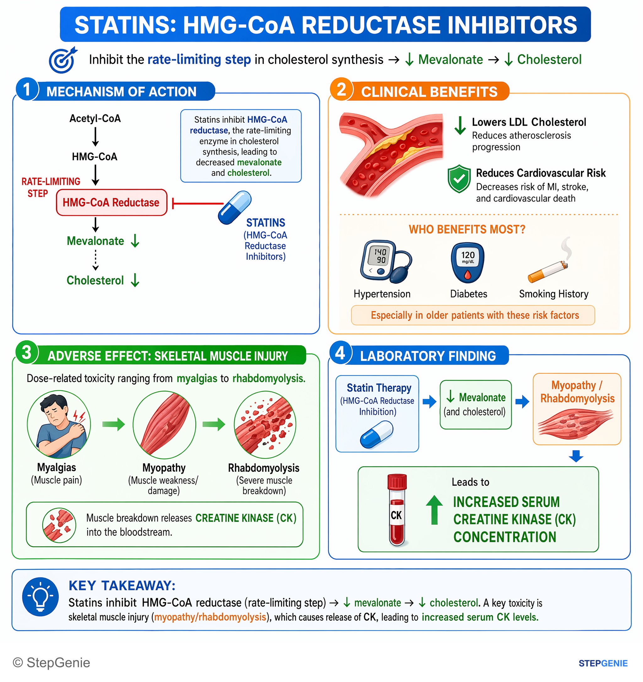
Statin therapy (HMG-CoA reductase inhibition → ↓mevalonate) can cause myopathy/rhabdomyolysis, leading to elevated serum creatine kinase.
USMLE Step 1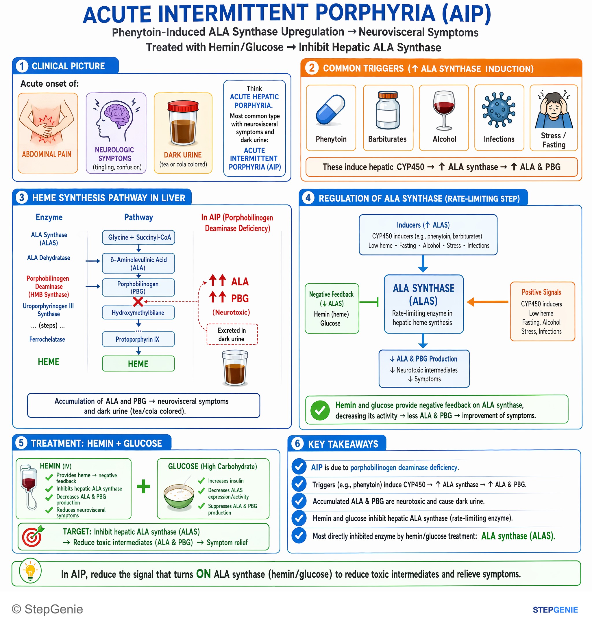
Acute intermittent porphyria: phenytoin-induced ALA synthase upregulation; treated with hemin/glucose, which inhibit hepatic ALA synthase…
USMLE Step 1Frequently Asked Questions
What is the rate-limiting enzyme of the urea cycle?
Carbamoyl phosphate synthetase I (CPS I) catalyzes the first and rate-limiting step of the urea cycle in liver mitochondria, using ammonia, bicarbonate and 2 ATP to make carbamoyl phosphate. It cannot work without its required allosteric activator, N-acetylglutamate, which N-acetylglutamate synthase makes from acetyl-CoA and glutamate; arginine stimulates that synthase. A defect in either enzyme causes hyperammonemia with low citrulline and normal or low orotic acid, and N-acetylglutamate synthase deficiency responds to carglumic acid.
What is the rate-limiting enzyme of ketogenesis?
Mitochondrial HMG-CoA synthase (HMGCS2) catalyzes the committed, rate-limiting step of ketogenesis in liver mitochondria, joining acetoacetyl-CoA and acetyl-CoA to form HMG-CoA, which HMG-CoA lyase splits into acetoacetate and acetyl-CoA. Ketogenesis speeds up when insulin is low and glucagon is high, as in fasting and diabetic ketoacidosis, because lipolysis and an open CPT I flood the liver with acetyl-CoA. Insulin is the main brake. The liver cannot use the ketones it makes because it lacks SCOT (thiophorase).
Is HMG-CoA synthase or HMG-CoA reductase the rate-limiting step?
Both are, in different pathways. Mitochondrial HMG-CoA synthase is rate-limiting for ketogenesis. HMG-CoA reductase, on the endoplasmic reticulum membrane, converts HMG-CoA to mevalonate and is rate-limiting for cholesterol synthesis; insulin, thyroid hormone and estrogen increase it, while cholesterol, glucagon and glucocorticoids decrease it, and statins competitively inhibit it. A separate cytosolic HMG-CoA synthase makes the HMG-CoA used for cholesterol, but its step is not the controlled one.
What are the rate-limiting enzymes of glycolysis, gluconeogenesis and the TCA cycle?
Phosphofructokinase-1 is rate-limiting for glycolysis, fructose-1,6-bisphosphatase for gluconeogenesis and isocitrate dehydrogenase for the TCA cycle. AMP and fructose-2,6-bisphosphate activate PFK-1 and inhibit fructose-1,6-bisphosphatase, while ATP and citrate inhibit PFK-1 and citrate activates the phosphatase. Insulin raises fructose-2,6-bisphosphate and glucagon lowers it, so the two pathways run in opposite directions. Isocitrate dehydrogenase is activated by ADP and calcium and slowed by ATP and NADH.
What is the rate-limiting step of fatty acid synthesis and beta-oxidation?
Acetyl-CoA carboxylase, a biotin-dependent cytosolic enzyme, catalyzes the rate-limiting step of fatty acid synthesis by making malonyl-CoA; citrate and insulin activate it, while palmitoyl-CoA, AMP-activated protein kinase and glucagon inhibit it. Carnitine palmitoyltransferase I, on the outer mitochondrial membrane, is the regulated step of beta-oxidation because it moves long-chain fatty acids into mitochondria. Malonyl-CoA inhibits CPT I, so the liver does not burn fatty acids while it is making them.
What is the rate-limiting enzyme of heme synthesis, and why does it matter in porphyria?
ALA synthase, a mitochondrial enzyme that needs vitamin B6 (pyridoxal phosphate), catalyzes the first and normally rate-controlling step of heme synthesis. Heme turns down the liver form, ALAS1, while CYP450-inducing drugs raise it. In acute intermittent porphyria the deficient enzyme is porphobilinogen deaminase, but attacks begin when drugs, alcohol, fasting or hormonal changes drive ALAS1 up. IV hemin and glucose turn ALAS1 down, and givosiran, an siRNA against ALAS1 mRNA, is approved for adults with acute hepatic porphyria to reduce attacks.