Allosteric regulation is control of a protein by a molecule that binds a site other than the active site (the allosteric, or regulatory, site) and changes the protein's shape, and with it substrate affinity and/or catalytic rate. In the two-state picture, activators (positive effectors) favor the high-affinity R (relaxed) state and inhibitors (negative effectors) favor the low-affinity T (tense) state. When the substrate itself is the effector, the regulation is homotropic, and positive cooperativity gives a sigmoidal curve instead of a Michaelis-Menten hyperbola. When the effector is a different molecule, it is heterotropic. The Step 1 prototypes are PFK-1 (AMP and fructose-2,6-bisphosphate activate; ATP and citrate inhibit) and hemoglobin (2,3-BPG, H⁺ and CO₂ lower its O₂ affinity).
Allosteric regulation vocabulary
| Term | Meaning | Step 1 example |
|---|---|---|
| Allosteric site | Binding site distinct from the active site; binding there changes activity through a conformational change | ATP's regulatory site on PFK-1 |
| Positive effector | Raises substrate affinity and/or catalysis | Fructose-2,6-bisphosphate on PFK-1; N-acetylglutamate on CPS I |
| Negative effector | Lowers substrate affinity and/or catalysis | Citrate on PFK-1; UTP on CPS II |
| Homotropic | The effector is the same ligand that is being bound | O₂ on hemoglobin |
| Heterotropic | A chemically different ligand at its own site | 2,3-BPG on hemoglobin; AMP or citrate on PFK-1 |
| T and R states | Tense (low affinity) and relaxed (high affinity) conformations | Deoxyhemoglobin is the T state |
Allosteric enzyme kinetics: sigmoid curves, K0.5, K and V systems
When the substrate itself acts as an allosteric effector, kinetics deviate from the Michaelis-Menten model, and positive cooperativity gives a sigmoidal rate curve. It is often described with the Hill equation:
A Hill coefficient n > 1 means positive cooperativity. , the substrate concentration at half-maximal velocity, replaces the Michaelis-Menten . Across the steep middle of the curve, modest changes in substrate or effector change the rate sharply.
Two models of the T-to-R switch. In the concerted (Monod-Wyman-Changeux) model, all subunits switch together from T to R. In the sequential (Koshland) model, each subunit changes as it binds ligand and raises its neighbors' affinity. Neither model in pure form fully explains hemoglobin.
K systems vs V systems. Allosteric effectors act on affinity, catalysis or both:
| K system | V system | |
|---|---|---|
| Effector changes | Apparent substrate affinity () | Catalysis (, ) |
| Activator | Lowers : curve shifts left | Raises the plateau |
| Inhibitor | Raises : curve shifts right | Lowers the plateau |
| in a pure system | Unchanged | Changed |
PFK-1's textbook effectors act on affinity (a K-type pattern). Three cautions:
- A sigmoid on its own does not tell you whether a system is K-type or V-type.
- A sigmoid does not prove multiple subunits: glucokinase, a single subunit with one active site, still responds sigmoidally to glucose.
- Shape depends on conditions: a high ATP concentration converts liver PFK-1's hyperbolic curve for fructose 6-phosphate into a sigmoidal one.
Allosteric vs competitive, noncompetitive and uncompetitive inhibition
The classic inhibitor patterns belong to Michaelis-Menten enzymes, whose Lineweaver-Burk plot (1/v against 1/[S]) is a straight line with y-intercept , x-intercept and slope .
| Inhibition | Binds | Apparent | Apparent | Lineweaver-Burk lines | Overcome by more substrate? |
|---|---|---|---|---|---|
| Competitive | Free enzyme, usually at the active site | ↑ (× (1 + [I]/Ki)) | Unchanged | Intersect on the y-axis | Yes |
| Pure noncompetitive | E and ES equally, away from the active site | Unchanged | ↓ | Intersect on the x-axis | No |
| Uncompetitive | ES complex only | ↓ | ↓ (same factor) | Parallel | No; potency rises as substrate builds up |
| Mixed | E and ES, unequal affinity | ↑ or ↓ | ↓ | Intersect off the y-axis | No |
| Allosteric, cooperative enzyme | Regulatory site | ↑ (K system) | ↓ (V system) | Not a straight line | K system: yes, at saturation; V system: no |
A cooperative enzyme breaks the plot because the Hill equation is linear in , not , so read and from the v-versus-[S] curve. Three points examiners exploit:
- "Binds an allosteric site" does not always mean noncompetitive. Depending on what the conformational change does, an allosteric inhibitor can show a competitive, noncompetitive or uncompetitive pattern.
- Pure noncompetitive inhibition is a Michaelis-Menten pattern, not the same thing as tipping a cooperative enzyme between T and R.
- Irreversible, time-dependent and tight-binding inhibitors can all look noncompetitive, whatever their true binding site.
Intercepts, slopes and graph-reading drills are in the Lineweaver-Burk plot guide.
Hemoglobin: the classic allosteric protein, and why myoglobin isn't one
Hemoglobin is not an enzyme, but it is the textbook allosteric protein: a tetramer of 2 α and 2 β subunits, each binding one O₂. Unliganded hemoglobin sits mostly in T; O₂ binding pushes the other subunits toward R, so later O₂ binds more easily and the curve is sigmoidal. Hemoglobin's physiologic heterotropic effectors favor T and shift the curve right:
- 2,3-BPG binds a pocket in the center of the tetramer that exists only in the T form, stabilizing deoxyhemoglobin. Its level rises with chronic hypoxia, including high altitude.
- H⁺ and CO₂ (the Bohr effect): protons favor T, and CO₂ forms carbamates on terminal amino groups that stabilize deoxyhemoglobin. Exercising muscle, rich in CO₂, lactic acid and heat, gets more O₂.
- Higher temperature also favors unloading.
Affinity rises (left shift) with alkalosis or low CO₂ and with three classics. Fetal hemoglobin (α₂γ₂) binds 2,3-BPG less tightly, so its P50 is about 19 mm Hg versus about 27 mm Hg for HbA. Carbon monoxide binds hemoglobin with roughly 200 to 250 times the affinity of O₂ and stabilizes the R state, so the remaining hemes hold on to their O₂ (a left-shifted, less sigmoidal curve) while O₂ content falls. In methemoglobin, Fe³⁺ heme cannot carry O₂ and induces allosteric changes that make the remaining Fe²⁺ sites hold on to their O₂.
| Hemoglobin | Myoglobin | |
|---|---|---|
| Structure | Tetramer (α₂β₂), 4 O₂ sites | Single chain, 1 O₂ site |
| Binding | Cooperative | Noncooperative |
| Curve | Sigmoidal | Hyperbolic (Michaelis-Menten-like) |
| O₂ affinity | Lower | Higher |
Every cause of a shift is covered in the oxygen-hemoglobin dissociation curve.
The high-yield allosterically regulated enzymes
| Pathway | Enzyme | Activators | Inhibitors | Other control |
|---|---|---|---|---|
| Glycolysis | Hexokinase | — | Glucose 6-phosphate (product) | Liver glucokinase is not inhibited by glucose 6-phosphate |
| Glycolysis (rate-limiting) | PFK-1 | AMP, fructose-2,6-bisphosphate | ATP, citrate, low pH | Insulin and glucagon set the F2,6BP level |
| Glycolysis | Pyruvate kinase | Fructose-1,6-bisphosphate | ATP, alanine | Liver form inactivated by PKA phosphorylation |
| Gluconeogenesis | Pyruvate carboxylase | Acetyl-CoA | — | Biotin-dependent |
| Gluconeogenesis (rate-limiting) | Fructose-1,6-bisphosphatase | Citrate | AMP, fructose-2,6-bisphosphate | Mirror image of PFK-1 |
| TCA cycle (rate-limiting) | Isocitrate dehydrogenase | ADP, Ca²⁺ | ATP, NADH | — |
| Glycogen synthesis (rate-limiting) | Glycogen synthase | Glucose 6-phosphate | — | Active when dephosphorylated (insulin, PP1) |
| Glycogenolysis | Glycogen phosphorylase | AMP, IMP, Pi (muscle) | Glucose 6-phosphate, ATP (muscle), UDP-glucose; glucose (liver) | Active when phosphorylated by phosphorylase kinase |
| Fatty acid synthesis | Acetyl-CoA carboxylase | Citrate | Palmitoyl-CoA | AMPK phosphorylation inhibits |
| Urea cycle (rate-limiting) | CPS I (mitochondria) | N-acetylglutamate (obligate) | — | NAG synthase stimulated by arginine |
| Pyrimidines, humans (rate-limiting) | CPS II (CAD, cytosol) | PRPP | UTP | PKA phosphorylation overcomes UTP inhibition |
| Pyrimidines, E. coli | Aspartate transcarbamoylase | ATP | CTP (UTP inhibits only together with CTP) | Textbook model enzyme |
Malonyl-CoA, made by acetyl-CoA carboxylase, inhibits carnitine acyltransferase I (CPT-1), so fatty acids are not made and burned at once. The full list of rate-limiting steps is in rate-limiting enzymes.
PFK-1 and the fructose-2,6-bisphosphate switch
Liver PFK-1 is a tetramer. ATP, its substrate, also binds a separate regulatory site and lowers the affinity for fructose 6-phosphate; AMP reverses this, citrate enhances it and falling pH adds inhibition. Fructose-2,6-bisphosphate raises the affinity and blunts ATP inhibition, and hormones set its level:
- Fed: insulin leaves the bifunctional enzyme dephosphorylated, so its kinase (PFK-2) makes fructose-2,6-bisphosphate, which activates PFK-1 and inhibits fructose-1,6-bisphosphatase: glycolysis.
- Fasting: glucagon raises cAMP and protein kinase A phosphorylates the enzyme. PFK-2 turns off and its phosphatase (FBPase-2) turns on, fructose-2,6-bisphosphate falls, and gluconeogenesis takes over. PKA also inactivates liver pyruvate kinase.
Feedback inhibition, from E. coli to humans
In feedback inhibition, a pathway's end product inhibits the enzyme of its first committed step. E. coli aspartate transcarbamoylase is the textbook case: a 12-chain complex with separable catalytic and regulatory parts, feedback-inhibited by pyrimidine nucleotides and activated by ATP, which helps balance purine and pyrimidine pools. Humans control pyrimidine synthesis at CPS II, part of the cytosolic enzyme CAD (UTP inhibits, PRPP activates). CPS I is a separate mitochondrial enzyme that feeds the urea cycle and requires N-acetylglutamate.
Allosteric control vs covalent modification vs induction
| Mechanism | What changes | Trigger | Example |
|---|---|---|---|
| Allosteric regulation | Activity of existing enzyme, via effector binding | Metabolite levels | Acetyl-CoA activates pyruvate carboxylase |
| Covalent modification | Activity of existing enzyme, most often by adding or removing phosphate | Hormones and energy status, via kinases and phosphatases (PKA, AMPK, PP1) | Phosphorylase is active when phosphorylated, glycogen synthase when dephosphorylated |
| Induction or repression | Amount of enzyme (gene expression) | Hormones, nutrients | Glucagon induces PEPCK |
| Proteolytic activation | An inactive precursor is cut | Location | Enterokinase turns trypsinogen into trypsin, which activates other pancreatic proteases |
The layers interlock: glucose 6-phosphate allosterically activates glycogen synthase and makes it a better substrate for phosphatases. In liver, glucose binding to phosphorylase a shifts it toward the T state, which lets PP1 dephosphorylate and inactivate it. For cleavage-based activation, see zymogen activation.
HMG-CoA reductase catalyzes the rate-limiting, committed step of cholesterol synthesis, yet it is not a classic allosteric control point: the familiar feedback by cholesterol works mainly through SREBP-driven transcription and sterol-accelerated degradation, and the enzyme is also controlled at translation and by phosphorylation.
- Transcription: in cholesterol-depleted cells, SREBPs are cleaved in the Golgi and the released fragment activates the reductase gene; high cholesterol keeps them in the ER.
- Degradation: accumulating sterols make the reductase bind Insig-1 and Insig-2, which recruits the ubiquitin ligase gp78; the tagged enzyme is destroyed by proteasomes.
- Phosphorylation and hormones: AMPK phosphorylation inactivates it; insulin and thyroxine upregulate it and glucagon inhibits it.
- Statins are reversible competitive inhibitors that resemble HMG-CoA, so apparent rises with unchanged; the fall in liver cholesterol activates SREBP and adds LDL receptors.
Drugs that work allosterically
| Drug | Target | Allosteric action | Exam hook |
|---|---|---|---|
| Benzodiazepines | GABA-A receptor, at the α/γ interface (GABA binds α/β) | Positive modulator: ↑ frequency of Cl⁻ channel opening; no effect without GABA | Zolpidem uses the same site; flumazenil competitively blocks it and can provoke seizures |
| Barbiturates | GABA-A receptor | Positive modulator: ↑ duration of channel opening | More respiratory depression than benzodiazepines |
| Cinacalcet | Calcium-sensing receptor | Positive modulator: ↑ sensitivity to extracellular Ca²⁺, so PTH falls | Watch for hypocalcemia; labeled for secondary hyperparathyroidism on dialysis and for hypercalcemia in parathyroid carcinoma or in primary hyperparathyroidism when parathyroidectomy is not possible (IV etelcalcetide allosterically modulates the same receptor) |
| Maraviroc | CCR5 co-receptor | Binds a transmembrane cavity and blocks gp120 binding allosterically | CCR5-tropic HIV-1 only; test tropism first |
| NNRTIs (efavirenz, nevirapine) | HIV-1 reverse transcriptase | Bind a hydrophobic pocket near, not at, the active site; noncompetitive | Not active against HIV-2 |
| Mavacamten | Cardiac myosin | Allosteric, reversible myosin inhibitor | Symptomatic obstructive hypertrophic cardiomyopathy |
Classic exam traps
- Allosteric is not a synonym for noncompetitive, and cannot be read off a sigmoid: use .
- Sigmoid does not equal oligomer. Glucokinase is monomeric yet sigmoidal; PFK-1's curve is hyperbolic at low ATP.
- Hemoglobin is allosteric but not an enzyme; myoglobin is a single chain with a hyperbolic curve.
- Insulin and glucagon are not allosteric effectors. Through receptors and second messengers they change phosphorylation, effector levels (fructose-2,6-bisphosphate) and gene expression.
- CPS I vs CPS II. N-acetylglutamate activates CPS I (urea cycle); UTP and PRPP regulate CPS II, the human pyrimidine control point (ATCase is the bacterial model).
- Statins are competitive inhibitors, not allosteric ones.
Summary: stem clue to answer
| Stem clue | Think |
|---|---|
| Sigmoidal velocity curve; "not Michaelis-Menten" | Cooperative allosteric enzyme; use |
| Sigmoid shifts left, same plateau | K-type activator |
| Plateau falls, midpoint unchanged | V-type inhibitor |
| Lineweaver-Burk lines meet on the y-axis / x-axis / never | Competitive / pure noncompetitive / uncompetitive |
| Right-shifted O₂ curve after days at altitude | More 2,3-BPG stabilizing T |
| Neonatal hyperammonemia; CPS I works once NAG is added | NAG synthase deficiency: carglumic acid |
| "Increases the frequency of Cl⁻ channel opening" | Benzodiazepine |
Worked examples
Vignette 1. A 3-day-old girl becomes lethargic and stops feeding. She is tachypneic with a respiratory alkalosis and markedly elevated plasma ammonia; urine orotic acid is not increased. Her hepatic carbamoyl phosphate synthetase I is fully active once N-acetylglutamate is added to the assay. Which drug most directly restores the blocked step?
Answer: Carglumic acid.
One-line reasoning: N-acetylglutamate synthase deficiency leaves CPS I without its obligate allosteric activator, and carglumic acid is a synthetic analog that activates CPS I; normal orotic acid argues against ornithine transcarbamylase deficiency.
Vignette 2. PFK-1 activity in liver extract is measured across fructose 6-phosphate concentrations with high ATP present, giving a sigmoidal curve. Adding fructose-2,6-bisphosphate shifts the curve left. Why?
- A. It competes with fructose 6-phosphate at the active site
- B. It binds an allosteric site, raising affinity for fructose 6-phosphate and blunting ATP inhibition
- C. It phosphorylates PFK-1
- D. It increases the amount of PFK-1 protein
Answer: B.
One-line reasoning: A left shift means a lower , the signature of a K-type activator; a competitive inhibitor would shift the curve right, and more enzyme would raise the plateau.
Key takeaways
- Allosteric effectors bind away from the active site and shift the T/R balance; cooperative enzymes give sigmoids, not Michaelis-Menten hyperbolas.
- Use , not ; K-type effectors move and V-type effectors move .
- PFK-1: AMP and fructose-2,6-bisphosphate activate; ATP, citrate and low pH inhibit. Insulin and glucagon set fructose-2,6-bisphosphate through the phosphorylation state of PFK-2/FBPase-2.
- Also know: acetyl-CoA activates pyruvate carboxylase, citrate activates acetyl-CoA carboxylase, malonyl-CoA inhibits CPT-1, N-acetylglutamate activates CPS I and UTP inhibits CPS II.
- HMG-CoA reductase is controlled through SREBP, degradation and AMPK; statins inhibit it competitively.
More enzyme and metabolism reviews are collected on the biochemistry topic hub.
Sources
- McCullagh M, et al. What is allosteric regulation? J Biol Chem. 2024 (PMC)
- Wang Y, et al. Enzyme kinetics by isothermal titration calorimetry. Front Mol Biosci. 2020 (PMC)
- Biochemistry, Proteins Enzymes (StatPearls, NCBI Bookshelf)
- Strelow J, et al. Mechanism of Action Assays for Enzymes. Assay Guidance Manual (NCBI Bookshelf)
- Berg JM, et al. Biochemistry, 5th ed. Section 8.5 (NCBI Bookshelf)
- Berg JM, et al. Biochemistry, 5th ed. Section 10.2 (NCBI Bookshelf)
- Berg JM, et al. Biochemistry, 5th ed. Section 16.2 (NCBI Bookshelf)
- Physiology, Oxyhemoglobin Dissociation Curve (StatPearls, NCBI Bookshelf)
- Physiology, Bohr Effect (StatPearls, NCBI Bookshelf)
- Biochemistry, Myoglobin (StatPearls, NCBI Bookshelf)
- Physiology, Fetal Hemoglobin (StatPearls, NCBI Bookshelf)
- Carbon Monoxide Poisoning (StatPearls, NCBI Bookshelf)
- Methemoglobinemia (StatPearls, NCBI Bookshelf)
- Biochemistry, Glycolysis (StatPearls, NCBI Bookshelf)
- Biochemistry, Gluconeogenesis (StatPearls, NCBI Bookshelf)
- Biochemistry, Citric Acid Cycle (StatPearls, NCBI Bookshelf)
- Physiology, Krebs Cycle (StatPearls, NCBI Bookshelf)
- Biochemistry, Glycogen (StatPearls, NCBI Bookshelf)
- Adeva-Andany MM, et al. Glycogen metabolism in humans. BBA Clin. 2016 (PMC)
- Tymoczko JL, Berg JM, Stryer L. Biochemistry: A Short Course, 3rd ed. Section 24.2 (Macmillan)
- Zu X, et al. Chemical genetics of acetyl-CoA carboxylases. Molecules. 2013 (PMC)
- Physiology, Urea Cycle (StatPearls, NCBI Bookshelf)
- Urea Cycle Disorders (StatPearls, NCBI Bookshelf)
- Carbaglu (carglumic acid) label (DailyMed)
- del Caño-Ochoa F, Ramón-Maiques S. Deciphering CAD. Protein Sci. 2021 (PMC)
- Kantrowitz ER. Allostery and cooperativity in E. coli aspartate transcarbamoylase. Arch Biochem Biophys. 2012 (PMC)
- Physiology, Digestion (StatPearls, NCBI Bookshelf)
- Biochemistry, Cholesterol (StatPearls, NCBI Bookshelf)
- DeBose-Boyd RA. Sterol-accelerated ubiquitination and degradation of HMG CoA reductase. Cell Res. 2008 (PubMed)
- Burg JS, Espenshade PJ. Regulation of HMG-CoA reductase in mammals and yeast. Prog Lipid Res. 2011 (PubMed)
- Berg JM, Tymoczko JL, Gatto GJ, Stryer L. Biochemistry, 8th ed. Section 26.3 (Macmillan)
- GABA Receptor Positive Allosteric Modulators (StatPearls, NCBI Bookshelf)
- Benzodiazepines (StatPearls, NCBI Bookshelf)
- GABA Receptor (StatPearls, NCBI Bookshelf)
- Sensipar (cinacalcet) label (DailyMed)
- Parsabiv (etelcalcetide) label (DailyMed)
- Garcia-Perez J, et al. Allosteric model of maraviroc binding to CCR5. J Biol Chem. 2011 (PubMed)
- Selzentry (maraviroc) label (DailyMed)
- Reverse Transcriptase Inhibitors (StatPearls, NCBI Bookshelf)
- Camzyos (mavacamten) label (DailyMed)
Visual explanations from the StepGenie QBank
Infographics StepGenie drew to explain questions in its own QBank, matched to this article's topic. Each caption is the takeaway of the question it explains.
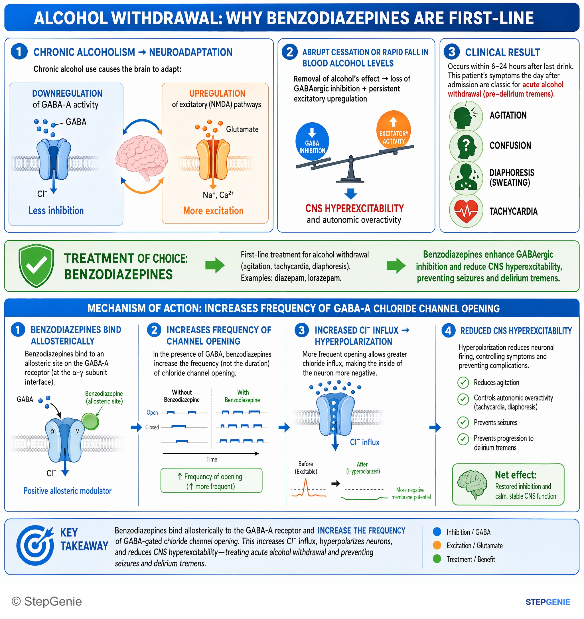
Alcohol withdrawal (agitation, tachycardia, diaphoresis) is treated with benzodiazepines, which increase frequency of GABA-A chloride…
USMLE Step 1Frequently Asked Questions
What is allosteric regulation?
Allosteric regulation is control of an enzyme or other protein by a molecule that binds a regulatory site distinct from the active site. Binding changes the protein's shape and, with it, its affinity for substrate and/or its catalytic rate. In the two-state model, activators favor the high-affinity R state and inhibitors the low-affinity T state. Classic Step 1 examples are PFK-1, activated by AMP and fructose-2,6-bisphosphate and inhibited by ATP and citrate, and hemoglobin, whose O2 affinity is lowered by 2,3-BPG, H+ and CO2.
What is the difference between allosteric inhibition and competitive inhibition?
A competitive inhibitor binds the free enzyme, usually at the active site, so enough substrate overcomes it: apparent Km rises, Vmax is unchanged and Lineweaver-Burk lines meet on the y-axis. An allosteric inhibitor binds a separate regulatory site and changes the enzyme's shape. On a cooperative enzyme it raises K0.5 (a K-type effect) or lowers Vmax (a V-type effect). Depending on the shape change, an allosteric inhibitor can even show a competitive, noncompetitive or uncompetitive pattern.
What is the difference between homotropic and heterotropic allosteric regulation?
In homotropic regulation the effector is the same molecule as the ligand being bound, so binding at one site changes affinity at the others; positive homotropic cooperativity, as when O2 binds hemoglobin, produces a sigmoidal curve. In heterotropic regulation a chemically different molecule binds its own site, such as 2,3-BPG, H+ and CO2 on hemoglobin or AMP, citrate and fructose-2,6-bisphosphate on PFK-1. Heterotropic effectors shift the curve or change its maximum.
Why do allosteric enzymes show sigmoidal kinetics instead of Michaelis-Menten kinetics?
When the substrate itself acts as an allosteric effector, binding at one subunit raises the affinity of the others, so the rate rises slowly at low substrate, then steeply, then plateaus. The Hill equation describes this, and a Hill coefficient above 1 means positive cooperativity. These enzymes are described by K0.5, the substrate concentration at half-maximal velocity, rather than Km, and their Lineweaver-Burk plots are not straight lines. A sigmoid does not require several subunits: monomeric glucokinase is sigmoidal too.
What are the allosteric regulators of PFK-1?
PFK-1, the rate-limiting enzyme of glycolysis, is activated by AMP, which reverses ATP inhibition, and by fructose-2,6-bisphosphate, which raises its affinity for fructose 6-phosphate. It is inhibited by ATP acting at a regulatory site, by citrate, which enhances the ATP effect, and by low pH. In the liver, insulin raises fructose-2,6-bisphosphate to favor glycolysis, while glucagon, through PKA phosphorylation of the bifunctional PFK-2/FBPase-2 enzyme, lowers it to favor gluconeogenesis.
Is hemoglobin an allosteric enzyme?
Hemoglobin is an allosteric protein but not an enzyme, because it carries oxygen rather than catalyzing a reaction. Its four subunits bind O2 cooperatively, shifting from the low-affinity T state toward the high-affinity R state, which gives a sigmoidal curve. 2,3-BPG, H+ and CO2 stabilize the T state and shift the curve right. Myoglobin is a single chain with one O2 site, so it binds O2 noncooperatively, has a hyperbolic curve and has higher O2 affinity.