Adenosine deaminase (ADA) deficiency is an autosomal recessive disorder of purine breakdown that causes severe combined immunodeficiency (SCID) with low T, B and NK cells. ADA normally deaminates adenosine to inosine and deoxyadenosine to deoxyinosine; without it, deoxyadenosine is phosphorylated to dATP, which inhibits ribonucleotide reductase, so developing lymphocytes cannot make or repair DNA and die. About 80% of patients present in infancy with SCID, ADA deficiency accounts for about 15% of all SCID, and treatment is hematopoietic stem cell transplantation (HSCT), enzyme replacement with PEGylated ADA, or gene therapy.
Mechanism: one missing enzyme, two toxic effects
ADA works in the purine breakdown pathway:
- Adenosine → inosine and 2′-deoxyadenosine → 2′-deoxyinosine.
- The next enzyme, purine nucleoside phosphorylase (PNP), turns deoxyinosine into hypoxanthine.
When ADA is missing:
ADA ↓ → deoxyadenosine ↑ → phosphorylated to dATP ↑ → ribonucleotide reductase inhibited → too few deoxyribonucleotides → DNA synthesis and repair fail → lymphocytes die.
Deoxyadenosine also inactivates S-adenosylhomocysteine (SAH) hydrolase, which blocks the methylation reactions lymphocytes need for activation. Lymphocytes take the biggest hit: ADA levels are highest in lymphoid tissue, particularly the thymus, and immature lymphocytes in the thymus are especially vulnerable to deoxyadenosine. But ADA is present in every cell, which is why the disease also has non-immune features. To place ADA among the purine pathways, see de novo vs salvage nucleotide synthesis.
How it presents
ADA-SCID (about 80%) typically starts within the first 6 months of life:
- Infections: thrush and systemic candidiasis, persistent viral infections, Pneumocystis jirovecii pneumonia and recurrent pneumonia.
- Gut and skin: persistent diarrhea and extensive dermatitis, leading to failure to thrive.
- Lymphoid tissue: a very small thymus (no thymic shadow on chest x-ray) and decreased or absent lymphoid tissue.
- Graft-versus-host disease can come from maternal lymphocytes or from a transfusion of non-irradiated blood.
- Without treatment, affected babies usually do not survive past age 2.
ADA-specific, non-immune clues (because ADA is in every cell):
- Skeletal: costochondral cupping (flared, cupped rib ends) and scapular spurring or squaring.
- Sensorineural hearing loss and neurologic or behavioral abnormalities.
- Abnormal liver tests, pulmonary alveolar proteinosis and hemolytic anemia.
- Dermatofibrosarcoma protuberans, a rare skin cancer, has been reported in several patients.
Delayed- or late-onset disease (15–20%) begins at 1–10 years of age or in adulthood as a milder combined immunodeficiency (ADA-CID), typically with recurrent upper respiratory and ear infections.
Diagnosis
| Test | Result in ADA deficiency |
|---|---|
| Newborn screen: TREC PCR on a dried blood spot | Low or absent T-cell receptor excision circles flag T-cell lymphopenia; milder delayed-onset cases can be missed |
| Lymphocyte subsets | T, B and NK cells all markedly reduced (T−B−NK−) |
| ADA enzyme activity | Less than 1% of normal in red cells in ADA-SCID |
| Red cell dATP or total deoxyadenosine nucleotides (dAXP) | Markedly elevated; with low ADA activity, establishes the diagnosis faster than gene testing |
| Adenosine and deoxyadenosine (tandem mass spectrometry) | Elevated |
| Molecular testing | Biallelic pathogenic variants in ADA (chromosome 20q13) |
Treatment
- HSCT. A transplant from an HLA-identical healthy sibling is the method of choice for all forms of SCID; survival with matched sibling or family donors is above 80%.
- Enzyme replacement therapy. PEGylated ADA is injected intramuscularly, typically once or twice a week. It is started soon after diagnosis to stabilize the patient while a transplant or gene therapy is planned, and continued in patients who receive neither. Elapegademase-lvlr (Revcovi), a recombinant PEGylated ADA based on the bovine enzyme and first approved by the FDA in 2018, has replaced the older product (Adagen).
- Gene therapy. The patient's own CD34+ cells are given a working ADA gene with a gammaretroviral vector (Strimvelis), authorized in the European Union in 2016 for patients without a suitable matched related donor.
- Supportive care (prevents infections but doesn't cure): immune globulin replacement, antibiotics including Pneumocystis prophylaxis, and antifungals; no live vaccines; blood products only if irradiated, leukoreduced and CMV-negative.
ADA-SCID vs look-alikes
| Disorder | Defect and inheritance | Lymphocytes | What separates it |
|---|---|---|---|
| ADA deficiency | ADA; autosomal recessive; dATP toxicity | T−B−NK− | Rib cupping, scapular changes, hearing loss, neurologic and liver problems |
| X-linked SCID | IL2RG (IL-2 receptor common gamma chain); X-linked recessive | T−B+NK− | Boys; the most common SCID, one-quarter to one-third of cases |
| PNP deficiency | PNP; autosomal recessive; dGTP toxic to T cells | Low T cells; B cells low in some | Neurologic problems in about two-thirds (ataxia, spasticity, developmental delay) and autoimmunity; about 4% of SCID |
| DiGeorge syndrome (22q11.2 deletion) | Maldevelopment of pharyngeal pouch structures (thymus, parathyroids); usually not inherited | T-cell deficiency, partial or complete | Hypocalcemic tetany in the first 24–48 hours, conotruncal heart defects, facial features |
For the full SCID picture, see the SCID visual hack; the thymic-aplasia look-alike is covered in the DiGeorge syndrome overview.
Exam traps
- Excess, not deficient, dATP. The toxic metabolite is dATP and the blocked enzyme is ribonucleotide reductase; in PNP deficiency the toxic nucleotide is dGTP.
- B cells present? Think X-linked SCID (IL2RG); ADA-SCID is T−B−NK−.
- Flared, cupped rib ends on the chest film of an infant with SCID point to ADA deficiency.
- Neurologic signs plus autoimmunity with T-cell lymphopenia suggest PNP deficiency; ADA deficiency can affect the nervous system too, but absent B and NK cells and cupped ribs favor ADA.
- Hypocalcemia plus a conotruncal heart defect is a 22q11.2 deletion, not SCID.
- No live vaccines, and only irradiated blood; non-irradiated blood can cause graft-versus-host disease.
- Not only babies: 15–20% of patients present later in childhood or as adults.
Worked example
Vignette. A 3-month-old girl born to first-cousin parents has had watery diarrhea since 6 weeks of age, oral thrush that returns after every course of nystatin and a diffuse scaly rash. Her weight has fallen below the 3rd percentile. She is now hypoxemic with bilateral interstitial infiltrates, and bronchoalveolar lavage shows Pneumocystis jirovecii. The chest x-ray shows no thymic shadow and cupped, flared anterior rib ends. Flow cytometry shows very low T, B and NK cells. Accumulation of which metabolite best explains her immunodeficiency?
Answer: dATP, formed from accumulated deoxyadenosine. It inhibits ribonucleotide reductase and starves developing lymphocytes of deoxyribonucleotides: ADA deficiency.
Why the distractors fail:
- dGTP (PNP deficiency): mainly low T cells, often with neurologic problems; absent B and NK cells and rib cupping fit ADA instead.
- A defective IL-2 receptor common gamma chain (X-linked SCID): B cells would be present, and the X-linked recessive disease is typically seen in boys.
- A 22q11.2 deletion: thymic and parathyroid hypoplasia cause a T-cell deficiency with hypocalcemia, often with a conotruncal heart defect; it doesn't explain absent B and NK cells or cupped ribs.
Key takeaways
- ADA deficiency: autosomal recessive; ADA on 20q13; adenosine → inosine and deoxyadenosine → deoxyinosine are blocked.
- Excess dATP inhibits ribonucleotide reductase, so DNA synthesis fails and lymphocytes die; deoxyadenosine also inhibits SAH hydrolase.
- T−B−NK− SCID in about 80%, usually in the first months of life; 15–20% present later.
- Non-immune clues: costochondral cupping, hearing loss, neurologic and liver abnormalities, pulmonary alveolar proteinosis.
- Diagnosis: low TRECs → lymphocyte subsets → ADA activity under 1% and high red cell dATP → ADA gene testing.
- Treatment: HSCT (HLA-identical sibling first), PEG-ADA enzyme replacement (elapegademase), gene therapy; no live vaccines; irradiated blood.
Sources
- Adenosine deaminase deficiency — MedlinePlus Genetics
- Severe Combined Immunodeficiency (SCID) — Merck Manual Professional Edition
- Secord E, Hartog NL. Review of Treatment for Adenosine Deaminase Deficiency (ADA) Severe Combined Immunodeficiency (SCID) — Therapeutics and Clinical Risk Management (2022)
- Alsukhon J, et al. Narrative review of severe combined immunodeficiency—a purine metabolism disorder — Pediatric Medicine (2018)
- Dąbrowska-Leonik N, et al. National experience with adenosine deaminase deficiency related SCID in Polish children — Frontiers in Immunology (2023)
- Strimvelis: EPAR — European Medicines Agency
- Revcovi (elapegademase-lvlr) prescribing information — U.S. Food and Drug Administration (2018)
- X-linked severe combined immunodeficiency — MedlinePlus Genetics
- Purine nucleoside phosphorylase deficiency — MedlinePlus Genetics
- PNP gene — MedlinePlus Genetics
- DiGeorge Syndrome — Merck Manual Professional Edition
- 22q11.2 deletion syndrome — MedlinePlus Genetics
Visual explanations from the StepGenie QBank
Infographics StepGenie drew to explain questions in its own QBank, matched to this article's topic. Each caption is the takeaway of the question it explains.
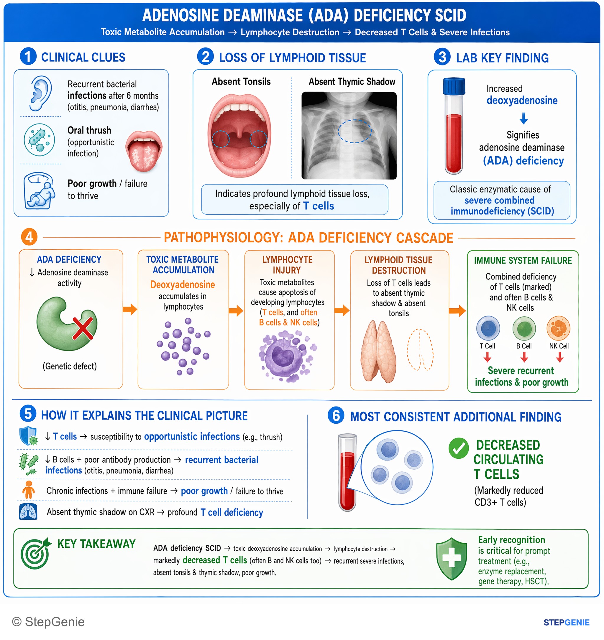
Adenosine deaminase–deficiency SCID → toxic deoxyadenosine accumulation → lymphocyte destruction → markedly decreased T cells (often B and…
USMLE Step 1Frequently Asked Questions
Why does adenosine deaminase deficiency cause SCID?
Without ADA, deoxyadenosine is not converted to deoxyinosine, so it builds up and is phosphorylated to dATP. Excess dATP inhibits ribonucleotide reductase, the enzyme that supplies deoxyribonucleotides, so lymphocytes cannot synthesize or repair DNA. Deoxyadenosine also inactivates S-adenosylhomocysteine hydrolase. ADA is most highly expressed in lymphocytes and immature thymocytes are especially vulnerable, so T, B and NK cells are all lost.
How is ADA-SCID different from X-linked SCID?
ADA-SCID is autosomal recessive and removes T, B and NK cells (T−B−NK−); because ADA is present in every cell, it can also cause rib cupping, hearing loss and neurologic or liver problems. X-linked SCID is caused by variants in IL2RG, which encodes the IL-2 receptor common gamma chain. It affects boys, has a T−B+NK− pattern with B cells present, and is the most common form of SCID.
How is ADA deficiency diagnosed?
Many infants are flagged by newborn screening that finds low or absent T-cell receptor excision circles (TRECs). Lymphocyte subsets then show low T, B and NK cells. ADA activity below 1% of normal in red cells plus markedly elevated red cell deoxyadenosine nucleotides (dATP) establishes the diagnosis faster than gene testing, which then finds biallelic pathogenic variants in the ADA gene.
What is the treatment for ADA deficiency?
Hematopoietic stem cell transplantation from an HLA-identical sibling is the preferred treatment for all forms of SCID. Enzyme replacement with PEGylated ADA injections (elapegademase-lvlr, which replaced the older Adagen) is started at diagnosis as a bridge and continued in patients who are not transplanted. Gene therapy with the patient's own corrected stem cells (Strimvelis in the European Union) is another option. Supportive care includes infection prophylaxis, immune globulin, no live vaccines and only irradiated blood products.
What non-immune problems occur in ADA deficiency?
Because ADA is expressed in all cells, patients can have skeletal changes such as costochondral cupping and scapular spurring, sensorineural hearing loss, neurologic or behavioral abnormalities, abnormal liver tests, pulmonary alveolar proteinosis and hemolytic anemia. Dermatofibrosarcoma protuberans, a rare skin cancer, has also been reported. On a board question, flared rib ends in an infant with SCID are the classic hint that ADA deficiency is the cause.