Fat-soluble vitamins are classic Step 1 “gotcha” territory: they’re absorbed with dietary fat, stored in the body (so toxicity is real), and deficiencies show up in predictable organ systems (eyes, bones, nerves, coagulation). If you can tie each vitamin to its key biochemical role → risk factors for deficiency → hallmark symptoms → best test → treatment/toxicity, you’ll crush a whole category of questions.
Big Picture: What Makes Vitamins A, D, E, K “Fat-Soluble”?
Core concept: Fat-soluble vitamins require bile acids and pancreatic enzymes for absorption in the small intestine and are transported in chylomicrons.
High-yield implications
- Deficiency risks increase with fat malabsorption, including:
- Cholestasis/obstructive jaundice (no bile → no micelles)
- Pancreatic insufficiency (e.g., cystic fibrosis, chronic pancreatitis)
- Celiac disease, Crohn disease (esp. ileal)
- Bariatric surgery (esp. bypass procedures)
- Orlistat (fat absorption inhibitor)
- Toxicity is more likely than with water-soluble vitamins because these are stored (liver/adipose).
Quick absorption & storage table (Step-friendly)
| Vitamin | Absorption needs | Stored where? | Toxicity risk? |
|---|---|---|---|
| A | Fat + bile | Liver | Yes |
| D | Fat + bile | Fat/liver | Yes |
| E | Fat + bile | Adipose | Uncommon but possible |
| K | Fat + bile (plus gut flora) | Limited storage | Toxicity rare; deficiency common in newborns |
First Aid cross-ref: Biochemistry → Vitamins; GI → Malabsorption; Heme/Onc → Coagulation pathways (vit K); MSK/Endo → Bone metabolism (vit D).
Vitamin A (Retinol): Vision, Epithelium, Immunity
Definition & functions
Vitamin A includes:
- Retinol (storage/transport form)
- Retinal (vision—rhodopsin)
- Retinoic acid (gene transcription, epithelial differentiation)
High-yield roles
- Vision (especially night vision): 11-cis-retinal combines with opsin → rhodopsin
- Epithelial differentiation: maintains mucosal surfaces (resp/GI/GU)
- Immune function: supports barrier + immune signaling
Pathophysiology of deficiency
Low vitamin A → impaired rhodopsin regeneration + impaired epithelial differentiation → xerosis and keratinization, higher infection risk.
Clinical presentation (classic Step findings)
- Night blindness (nyctalopia) = early clue
- Xerophthalmia (dry eyes), Bitot spots (foamy plaques on conjunctiva)
- Keratomalacia (corneal softening) → blindness
- Follicular hyperkeratosis (“gooseflesh” skin)
- Increased susceptibility to infections (esp. measles severity)
Diagnosis (what NBME likes)
- Often clinical in the right context (malnutrition, malabsorption)
- Low serum retinol may support diagnosis (not always needed for exam logic)
Treatment
- Vitamin A supplementation
- Address underlying cause (e.g., treat cholestasis/malabsorption)
- In measles: vitamin A supplementation reduces morbidity (a common board association)
Toxicity (very high yield)
Usually from supplements (not food).
- Acute toxicity: nausea, vomiting, vertigo, blurred vision, increased ICP
- Chronic toxicity:
- Hepatotoxicity
- Alopecia, dry skin
- Bone pain
- Pseudotumor cerebri (increased ICP)
- Teratogenicity: retinoids (e.g., isotretinoin) → severe fetal defects
First Aid cross-ref: Derm → Retinoids (isotretinoin); Biochem → Vitamin A deficiency/toxicity; Neuro → Increased intracranial pressure.
HY association: Zinc deficiency can mimic vitamin A issues because zinc is needed for retinol-binding protein synthesis and retinal metabolism (Step 1 loves “indirect deficiency”).
Vitamin D (Cholecalciferol/Ergocalciferol): Calcium & Phosphate Homeostasis
Definition & synthesis pathway (must-know)
Vitamin D sources:
- D3 (cholecalciferol): skin (UVB converts 7-dehydrocholesterol) + animal diet
- D2 (ergocalciferol): plant sources
Activation steps:
- Liver: vitamin D → 25-hydroxyvitamin D (calcidiol)
- Kidney (proximal tubule): 25-OH D → 1,25-dihydroxyvitamin D (calcitriol) via 1α-hydroxylase
Regulation of 1α-hydroxylase
- Upregulated by: PTH, low phosphate
- Downregulated by: high phosphate, high calcium, FGF23
Physiology (what it does)
Calcitriol increases:
- Intestinal absorption of Ca²⁺ and PO₄³⁻
- Bone mineralization (at physiologic levels)
- Can increase bone resorption indirectly when needed (via RANKL signaling in severe deficiency/high PTH states)
Deficiency: pathophysiology
Low vitamin D → decreased Ca/PO₄ absorption → secondary hyperparathyroidism → bone demineralization.
Common causes
- Low sunlight exposure
- Darker skin (reduced UVB penetration)
- Malabsorption (cholestasis, CF)
- Chronic kidney disease (↓ 1α-hydroxylation)
- Liver disease (↓ 25-hydroxylation)
Clinical presentation
- Rickets (children):
- Delayed closure of fontanelles
- Bowed legs
- Rachitic rosary (costochondral swelling)
- Osteomalacia (adults):
- Bone pain, fragility fractures
- Proximal muscle weakness
Diagnosis (exam-style)
- Best screening marker of vitamin D stores: 25-hydroxyvitamin D
- Expected labs in deficiency (classic pattern):
- ↓ 25-OH D
- ↓ or normal Ca²⁺ (often low-normal early)
- ↓ phosphate
- ↑ PTH
- ↑ alkaline phosphatase
Treatment
- Vitamin D supplementation (D2 or D3)
- Add calcium if needed
- In CKD: consider calcitriol (active form) or analogs, plus phosphate management
Toxicity (boards love the consequence)
Usually from excessive supplementation:
- Hypercalcemia → stones, bones, groans, psychiatric overtones
- Polyuria, dehydration
- Can cause soft tissue calcification
First Aid cross-ref: Renal → CKD mineral bone disease; Endo → PTH regulation; MSK → Rickets/osteomalacia.
Vitamin E (α-Tocopherol): Antioxidant & RBC/Neuronal Protection
Definition & function
Vitamin E is a major antioxidant that protects cell membranes from oxidative damage by preventing lipid peroxidation.
Where it matters most
- RBC membranes (prevent hemolysis)
- Neurons (long axons are sensitive to oxidative injury)
Deficiency: pathophysiology
Often due to fat malabsorption or impaired lipoprotein assembly/transport:
- Cystic fibrosis (pancreatic insufficiency)
- Cholestasis
- Abetalipoproteinemia (can’t form ApoB-containing lipoproteins → can’t transport fat-soluble vitamins)
Clinical presentation (high yield)
- Hemolytic anemia (especially in premature infants)
- Acanthocytosis (spur cells) classically in abetalipoproteinemia
- Neurologic deficits:
- Peripheral neuropathy
- Ataxia
- Loss of vibration/proprioception (posterior columns)
Diagnosis
- Clinical + risk factors; low serum α-tocopherol may be used
- Hemolysis markers if anemia present (↑ LDH, ↑ indirect bilirubin, ↓ haptoglobin)
Treatment
- Vitamin E supplementation
- Treat the underlying malabsorption issue
First Aid cross-ref: Biochem → Vitamin E deficiency; GI → Abetalipoproteinemia; Heme → Hemolytic anemia patterns.
HY association: Abetalipoproteinemia presents with fat malabsorption, steatorrhea, failure to thrive, neurologic symptoms, acanthocytes, and deficiencies of A, D, E, K—vitamin E-related neuro findings often stand out.
Vitamin K: γ-Carboxylation for Clotting
Definition & function
Vitamin K is a cofactor for γ-glutamyl carboxylase, which γ-carboxylates glutamate residues on clotting proteins, enabling them to bind calcium and function properly.
Vitamin K–dependent proteins
- II, VII, IX, X
- Protein C and Protein S
Pathophysiology of deficiency
Without vitamin K → impaired activation of clotting factors → bleeding tendency.
Who gets deficient? (classic Step scenarios)
- Newborns (sterile gut + low vitamin K in breast milk) → hemorrhagic disease of the newborn
- Broad-spectrum antibiotics (wipe out gut flora)
- Fat malabsorption (cholestasis, CF)
- Warfarin therapy (vitamin K antagonist)
Clinical presentation
- Easy bruising, mucosal bleeding, GI bleeding
- Oozing from venipuncture sites
- In newborns: intracranial hemorrhage risk
Diagnosis: the coag labs you must interpret
- Increased PT/INR (factor VII has the shortest half-life → PT rises first)
- If severe: can also prolong aPTT (due to IX, X, II)
- Platelets are typically normal (this is not thrombocytopenia)
PT vs PTT high-yield table
| Problem | PT | PTT | Platelets | Classic clue |
|---|---|---|---|---|
| Vitamin K deficiency / warfarin | ↑ | normal or ↑ | Normal | Antibiotics, newborn, cholestasis |
| Hemophilia A/B | Normal | ↑ | Normal | Hemarthroses |
| DIC | ↑ | ↑ | ↓ | Schistocytes, sick patient |
Treatment
- Vitamin K (phytonadione) for deficiency
- In serious bleeding or warfarin-associated major bleed: PCC (prothrombin complex concentrate) ± FFP, plus vitamin K
- Newborn prophylaxis: vitamin K injection at birth
Warfarin tie-in (Step 1 classic)
- Warfarin inhibits vitamin K epoxide reductase → decreases activation of II, VII, IX, X, C, S
- Early warfarin therapy can cause transient hypercoagulability due to Protein C drop → “warfarin skin necrosis” (heparin bridge in high-risk patients)
First Aid cross-ref: Pharm → Anticoagulants (warfarin, heparin, PCC/FFP); Biochem → Vitamin K; Peds → Newborn prophylaxis.
Putting It Together: Rapid Fire High-Yield Comparison
| Vitamin | Main job | Deficiency clue | Toxicity clue | Classic association |
|---|---|---|---|---|
| A | Vision, epithelial differentiation | Night blindness, xerophthalmia, Bitot spots | Teratogenicity, hepatotoxicity, ↑ ICP | Isotretinoin effects |
| D | ↑ Ca/PO₄ absorption | Rickets/osteomalacia, ↑ PTH, ↑ ALP | Hypercalcemia, stones | CKD → low calcitriol |
| E | Antioxidant | Hemolytic anemia + neuro deficits | Rare; bleeding risk at very high doses | Abetalipoproteinemia, CF |
| K | γ-carboxylation of II, VII, IX, X, C, S | ↑ PT/INR, bleeding | Rare | Newborns, antibiotics, warfarin |
Test-Day Patterns (How NBME Writes These)
Pattern 1: Cholestasis → multiple deficiencies
A patient with pale stools, dark urine, pruritus → cholestasis → can’t absorb fat → deficiencies in A, D, E, K:
- Night blindness (A)
- Bone pain/fractures (D)
- Ataxia/hemolysis (E)
- Easy bruising and ↑ PT (K)
Pattern 2: Broad-spectrum antibiotics → bleeding
Antibiotics wipe gut flora → ↓ vitamin K production → ↑ PT/INR. Treat with vitamin K.
Pattern 3: Newborn bleed prevention
Newborn with bleeding or to prevent it → vitamin K injection at birth.
Pattern 4: Warfarin affects PT first
Warfarin patient with elevated INR: remember factor VII drops quickly → PT increases first.
Mini “First Aid Map” (What to flip to while studying)
- Biochemistry
- Vitamins A, D, E, K: functions, deficiency, toxicity
- GI
- Fat malabsorption syndromes (cholestasis, CF, celiac)
- Renal/Endocrine
- Vitamin D activation (liver/kidney), PTH regulation
- Heme/Onc
- Coagulation cascade, PT/INR interpretation, factor half-lives
- Pharmacology
- Warfarin mechanism, reversal (vit K, PCC/FFP), heparin bridge
Quick Self-Check (1-minute drill)
- Night blindness + dry eyes → A deficiency
- Bone pain + ↑ ALP + ↑ PTH → D deficiency
- Hemolysis + neuro deficits in malabsorption → E deficiency
- ↑ PT/INR after antibiotics or in newborn → K deficiency
- Toxicity worries (stored vitamins) → A and D lead the pack
Visual explanations from the StepGenie QBank
Infographics StepGenie drew to explain questions in its own QBank, matched to this article's topic. Each caption is the takeaway of the question it explains.
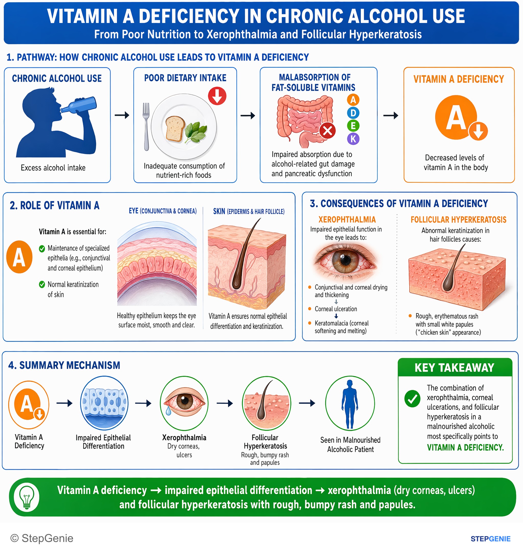
Vitamin A deficiency → impaired epithelial differentiation → xerophthalmia (dry corneas, ulcers) and follicular hyperkeratosis with rough…
USMLE Step 1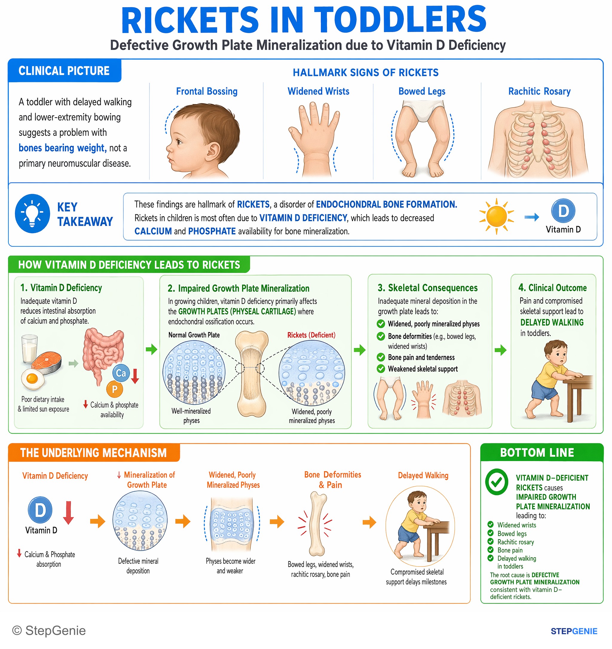
Vitamin D–deficient rickets causes impaired growth plate mineralization → widened wrists, bowed legs, rachitic rosary, delayed walking in…
USMLE Step 1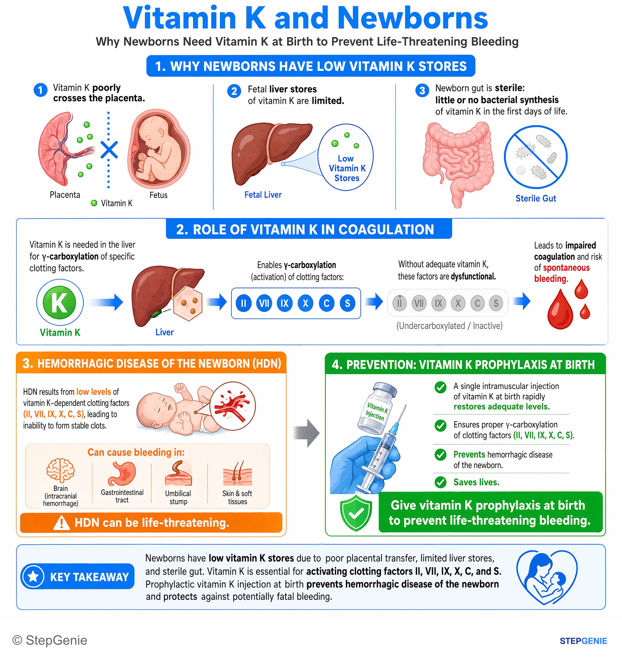
Hemorrhagic disease of the newborn results from low vitamin K–dependent clotting factors; give vitamin K prophylaxis to prevent…
USMLE Step 2Frequently Asked Questions
Why do fat-soluble vitamin deficiencies happen with cholestasis, pancreatic insufficiency, or bariatric surgery?
Fat-soluble vitamin deficiencies occur because vitamins A, D, E, and K need dietary fat, bile acids, and pancreatic enzymes for small-intestinal absorption. They are transported in chylomicrons, so disorders that impair fat absorption reduce vitamin uptake. Classic risk factors include cholestasis or obstructive jaundice, pancreatic insufficiency such as cystic fibrosis or chronic pancreatitis, celiac or Crohn disease, bariatric bypass procedures, and orlistat use.
Why does vitamin A deficiency cause night blindness and dry eyes?
Vitamin A deficiency causes night blindness because retinal is required to regenerate rhodopsin, the visual pigment used especially in dim light. It also causes dry eyes because vitamin A is needed for normal epithelial differentiation and maintenance of mucosal surfaces. As deficiency worsens, patients can develop xerophthalmia, Bitot spots, keratomalacia, follicular hyperkeratosis, and increased susceptibility to infections.
What is the best test for vitamin D deficiency on exams?
The best screening test for vitamin D stores is the serum 25-hydroxyvitamin D level. This reflects body vitamin D status better than the active 1,25-dihydroxy form for deficiency screening. The classic deficiency pattern also includes decreased 25-OH vitamin D, low or low-normal calcium, low phosphate, elevated PTH, and elevated alkaline phosphatase, which fits secondary hyperparathyroidism from reduced calcium and phosphate absorption.
How do you tell rickets from osteomalacia in vitamin D deficiency?
Rickets is the childhood form of vitamin D deficiency, while osteomalacia is the adult form. Rickets presents with delayed closure of fontanelles, bowed legs, and a rachitic rosary from costochondral swelling. Osteomalacia presents with bone pain, fragility fractures, and proximal muscle weakness. Both result from impaired calcium and phosphate absorption leading to poor bone mineralization.
What causes vitamin D deficiency in chronic kidney disease or liver disease?
Vitamin D deficiency occurs in chronic kidney disease because the kidney normally converts 25-hydroxyvitamin D to active 1,25-dihydroxyvitamin D using 1α-hydroxylase, and that step is reduced in CKD. It occurs in liver disease because the liver performs the 25-hydroxylation step needed to form calcidiol. When activation is impaired, calcium and phosphate absorption fall, promoting secondary hyperparathyroidism and bone demineralization.