Severe combined immunodeficiency (SCID) is a group of inherited defects in which T cells fail to develop, so B cells, even when present, cannot make useful antibody. Infants get thrush, Pneumocystis pneumonia, chronic diarrhea and failure to thrive in the first months of life, and every form is fatal in infancy unless treated. The most common single form is X-linked SCID (IL2RG, the common γ chain), phenotype T−B+NK−; ADA deficiency (T−B−NK−) is second. All US newborns are screened by measuring T-cell receptor excision circles (TRECs), and the cure is a hematopoietic stem cell transplant (HSCT), best done before 3.5 months; until then, no live vaccines and only irradiated, CMV-negative blood.
The visual hack: the "T-minus" grid
In typical SCID the T-cell box is always empty, so every phenotype starts with T−. Then ask two questions: are B cells there, and are NK cells there? Put the answers on a 2 × 2 grid and the gene falls out.
| T− in every box | NK− (IL-15 signal lost, or every lymphocyte poisoned) | NK+ (NK cells unaffected) |
|---|---|---|
| B+ (present but useless) | Common γ chain (IL2RG), X-linked; JAK3, autosomal recessive | IL-7Rα (IL7R); also CD3-chain defects |
| B− | ADA deficiency; reticular dysgenesis | RAG1/RAG2 and Artemis (DCLRE1C) |
Four rules explain every box:
- NK cells run on IL-15, which signals through the common γ chain (γc) and its kinase JAK3. Break either and NK cells vanish; an IL-7Rα defect spares them.
- Human B cells don't need IL-7, so B cells are present in γc, JAK3 and IL-7Rα SCID. They still fail, because B cells cannot function without T cells.
- Only T and B cells rearrange their receptor genes. RAG1/RAG2 make the V(D)J cuts and Artemis helps repair them, so a defect in either leaves no T or B cells. NK receptors aren't rearranged: T−B−NK+.
- ADA is a poison, not a signal. Deoxyadenosine builds up and kills precursors of all three lines (T−B−NK−). The dATP biochemistry and enzyme therapy are in the adenosine deaminase deficiency guide.
Sticky lines: "γc and JAK3 junk the NK cells." "RAG and Artemis can't rearrange: T and B gone, NK stays." "ADA: All Die Alike." "IL-7R: just the T cells."
Same phenotype, different inheritance. γc and JAK3 both give T−B+NK−, because JAK3 is the kinase γc signals through. A boy with an affected maternal uncle points to IL2RG (X-linked recessive); a girl, or related parents, points to JAK3 (autosomal recessive).
Why "combined": the empty school
Picture the thymus as a school, T cells as teachers and B cells as students. In SCID the school is empty: students may turn up, but with no teachers they never learn (no class switching, no germinal centers, no protective antibody). Both arms fail, so every class of pathogen gets in.
Genes and inheritance: which form is most common?
| Form | Gene, inheritance | Phenotype | Exam clue |
|---|---|---|---|
| X-linked SCID | IL2RG (γc, shared by the IL-2, -4, -7, -9, -15 and -21 receptors), X-linked recessive | T−B+NK− | Boys; most common single form, about a quarter to a third of cases |
| ADA deficiency | ADA, autosomal recessive (AR) | T−B−NK− | Second most common (about 15% of SCID); bone abnormalities; enzyme replacement |
| IL-7Rα deficiency | IL7R, AR | T−B+NK+ | NK cells spared |
| JAK3 deficiency | JAK3, AR | T−B+NK− | X-linked SCID look-alike in a girl |
| RAG1/RAG2 deficiency | RAG1, RAG2, AR | T−B−NK+ | No V(D)J recombination |
| Artemis deficiency | DCLRE1C, AR | T−B−NK+ | Mainly Navajo and Apache children |
| Omenn syndrome | "Leaky" SCID, usually partly working RAG, AR | T−B−NK+, leaky | Erythroderma, alopecia, eosinophilia, high IgE, hepatosplenomegaly, lymphadenopathy |
X-linked SCID is the single most common form, but autosomal recessive forms are more common as a group (NIAID: SCID is most often inherited in an autosomal recessive pattern), and no gene defect is found in about 15% of screen-detected infants.
Presentation: what the stem looks like
- Onset in the first months: most infants develop opportunistic infections within the first 3 months of life (Merck).
- Infections: persistent oral thrush, Pneumocystis jirovecii pneumonia (see the PJP guide), persistent viral infections, and chronic diarrhea with failure to thrive.
- Live-vaccine infections: cases of vaccine-acquired rotavirus infection in SCID infants are why SCID is a listed rotavirus vaccine contraindication (CDC).
- GVHD-like rash from engrafted maternal lymphocytes or a non-irradiated transfusion.
- Exam and imaging: an extremely small thymus (no thymic shadow on chest x-ray) and scant lymphoid tissue (tonsils, lymph nodes).
Diagnosis: from heel-prick to genotype
- Newborn screen (TRECs). TRECs are small circles of DNA cut out when developing T cells rearrange their receptor genes in the thymus, so they mark newly made T cells. SCID joined the US Recommended Uniform Screening Panel in 2010, and all US newborns are now screened (NIAID). Low TRECs mean T-cell lymphopenia: SCID, or another cause such as complete DiGeorge syndrome.
- CBC: lymphopenia suggests a T-cell disorder, because about 70% of circulating lymphocytes are T cells. Infant normals are higher than adult ones (Merck flags under 2000/µL at birth and under 4500/µL at 9 months).
- Flow cytometry for T, B and NK cells places the infant on the grid; lymphocyte proliferation to mitogens is absent.
- Immunoglobulins are low, but a normal IgG in the first months doesn't exclude SCID: it is mostly maternal IgG that crossed the placenta.
- Genetic testing names the type, and ADA activity is measured in blood cells.
Management: protect now, transplant soon
| Step | What to do | Why |
|---|---|---|
| Protection | Reverse isolation | Any infection can be fatal |
| No live vaccines | Hold rotavirus (due at 2 months), MMR, varicella, intranasal influenza, BCG, yellow fever, oral polio | CDC: all live vaccines are contraindicated in SCID, and other vaccines are unlikely to work |
| Blood products | Irradiated, leukocyte-reduced, from CMV-negative donors | Donor lymphocytes cause transfusion-associated GVHD |
| Prophylaxis | TMP-SMX for Pneumocystis; antifungals | PJP and Candida strike early |
| IgG replacement | IVIG or SCIG | Supplies the antibody B cells can't make |
| HSCT | HLA-matched sibling, or a T-cell-depleted haploidentical parent graft | Curative; survival is best before 3.5 months (NIAID); Merck quotes 96% when SCID is diagnosed by 3 months |
- ADA-SCID extras: intramuscular enzyme replacement once or twice a week with elapegademase-lvlr (Revcovi), a recombinant PEGylated ADA (PEG-ADA) that replaced the now-discontinued bovine product, pegademase bovine (Adagen), and Strimvelis gene therapy, authorized in the EU since 2016 for patients without a matched related donor.
- Gene therapy status (Oct 2026): no SCID gene therapy is on the FDA's list of approved cellular and gene therapy products. Early X-linked SCID gene therapy restored T-cell function, but about a quarter of the children developed leukemia 2-5 years later, probably because the vectors switched on growth-control genes; newer modified vectors appear effective and safe (NIAID). The FDA did approve one for Wiskott-Aldrich syndrome (Waskyra) in December 2025.
- Complete DiGeorge syndrome (congenital athymia) is the low-TREC infant who needs a thymus: the FDA approved processed thymus tissue (Rethymic) in 2021.
How USMLE tests it: traps and look-alikes
| Condition | Defect | Cells | What separates it |
|---|---|---|---|
| SCID | T-cell development (IL2RG, ADA, RAG...) | T−; B and NK vary | Infections of every type from the first months; low TRECs |
| Complete DiGeorge (22q11.2 deletion) | No thymus (3rd/4th pharyngeal pouches) | T−B+NK+ | Hypocalcemic tetany in the first 48 hours; truncus arteriosus, interrupted aortic arch, tetralogy of Fallot |
| X-linked agammaglobulinemia (Bruton) | BTK; B cells can't mature | B absent, T normal | Bacterial sinopulmonary infections after about 6 months, as maternal IgG wanes |
| X-linked hyper-IgM (CD40L) | No class switching | T and B present | Normal or high IgM, low IgG, IgA and IgE; PJP |
| Wiskott-Aldrich (WAS) | WAS protein | Variable | Eczema, small platelets with bleeding, infections |
- Chronic diarrhea weeks after the 2-month vaccines: vaccine-strain rotavirus in undiagnosed SCID.
- Rash, diarrhea and pancytopenia after a transfusion: transfusion-associated GVHD, prevented by irradiating blood.
- "Normal IgG" at 6 weeks doesn't rule out SCID; it is maternal.
Practice question
A 9-week-old boy is evaluated because his newborn screen showed undetectable T-cell receptor excision circles. He has had oral thrush since 3 weeks of age and has dropped from the 50th to the 10th percentile for weight. His mother's brother died of pneumonia at 6 months of age. Flow cytometry shows CD3+ T cells of 40/µL, a normal number of CD19+ B cells and almost no CD16/CD56+ NK cells. A defect in which of the following is the most likely cause?
A. Adenosine deaminase B. Common gamma chain of the IL-2 receptor C. Recombination-activating gene 1 (RAG1) D. IL-7 receptor alpha chain E. CD40 ligand F. Bruton tyrosine kinase
Answer: B. The grid reads T−B+NK−, and an affected maternal uncle points to X-linked inheritance: X-linked SCID from an IL2RG (γc) mutation. γc is shared by the IL-2, -4, -7, -9, -15 and -21 receptors, so T-cell (IL-7) and NK-cell (IL-15) development both fail, while B cells are present but useless without T-cell help.
- A: ADA deficiency kills all lymphocyte lines (T−B−NK−) and is autosomal recessive; B cells would be absent.
- C: RAG1 defects block V(D)J recombination (T−B−NK+); B cells would be absent and NK cells normal.
- D: IL-7Rα deficiency is T−B+NK+; NK cells depend on IL-15, not IL-7, so they would be present.
- E: CD40 ligand deficiency (X-linked hyper-IgM) leaves T-cell numbers and TRECs normal; the defect is class switching.
- F: BTK deficiency (X-linked agammaglobulinemia) removes B cells, spares T cells, and causes bacterial infections once maternal IgG wanes.
Next steps: no rotavirus or other live vaccines, only irradiated, CMV-negative, leukocyte-reduced blood, TMP-SMX, IgG replacement and urgent referral for HSCT.
Key takeaways
- SCID = no T cells, so B cells can't work: infections of every kind in the first months, fatal in infancy untreated.
- The grid: γc or JAK3 = T−B+NK−; IL-7Rα = T−B+NK+; RAG1/2 or Artemis = T−B−NK+; ADA = T−B−NK−.
- X-linked IL2RG is the most common single form; ADA is about 15%; autosomal recessive forms are the majority as a group.
- All US newborns are screened with TRECs; confirm with flow cytometry and genetics.
- Protect: no live vaccines (rotavirus at 2 months is the classic trap), irradiated CMV-negative blood, PJP prophylaxis, IgG replacement.
- Cure: HSCT, best before 3.5 months. ADA-SCID adds elapegademase and EU-authorized Strimvelis; no SCID gene therapy is FDA-approved as of Oct 2026.
Sources
- Severe Combined Immunodeficiency (SCID) — Merck Manual Professional Edition (2026)
- Approach to the Patient With Suspected Immunodeficiency — Merck Manual Professional Edition (2026)
- Severe Combined Immunodeficiency (SCID) — National Institute of Allergy and Infectious Diseases (NIH)
- X-linked severe combined immunodeficiency — MedlinePlus Genetics (2022)
- Adenosine deaminase deficiency — MedlinePlus Genetics (2024)
- Severe Combined Immunodeficiency — StatPearls, NCBI Bookshelf (2023)
- Altered Immunocompetence: Vaccines and Immunizations — CDC (2024)
- Rotavirus: The Pink Book, Chapter 19 — CDC (2024)
- REVCOVI (elapegademase-lvlr) injection label — DailyMed, NLM (2026)
- Strimvelis: EPAR — European Medicines Agency (2026)
- Approved Cellular and Gene Therapy Products — US Food and Drug Administration (2026)
- RETHYMIC (allogeneic processed thymus tissue–agdc) — US Food and Drug Administration (2021)
Visual explanations from the StepGenie QBank
Infographics StepGenie drew to explain questions in its own QBank, matched to this article's topic. Each caption is the takeaway of the question it explains.
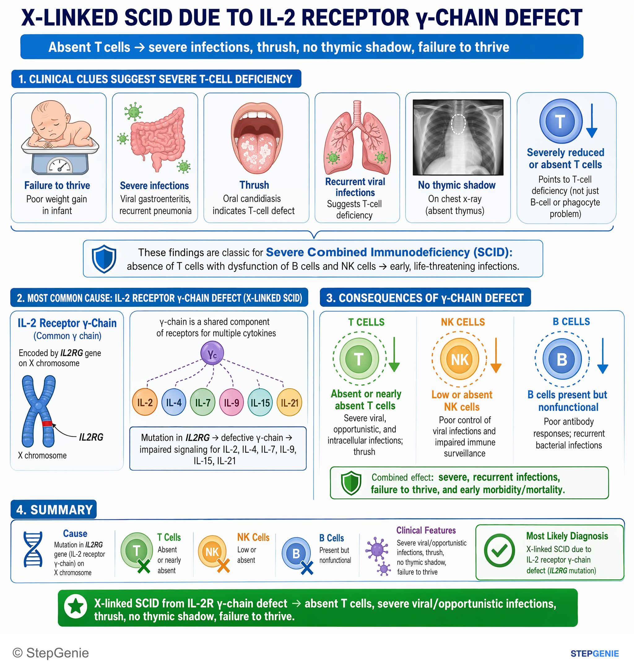
X-linked SCID from IL-2R γ-chain defect → absent T cells, severe viral/opportunistic infections, thrush, no thymic shadow, failure to…
USMLE Step 1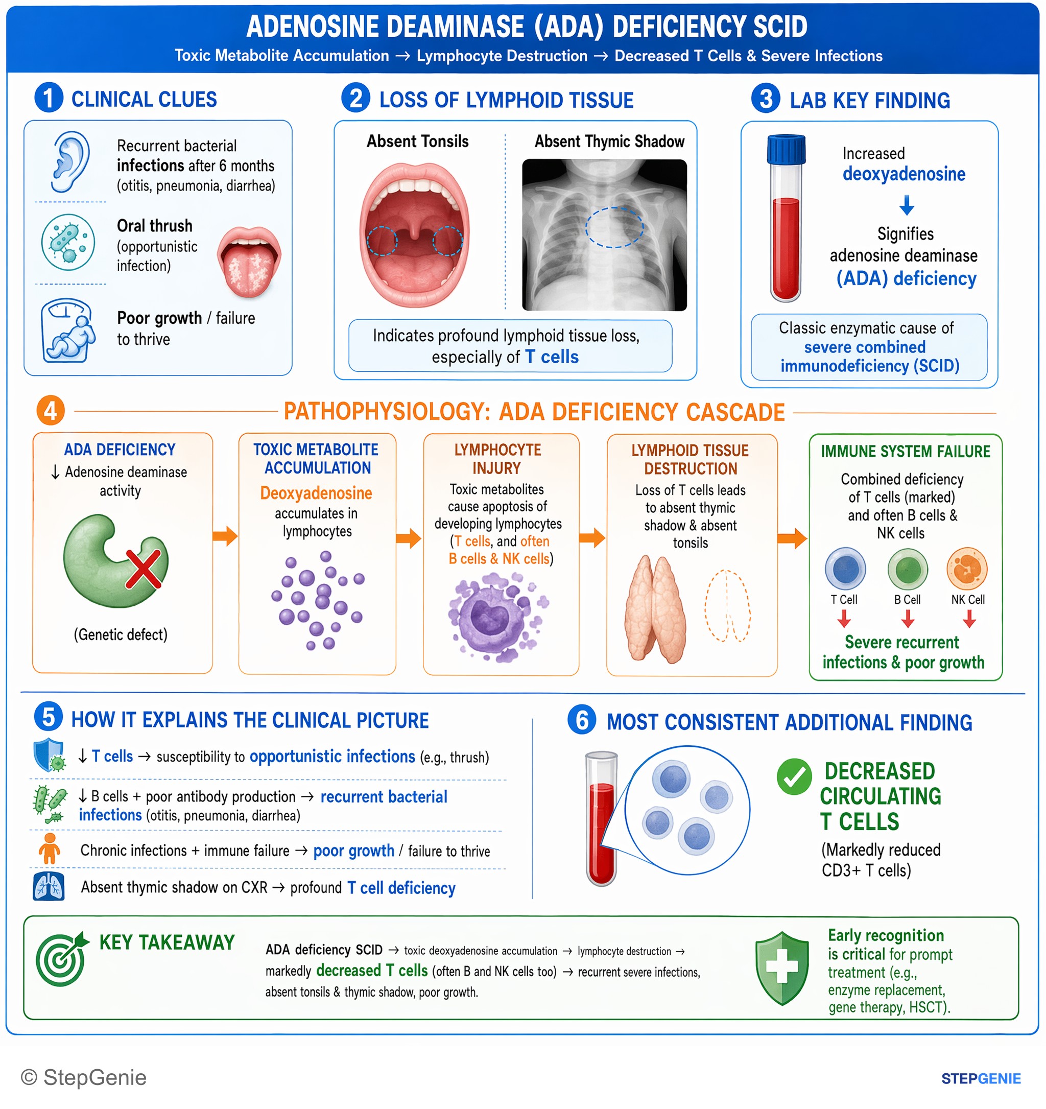
Adenosine deaminase–deficiency SCID → toxic deoxyadenosine accumulation → lymphocyte destruction → markedly decreased T cells (often B and…
USMLE Step 1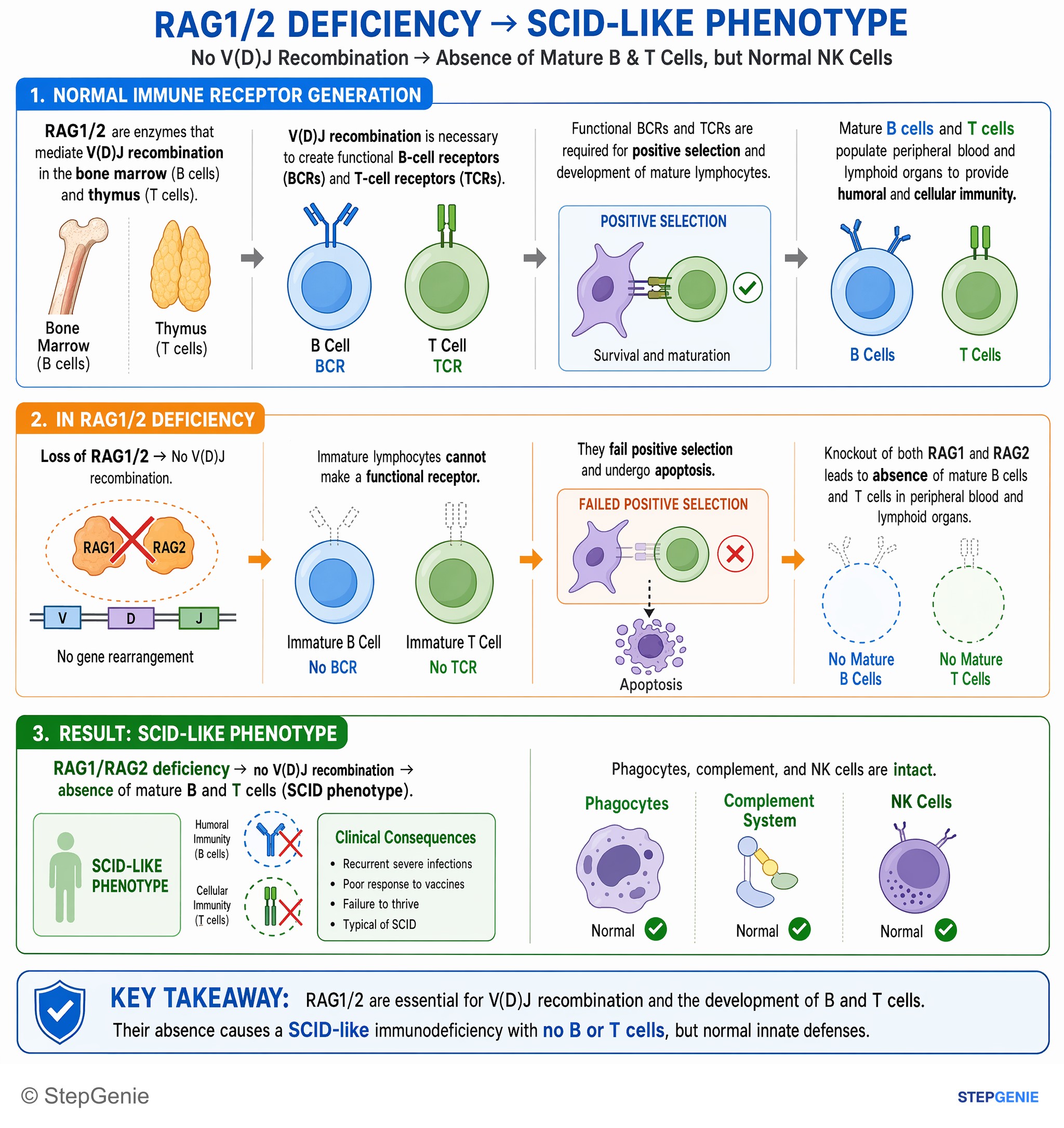
RAG1/RAG2 deficiency → no V(D)J recombination → absence of mature B and T cells (SCID phenotype), but normal NK cells.
USMLE Step 1Frequently Asked Questions
What is the most common cause of SCID?
The most common single cause is X-linked SCID, from variants in IL2RG, the gene for the common gamma chain shared by the IL-2, IL-4, IL-7, IL-9, IL-15 and IL-21 receptors. It accounts for roughly a quarter to a third of SCID cases, affects boys, and gives a T-minus, B-plus, NK-minus pattern. Adenosine deaminase deficiency is second at about 15%. Taken together, autosomal recessive forms such as ADA, RAG1, RAG2, IL7R, JAK3 and Artemis deficiency are more common than the X-linked form.
What does T-B+NK- mean in SCID?
It is shorthand for the flow cytometry pattern: T cells absent, B cells present and NK cells absent. That pattern points to a broken cytokine signal through the common gamma chain (X-linked IL2RG) or its kinase JAK3, because NK cells need IL-15 while human B cells do not need IL-7. IL-7 receptor alpha deficiency is T-B+NK+, RAG1, RAG2 and Artemis defects are T-B-NK+, and ADA deficiency is T-B-NK-. B cells that are present still cannot work without T-cell help.
How does newborn screening detect SCID?
Newborn screening measures T-cell receptor excision circles (TRECs) in the dried blood spot from the heel-prick card. TRECs are small circles of DNA cut out when developing T cells rearrange their receptor genes in the thymus, so very low or absent TRECs mean few new T cells are being made. SCID was added to the US Recommended Uniform Screening Panel in 2010, and all newborns in the United States are now screened. A positive screen is confirmed with flow cytometry of T, B and NK cells.
Why are live vaccines and blood transfusions dangerous in SCID?
Without T cells an infant cannot control weakened vaccine organisms, so live vaccines such as rotavirus, MMR, varicella, intranasal influenza and BCG can cause severe infection. CDC lists all live vaccines as contraindicated in SCID, and cases of vaccine-acquired rotavirus infection are why SCID is a specific rotavirus vaccine contraindication. Donor lymphocytes in blood products can engraft and attack the infant, causing transfusion-associated graft-versus-host disease, so only irradiated, leukocyte-reduced blood from CMV-negative donors is used.
How is SCID treated?
Hematopoietic stem cell transplant is the standard, curative treatment, ideally from a matched sibling, and survival is best when it is done before 3.5 months of age. Until then infants are kept in protective isolation and receive immunoglobulin replacement, trimethoprim-sulfamethoxazole to prevent Pneumocystis pneumonia, antifungals, no live vaccines and only irradiated blood products. ADA deficiency can also be treated with elapegademase, a recombinant PEG-ADA enzyme injected into muscle once or twice a week, and in the European Union with the gene therapy Strimvelis.
Is there an FDA-approved gene therapy for SCID?
Not as of October 2026: no SCID gene therapy appears on the FDA list of approved cellular and gene therapy products. Strimvelis has been authorized in the European Union since 2016 for ADA-SCID when no matched related donor is available. Early gene therapy for X-linked SCID restored T-cell function, but about a quarter of the children later developed leukemia, and newer modified vectors are still being studied. The FDA did approve a gene therapy for Wiskott-Aldrich syndrome in December 2025.