Reversible cell injury is what ATP depletion does before the damage becomes permanent: cellular (hydropic) swelling, fatty change, membrane blebs, loss of microvilli, ribosome detachment and chromatin clumping, all of which recover if oxygen returns. Injury turns irreversible when mitochondria can't restore ATP production even after the insult ends and membranes (plasma, lysosomal, mitochondrial) are severely damaged. Its markers are pyknosis → karyorrhexis → karyolysis, large amorphous densities in mitochondria and leakage of intracellular proteins such as troponin into the blood.
Reversible vs irreversible injury at a glance
| Feature | Reversible injury | Irreversible injury |
|---|---|---|
| Driving problem | ↓ ATP (pump failure, anaerobic glycolysis) | Persistent mitochondrial failure plus severe membrane damage |
| Cell size | Swelling (hydropic change) | Swelling → rupture (necrosis) |
| Plasma membrane | Blebs; blunted or lost microvilli | Breaks; intracellular proteins leak (troponin, CK-MB, AST/ALT) |
| Mitochondria | Swelling | Marked swelling with large amorphous (flocculent) densities |
| ER and ribosomes | ER dilation; ribosomes detach → ↓ protein synthesis | Organelle membranes break down |
| Cytoplasm | Pale, vacuolated; lipid droplets (fatty change) | Deeply eosinophilic |
| Nucleus | Chromatin clumping (low pH) | Pyknosis → karyorrhexis → karyolysis |
| Lysosomes | Intact | Rupture → autolysis |
| Myelin figures | Present | More prominent |
| If the insult stops | Full recovery | Cell death (necrosis, sometimes apoptosis) |
The hallmark question. Under the light microscope, the two features that announce reversible injury are cellular swelling and fatty change. The features that define irreversible injury are functional and structural at once: mitochondria that cannot recover and membranes that have failed.
What you can see: light vs electron microscopy
Boards like to ask which finding appears with which tool, because electron microscopy (EM) shows injury long before the light microscope (LM) does.
| Stage | Light microscopy | Electron microscopy |
|---|---|---|
| Reversible | Swollen, pale cells (hydropic or vacuolar change); small lipid vacuoles (fatty change) | Plasma membrane blebs, loss of microvilli, swollen mitochondria, dilated and vesiculated ER, detached ribosomes, clumped chromatin, myelin figures |
| Irreversible | Increased cytoplasmic eosinophilia; nuclear pyknosis, karyorrhexis or karyolysis | Plasma membrane breaks, large amorphous densities in swollen mitochondria, ruptured lysosomes, prominent myelin figures |
| After death | Necrosis pattern visible (coagulative, liquefactive and so on); inflammation | Cell debris |
In the heart, ultrastructural changes begin within 10–15 minutes of ischemia, but at time zero there is no light-microscopic change at all. That is why a patient who dies within an hour of an infarct can have a normal-looking heart on routine histology.
The mechanism chain, with ischemia as the model
- O₂ falls → oxidative phosphorylation stops → ATP falls. This is the first biochemical change.
- Na⁺/K⁺-ATPase fails → Na⁺ and water enter → cell and organelle swelling, blebs, loss of microvilli. Cellular swelling is the first visible consequence of ATP depletion and the most common expression of cell injury.
- Anaerobic glycolysis burns glycogen and produces lactic acid → ↓ pH → chromatin clumping.
- Ribosomes detach from the RER, and injured cells that cannot export lipid as lipoprotein accumulate it (fatty change, most often in the liver).
- Ca²⁺ pumps fail → Ca²⁺ floods the cytosol and activates phospholipases (membranes), proteases (cytoskeleton), endonucleases (chromatin) and ATPases (more ATP loss). It also opens the mitochondrial permeability transition pore.
- Point of no return: mitochondria can't recover and membranes fail. Lysosomal enzymes digest the cell, cytosolic proteins leak into the blood and the nucleus dies. Necrosis follows; cytochrome c release can also trigger apoptosis.
Why ischemia is worse than pure hypoxia. Ischemia cuts off nutrients such as glucose as well as oxygen, so anaerobic glycolysis cannot rescue ATP production. Cyanide blocks cytochrome oxidase (complex IV), so cells cannot use oxygen even when it is delivered normally.
How fast tissues cross the line
Different cells tolerate ischemia for very different times, and stems often hinge on that difference.
| Tissue | Timing to remember | High-yield detail |
|---|---|---|
| Neurons | Among the most vulnerable cells; injury starts within minutes of anoxia | Hippocampal CA1 pyramidal neurons, cortical layers 3, 5 and 6, Purkinje cells and the basal ganglia are the most vulnerable after global ischemia; watershed zones infarct with global hypoperfusion; "red neurons" (shrunken, eosinophilic) mark hypoxic death |
| Myocardium | Infarction follows about 20–40 minutes of severe ischemia | Necrosis spreads from subendocardium to subepicardium over hours; early reperfusion limits infarct size |
| Skeletal muscle | Prolonged limb ischemia (embolism, trauma) | After revascularization, reperfusion raises capillary permeability and edema, which can cause acute compartment syndrome |
Causes boards use to test the same pathway
- Hypoxia and ischemia: MI, stroke, shock, torsion.
- Free radicals: acetaminophen overdose makes NAPQI through cytochrome P450 (especially CYP2E1), which depletes hepatic glutathione and causes centrilobular (zone 3) necrosis; N-acetylcysteine is the antidote. Radiation and reperfusion work through reactive oxygen species as well; see free radical injury.
- Others: mitochondrial poisons (cyanide), infections, immune reactions, genetic defects and nutritional imbalances.
Fatty change as a model of reversibility. Alcohol metabolism raises the NADH/NAD⁺ ratio, which favors triglyceride synthesis in hepatocytes. Alcohol-related fatty liver is generally reversible when drinking stops; continued drinking can progress to steatohepatitis and cirrhosis.
Enzyme leak by organ: irreversible injury on the lab sheet
Once plasma membranes fail, intracellular proteins enter the blood. The marker tells you which cells died:
| Tissue | Marker released | Classic setting |
|---|---|---|
| Myocardium | Troponin I and T (also CK-MB) | Myocardial infarction |
| Liver | ALT and AST | Ischemic, viral or toxic hepatitis |
| Skeletal muscle | CK, myoglobin | Rhabdomyolysis |
| Pancreas | Lipase, amylase | Acute pancreatitis |
Apoptosis keeps the membrane intact, so it causes little enzyme leak and no inflammation. For the timing of each cardiac marker, see cardiac biomarkers.
What irreversible injury becomes: necrosis patterns
| Pattern | Typical setting | What it looks like |
|---|---|---|
| Coagulative | Ischemia in most organs except the brain | Tissue architecture and cell outlines preserved; nuclei lost |
| Liquefactive | Brain infarcts; focal bacterial and some fungal infections (abscess) | Tissue digested into fluid; pus in infections |
| Caseous | Tuberculosis (granulomas) | Crumbly, cheese-like center; amorphous eosinophilic debris |
| Fat | Acute pancreatitis; breast tissue | Fat saponification |
| Fibrinoid | Immune complex deposition in blood vessel walls | Fibrin-like pink material in the vessel wall |
| Gangrenous | Ischemic limbs | Dry (coagulative) or wet (liquefactive with infection) |
The pathways and the apoptosis comparison are covered in apoptosis vs necrosis.
When irreversible injury ends in apoptosis
Irreversibly injured cells usually die by necrosis, but damaged mitochondria can also release cytochrome c, which activates caspases and switches the cell into apoptosis. Apoptosis is a controlled death: the cell shrinks, chromatin condenses (pyknosis), the membrane stays intact, apoptotic bodies form and no inflammation follows. It also occurs normally, for example in the formation of fingers and in T-cell development, whereas necrosis is always pathological. On a slide, inflammation around dead cells points to necrosis; isolated shrunken cells in otherwise normal tissue point to apoptosis.
Ischemia–reperfusion injury
Restoring flow is essential, but it can kill cells that were only reversibly injured:
- Reactive oxygen species from xanthine oxidase, NADPH oxidase and nitric oxide synthase overwhelm antioxidant defenses.
- Ca²⁺ overload through the Na⁺/Ca²⁺ exchanger and opening of the mitochondrial permeability transition pore.
- Neutrophils and complement: neutrophils are recruited by cytokines, and complement (activated by natural IgM and necrotic debris in ischemic tissue) damages cells and recruits more neutrophils.
- Microvascular dysfunction: vasoconstriction and endothelial damage limit flow.
In reperfused myocardium, ATP returns while Ca²⁺ is still high, so myofibrils hypercontract, producing contraction band necrosis (dense eosinophilic bands). Typical settings are PCI or thrombolysis after MI (with stunning and arrhythmias), stroke reperfusion (hemorrhagic transformation and edema), organ transplantation and limb revascularization (compartment syndrome).
Reversible dysfunction in the heart: stunning and hibernation
Not every non-contracting segment of myocardium is dead, and Step 2 CK questions on revascularization depend on that distinction.
| State | What happened | Is the muscle viable? | What brings function back |
|---|---|---|---|
| Stunned myocardium | Acute ischemia followed by reperfusion; contraction stays depressed after flow returns | Yes | Time: recovery is often near-normal within about 2 weeks of a reperfused infarct |
| Hibernating myocardium | Persistent, chronic ischemia; the muscle downregulates contraction | Yes | Timely revascularization (PCI or bypass) |
| Infarcted (necrotic) myocardium | Irreversible injury followed by scar | No | Nothing; it scars |
Viability is assessed with dobutamine stress echocardiography (a biphasic response, better at low dose and worse at high dose, means viable but ischemic muscle), PET, perfusion imaging or cardiac MRI, where the absence of late contrast enhancement typically indicates viable myocardium.
Troponin on Step 2. Serial troponins (for example at 0, 3 and 6 hours) look for a rise and/or fall with at least one value above the sex-specific 99th percentile upper reference limit (acute myocardial injury); with symptoms, new ischemic ECG changes, pathological Q waves or imaging evidence of ischemia, that pattern indicates acute myocardial infarction. In MI, troponin leaks from myocytes with failed membranes, but a raised troponin does not always mean necrosis (after strenuous exercise, release likely reflects reversible injury).
Classic vignette clues
- Electron microscopy showing large amorphous densities in mitochondria or breaks in the plasma membrane → irreversible.
- Rising troponin or CK-MB with ischemic symptoms or ECG changes → myocyte membranes have ruptured, meaning infarction (irreversible injury, necrosis).
- Hepatocytes filled with lipid vacuoles in a heavy drinker → fatty change, reversible with abstinence.
- Pale, swollen cells with intact nuclei after a brief ischemic episode → hydropic change, reversible.
- Sudden death within an hour of an MI with a normal-looking heart on light microscopy → simply too early to see. Wavy fibers appear from about 30 minutes to 4 hours, and coagulative necrosis from about 4–12 hours.
Exam traps: how to tell A from B
- Chromatin clumping vs pyknosis. Clumping, caused by low pH, is reversible. Pyknosis (a shrunken, dark nucleus) is the first step of nuclear death.
- Mitochondrial swelling vs mitochondrial densities. Swelling alone is reversible; large amorphous densities mark irreversibility.
- Myelin figures are whorls of phospholipid from damaged membranes. They appear in both reversible and irreversible injury (more prominent in necrosis), so they don't settle the question on their own.
- Necrosis vs apoptosis. Necrosis means swelling, membrane rupture, enzyme leak and inflammation. Apoptosis means cell shrinkage, an intact membrane, apoptotic bodies and no inflammation.
- Earliest change: biochemical vs morphologic. ATP depletion comes first; cellular swelling is the earliest change you can see.
- Liquefactive vs coagulative. A brain infarct liquefies; an infarct almost anywhere else coagulates.
Worked examples
Vignette 1. A 61-year-old man dies 5 hours after an anterior STEMI. Electron microscopy of his left ventricular myocytes is reviewed. Which finding indicates that the injury had become irreversible?
- A. Cell swelling with dilated endoplasmic reticulum
- B. Plasma membrane blebs
- C. Detachment of ribosomes from the rough ER
- D. Clumping of nuclear chromatin
- E. Large amorphous densities within swollen mitochondria
Answer: E.
One-line reasoning: A–D are ATP-depletion changes that reverse if perfusion returns, whereas amorphous mitochondrial densities (like membrane breaks and pyknosis) mean the mitochondria can no longer recover, which is the point of no return.
Vignette 2. A 70-year-old woman is resuscitated after a 12-minute cardiac arrest but never regains consciousness and dies 3 days later. Which brain region is most likely to show shrunken, intensely eosinophilic neurons?
Answer: The CA1 sector of the hippocampus (also cortical layers 3, 5 and 6 and cerebellar Purkinje cells).
One-line reasoning: Global ischemia kills the most vulnerable neurons first, and "red neurons" are the histologic signature of irreversible hypoxic injury.
Vignette 3. A 54-year-old man has a large anterior STEMI treated with primary PCI 90 minutes after symptom onset. On day 2, echocardiography shows severe anterior hypokinesis; by 2 weeks, wall motion is nearly normal. What explains the early dysfunction?
Answer: Myocardial stunning.
One-line reasoning: Reperfused myocardium that was only reversibly injured can stay hypocontractile for days to weeks after flow returns, then recover; infarcted muscle would have scarred instead.
Key takeaways
- The first biochemical change in cell injury is ATP depletion; the first visible change is cellular swelling.
- Reversible: swelling, fatty change, blebs, loss of microvilli, ER dilation, ribosome detachment, chromatin clumping.
- Irreversible means two failures: mitochondria can't restore ATP, and membranes (plasma, lysosomal) are badly damaged.
- Markers of irreversibility: pyknosis → karyorrhexis → karyolysis, amorphous mitochondrial densities, lysosomal rupture and leakage of intracellular proteins (troponin).
- Neurons cross the line in minutes, myocardium in about 20–40 minutes of severe ischemia; EM changes precede light-microscopic necrosis by hours.
- Ca²⁺ influx switches on phospholipases, proteases, endonucleases and ATPases; reperfusion adds ROS, Ca²⁺ overload, hypercontraction and inflammation.
Find more general pathology reviews on the pathology topic hub.
Sources
- Histology, Cell Death (StatPearls, NCBI Bookshelf)
- Necrosis Pathology (StatPearls, NCBI Bookshelf)
- Acute Myocardial Infarction (StatPearls, NCBI Bookshelf)
- Myocardial Infarction (StatPearls, NCBI Bookshelf)
- Fifth Universal Definition of Myocardial Infarction (2026), Global Heart (open access)
- Myocardial Stunning and Hibernation (StatPearls, archived chapter, NCBI Bookshelf)
- Hypoxic Brain Injury (StatPearls, archived chapter, NCBI Bookshelf)
- Vascular Reperfusion Injury (StatPearls, NCBI Bookshelf)
- Acetaminophen Toxicity (StatPearls, NCBI Bookshelf)
- Alcohol-Associated Liver Disease (StatPearls, NCBI Bookshelf)
- Liver Function Tests (StatPearls, NCBI Bookshelf)
- Rhabdomyolysis (StatPearls, NCBI Bookshelf)
- Yusof et al. The contribution of cardiomyocyte hypercontracture to the burden of acute myocardial infarction: an update. Basic Res Cardiol. 2025 (PubMed)
Visual explanations from the StepGenie QBank
Infographics StepGenie drew to explain questions in its own QBank, matched to this article's topic. Each caption is the takeaway of the question it explains.
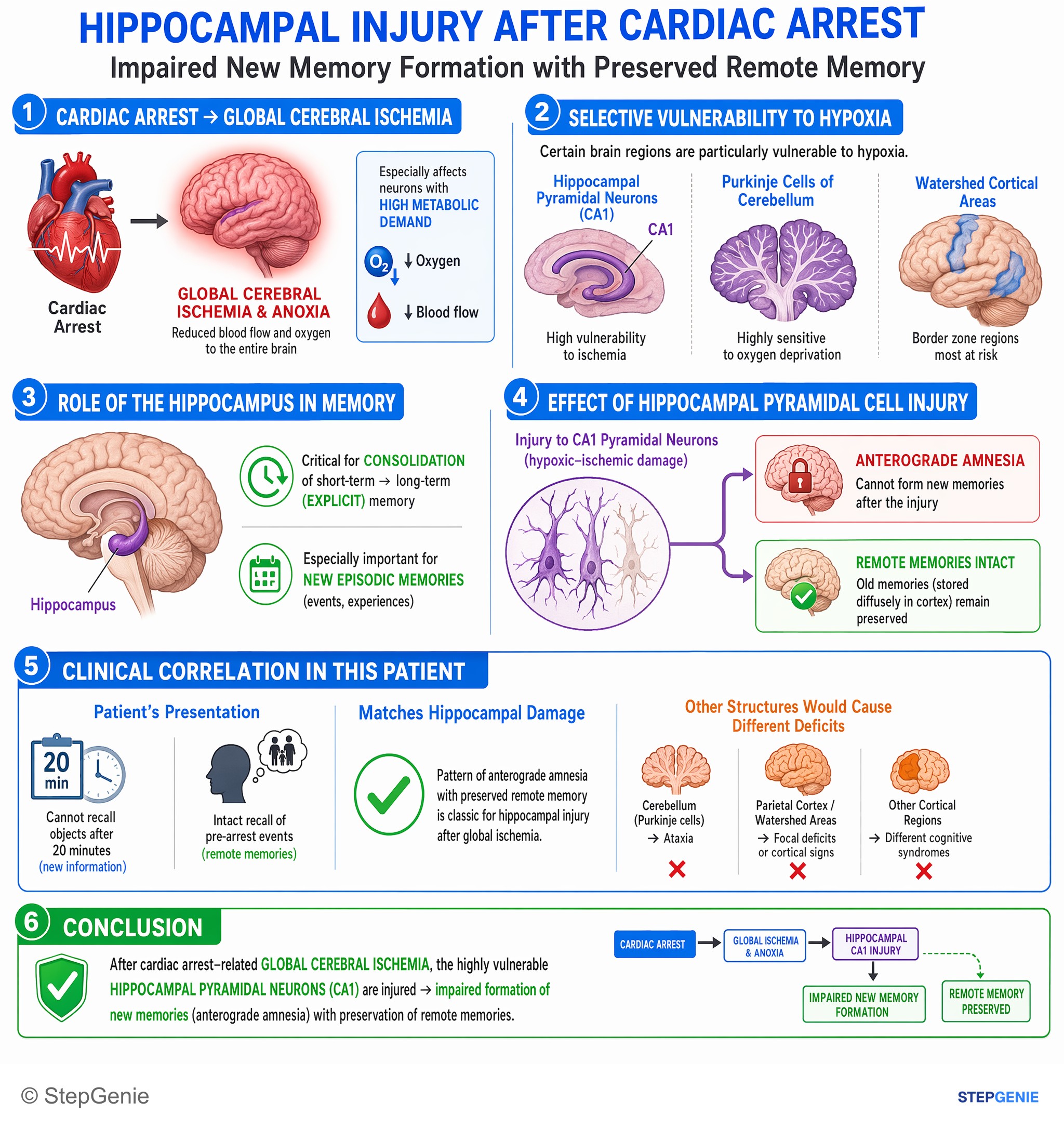
Anoxic injury of hippocampal pyramidal neurons → impaired new memory formation with preserved remote memory after cardiac arrest–related…
USMLE Step 1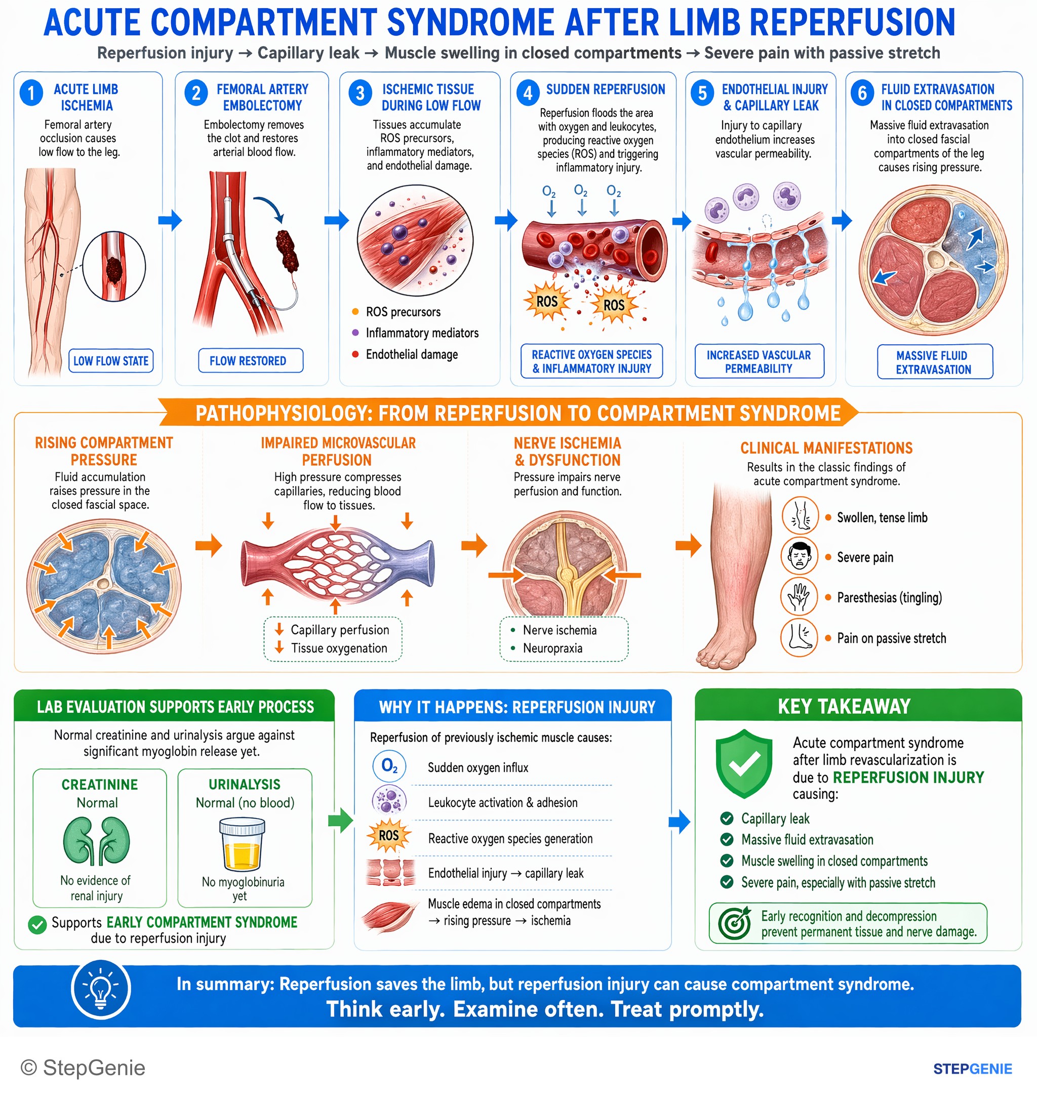
Acute compartment syndrome after limb revascularization is due to reperfusion injury causing capillary leak, muscle swelling, and severe…
USMLE Step 2Frequently Asked Questions
What is the hallmark of reversible cell injury?
Cellular (hydropic) swelling is the hallmark and the first visible change under the light microscope, and fatty change is the other classic light-microscopic sign. Swelling comes from ATP depletion (the sodium pump fails, so water enters the cell); fatty change follows hypoxic, toxic or metabolic injury (alcohol, for example) when cells cannot process or export lipid normally. Under the electron microscope the same stage shows blebs, loss of microvilli, swollen mitochondria and endoplasmic reticulum, detached ribosomes and clumped chromatin.
What is the point of no return in cell injury?
Two events mark it: mitochondria can no longer restore oxidative phosphorylation and ATP production even after the insult is removed, and membranes (plasma, lysosomal and mitochondrial) are severely damaged. Once that happens, lysosomal enzymes digest the cell and cytosolic proteins leak into the blood, so the cell is committed to death.
What is the earliest change in cell injury?
The first biochemical change is a fall in ATP, usually from loss of oxidative phosphorylation. The first visible change is cellular (hydropic) swelling: the ATP-dependent Na⁺/K⁺ pump fails, so sodium and water enter the cell and its organelles. Both are reversible if oxygen and ATP are restored in time; the injury becomes irreversible when mitochondrial function cannot be restored and membranes are severely damaged.
Is fatty change reversible or irreversible?
Fatty change (steatosis) is a form of reversible injury, seen most often in the liver, in which lipid builds up because injured cells can't process or export it normally. Alcohol-related fatty liver is generally reversible if drinking stops. If the injury continues, it can progress to steatohepatitis, cell death and cirrhosis.
Are myelin figures a sign of reversible or irreversible injury?
They can be seen in both. Myelin figures are whorled masses of phospholipid derived from damaged cell membranes; they appear in reversible injury and become more prominent in necrotic cells. To call injury irreversible on an exam, look for pyknosis, karyorrhexis or karyolysis, large amorphous densities in mitochondria, or membrane rupture.
Why is troponin a marker of irreversible myocardial injury?
Troponin is an intracellular protein that leaks into the blood when myocyte plasma membranes break down, so in a myocardial infarction a rising troponin signals that heart muscle cells have crossed into irreversible injury. This happens well before necrosis is visible: at the moment of death there is no light-microscopic change, wavy fibers appear from about 30 minutes to 4 hours and coagulative necrosis from about 4 to 12 hours. Serial measurements look for a rise and/or fall with at least one value above the sex-specific 99th percentile; troponin can also rise without necrosis, for example after strenuous exercise.