Necrosis and apoptosis are two words that show up everywhere in Step-style stems—MI, stroke, hepatitis, toxins, tumors, autoimmune disease, even embryology. The trick isn’t just “which one is programmed?” It’s recognizing mechanism + morphology + clinical context, and then matching them to the specific necrosis pattern (coagulative, liquefactive, caseous, fat, fibrinoid, gangrenous). If you can do that, a lot of pathology questions become pattern-recognition instead of memorization.
Big picture: how they differ (Step 1 must-know)
Core idea
- Apoptosis = regulated, energy-dependent cell death → minimal inflammation
- Necrosis = unregulated cell death due to injury → membrane rupture + inflammation
Apoptosis vs Necrosis (rapid comparison table)
| Feature | Apoptosis | Necrosis |
|---|---|---|
| Trigger | Physiologic (normal) or pathologic but regulated | Always pathologic injury (ischemia, toxins, infection, trauma) |
| Energy | ATP-dependent | Often associated with ATP depletion |
| Cell membrane | Intact (blebs → apoptotic bodies) | Disrupted → contents spill |
| DNA | Fragmentation (laddering) | Random degradation (smear) |
| Cell size | Shrinkage | Swelling (oncosis) |
| Inflammation | Minimal/none | Prominent |
| Key histology | Apoptotic bodies, pyknotic nuclei | Eosinophilic “ghost cells,” karyolysis |
| Clinical tie-in | Development, immune tolerance, cancer therapy | MI, stroke, abscess, pancreatitis, vasculitis |
First Aid cross-reference: General Pathology → Cell injury and death (Apoptosis & Necrosis patterns).
Apoptosis: the “clean” death
Definition
Apoptosis is a controlled cellular suicide program that eliminates unwanted cells without spilling intracellular contents, so it doesn’t recruit much inflammation.
When it happens (high-yield physiology + pathology)
Physiologic (normal):
- Embryogenesis (digit separation)
- Hormone withdrawal (endometrial shedding during menses)
- Involution (post-lactational breast involution)
- Immune regulation
- Negative selection of self-reactive lymphocytes
- Termination of immune responses (removing activated lymphocytes)
Pathologic but regulated:
- DNA damage (e.g., radiation, chemotherapy) via p53
- Viral infections → cytotoxic T-cell killing (e.g., hepatitis)
- Protein misfolding/ER stress (neurodegenerative diseases)
Pathophysiology (know these pathways cold)
1) Intrinsic (mitochondrial) pathway — “internal stress”
Key triggers: DNA damage, growth factor withdrawal, ER stress
Key proteins:
- Bcl-2 family regulates mitochondrial permeability
- Anti-apoptotic: Bcl-2, Bcl-xL
- Pro-apoptotic: Bax, Bak (promote cytochrome c release)
Core sequence:
- Stress → Bax/Bak open mitochondrial pores
- Cytochrome c released → activates Apaf-1
- Activates initiator caspase-9
- Activates executioner caspases (3, 6, 7) → cell dismantling
Step association: p53 pushes damaged cells toward apoptosis; loss of p53 → cancer risk.
2) Extrinsic (death receptor) pathway — “external signal”
Key receptors: Fas (CD95), TNF receptor
Key ligands: FasL (on cytotoxic T cells), TNF-α
Core sequence:
- FasL binds Fas → recruits adaptor proteins (e.g., FADD)
- Activates initiator caspase-8
- Executioner caspases (3, 6, 7) → apoptosis
High-yield disease tie-in:
- Autoimmune lymphoproliferative syndrome (ALPS): defective Fas → failure of lymphocyte apoptosis → lymphadenopathy, autoimmune cytopenias.
Morphology + lab clues
What you see:
- Cell shrinkage
- Chromatin condensation (pyknosis) and nuclear fragmentation (karyorrhexis)
- Apoptotic bodies (membrane-bound fragments)
Why minimal inflammation?
Apoptotic bodies expose “eat-me” signals (e.g., phosphatidylserine) → quickly phagocytosed.
Diagnostic hint:
- TUNEL stain detects DNA fragmentation (often taught as apoptosis-associated)
Clinical presentation (how it appears in vignettes)
- Often silent clinically unless widespread
- In hepatitis: Councilman bodies (apoptotic hepatocytes)
- After chemo/radiation: tumor cell apoptosis is a desired effect
Treatment (how Step frames it)
You typically don’t treat apoptosis directly; you treat the condition driving it. But boards love therapy mechanisms that induce apoptosis:
- Many chemotherapy agents and radiation → DNA damage → p53-mediated apoptosis
- Glucocorticoids can induce apoptosis in lymphoid cells (relevant in some hematologic malignancies and inflammation control)
Necrosis: the “messy” death (injury → inflammation)
Definition
Necrosis is uncontrolled cell death due to severe injury → membrane rupture → enzyme leakage + inflammation.
Core pathogenesis (Step-level)
Common upstream events:
- ATP depletion (ischemia) → failure of ion pumps → cell swelling
- Ca²⁺ influx → activates phospholipases, proteases, endonucleases
- Reactive oxygen species → lipid peroxidation, DNA/protein damage
- Membrane damage → lysosomal enzyme leakage → autodigestion
First Aid cross-reference: Cell injury: ATP depletion, Ca²⁺ influx, ROS, reperfusion injury.
Clinical/lab clues
- Inflammation (pain, swelling, fever depending on extent/site)
- Elevated serum enzymes from cell leakage:
- MI: troponin, CK-MB
- Pancreatitis: lipase
- Hepatocellular injury: AST/ALT
Types of necrosis (the classic Step 1 patterns)
1) Coagulative necrosis — “architecture preserved”
Mechanism: protein denaturation dominates over enzymatic digestion
Where: ischemic infarcts in most solid organs
Classic locations: heart, kidney, spleen (NOT brain)
Histology buzzwords:
- Preserved tissue outline (“ghost cells”)
- Increased eosinophilia
- Loss of nuclei (karyolysis)
High-yield exception:
- Brain infarct → liquefactive, not coagulative
Clinical vignette:
Chest pain → STEMI → infarcted myocardium shows coagulative necrosis.
First Aid cross-reference: Infarction → coagulative necrosis (except brain).
2) Liquefactive necrosis — “liquid goo”
Mechanism: enzymatic digestion dominates
Where:
- Brain infarcts (high lipid content + enzymes)
- Abscesses from bacterial/fungal infections (neutrophil enzymes)
Gross: soft, cystic cavity filled with debris/pus
Histology: loss of architecture, many neutrophils (if infection)
Clinical vignette:
Stroke → CT later shows encephalomalacia/cystic change; or fluctuance with pus in abscess.
First Aid cross-reference: Brain infarct = liquefactive; abscess = liquefactive.
3) Caseous necrosis — “cheesy”
Mechanism: granulomatous inflammation; mixture of coagulative + liquefactive features
Where: TB (classic), some systemic fungi (e.g., Histoplasma)
Gross: white, friable “cheese-like”
Histology: amorphous granular debris with surrounding granulomas (epithelioid histiocytes, giant cells)
Clinical vignette:
Chronic cough, weight loss, night sweats; apical lung lesion + granulomas with caseation.
First Aid cross-reference: Granulomatous disease → caseous necrosis (TB).
4) Fat necrosis — “saponification”
Mechanism: lipase-mediated fat breakdown → free fatty acids bind calcium → chalky deposits
Where:
- Acute pancreatitis (enzymes leak into peripancreatic fat)
- Traumatic fat necrosis (e.g., breast)
Key buzzwords:
- Chalky white areas
- Saponification
- Hypocalcemia can occur in severe pancreatitis (calcium consumption)
Clinical vignette:
Epigastric pain radiating to back + elevated lipase + chalky fat deposits around pancreas.
First Aid cross-reference: Pancreatitis → fat necrosis; saponification.
5) Fibrinoid necrosis — “bright pink vessel walls”
Mechanism: immune-mediated vascular injury → leakage of proteins (including fibrin) into vessel wall
Where:
- Vasculitides (e.g., polyarteritis nodosa)
- Malignant hypertension
- Preeclampsia/HELLP (placental vessels can show fibrinoid change)
Histology: eosinophilic (“fibrin-like”) material in vessel wall
Clinical vignette:
Systemic symptoms + vessel inflammation; or very high BP with end-organ damage.
First Aid cross-reference: Vasculitis/malignant HTN → fibrinoid necrosis.
6) Gangrenous necrosis — “clinical pattern label”
Not a microscopic pattern by itself—think of it as a gross description.
- Dry gangrene: ischemic limb → essentially coagulative necrosis
- Wet gangrene: ischemia + superimposed infection → liquefactive components, foul smell, rapid spread
- Gas gangrene: Clostridium perfringens → myonecrosis + gas in tissue (crepitus)
Clinical vignette:
Diabetic patient with peripheral vascular disease: black, mummified toe (dry) vs swollen, infected, oozing foot (wet).
First Aid cross-reference: Gangrene: dry vs wet; Clostridium → gas gangrene.
Nuclear changes you should recognize (common Step question)
| Nuclear change | Meaning | Seen in |
|---|---|---|
| Pyknosis | Nuclear shrinkage/condensation | Apoptosis, necrosis |
| Karyorrhexis | Nuclear fragmentation | Apoptosis, necrosis |
| Karyolysis | Nuclear fading (DNase activity) | Necrosis (classically) |
Apoptosis vs necrosis in real clinical contexts (classic vignettes)
Myocardial infarction (MI)
- Primary pattern: coagulative necrosis
- Labs: troponin/CK-MB rise
- Inflammation follows necrosis (neutrophils early)
Ischemic stroke
- Primary pattern: liquefactive necrosis
- Expect cystic space later
Viral hepatitis
- Prominent apoptosis of hepatocytes (Councilman bodies)
- Immune-mediated killing (extrinsic pathway via Fas)
TB lung lesion
- Caseous necrosis within granulomas
Acute pancreatitis
- Fat necrosis with saponification (chalky deposits), possible hypocalcemia
Vasculitis / malignant HTN
- Fibrinoid necrosis in vessel walls
High-yield associations & “exam traps”
Traps that cost points
- Brain infarcts are liquefactive, not coagulative.
- Gangrene is a gross term; dry = coagulative, wet = infection + liquefactive.
- Apoptosis can be pathologic (DNA damage, viral infections), not just physiologic.
- Caspases are central: initiator (8/9) → executioner (3/6/7).
- Bcl-2 is anti-apoptotic; overexpression helps cancers survive (classically in some lymphomas).
Quick memory hooks (board-friendly)
- Coagulative = Cardiac (and most organs) infarct
- Liquefactive = Lesion in brain / pus
- Caseous = Chronic cough (TB)
- Fat = Free fatty acids + Foamy/chalky
- Fibrinoid = Freaked-out vessels (vasculitis/HTN)
Diagnosis & management: what Step expects
Diagnosis
- Mostly histopath pattern recognition + clinical context
- Supportive labs (enzyme leakage) help identify necrosis in certain organs:
- MI: troponin, CK-MB
- Pancreatitis: lipase
- Hepatitis: AST/ALT (note: apoptosis itself doesn’t “leak enzymes” the same way necrosis does, but real diseases often have mixed injury patterns)
Treatment (principle-based)
You treat the underlying cause, because necrosis/apoptosis are endpoints:
- Restore perfusion when appropriate (MI/stroke pathways), but remember reperfusion injury can worsen damage via ROS.
- Antibiotics/drainage for abscesses (liquefactive necrosis from infection).
- Immunosuppression for vasculitis (fibrinoid necrosis contexts).
- Supportive care and cause-directed therapy for pancreatitis (fat necrosis).
Step-style “one-liners” to keep in your head
- Apoptosis = caspases + no inflammation.
- Necrosis = membrane rupture + inflammation + enzyme leakage.
- Ischemia → coagulative necrosis (except brain).
- Brain infarct/abscess → liquefactive necrosis.
- TB → caseous necrosis in granulomas.
- Pancreatitis/trauma → fat necrosis with saponification.
- Vasculitis/malignant HTN → fibrinoid necrosis.
Visual explanations from the StepGenie QBank
Infographics StepGenie drew to explain questions in its own QBank, matched to this article's topic. Each caption is the takeaway of the question it explains.
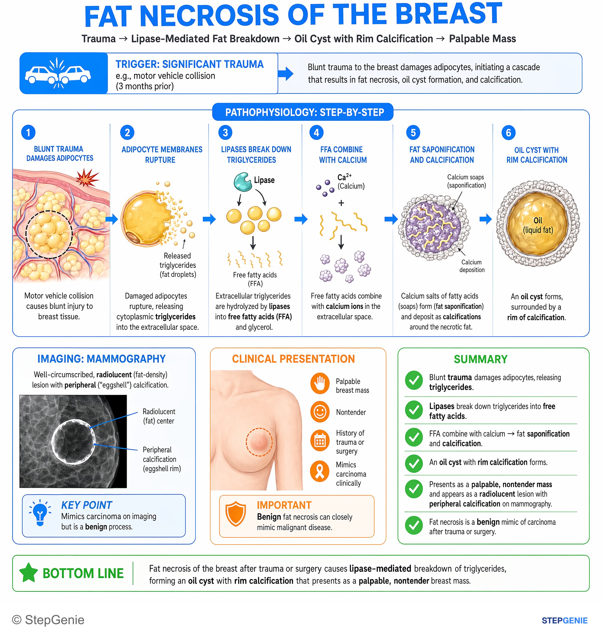
Fat necrosis of the breast after trauma or surgery causes lipase-mediated release of triglycerides, forming an oil cyst with rim…
USMLE Step 1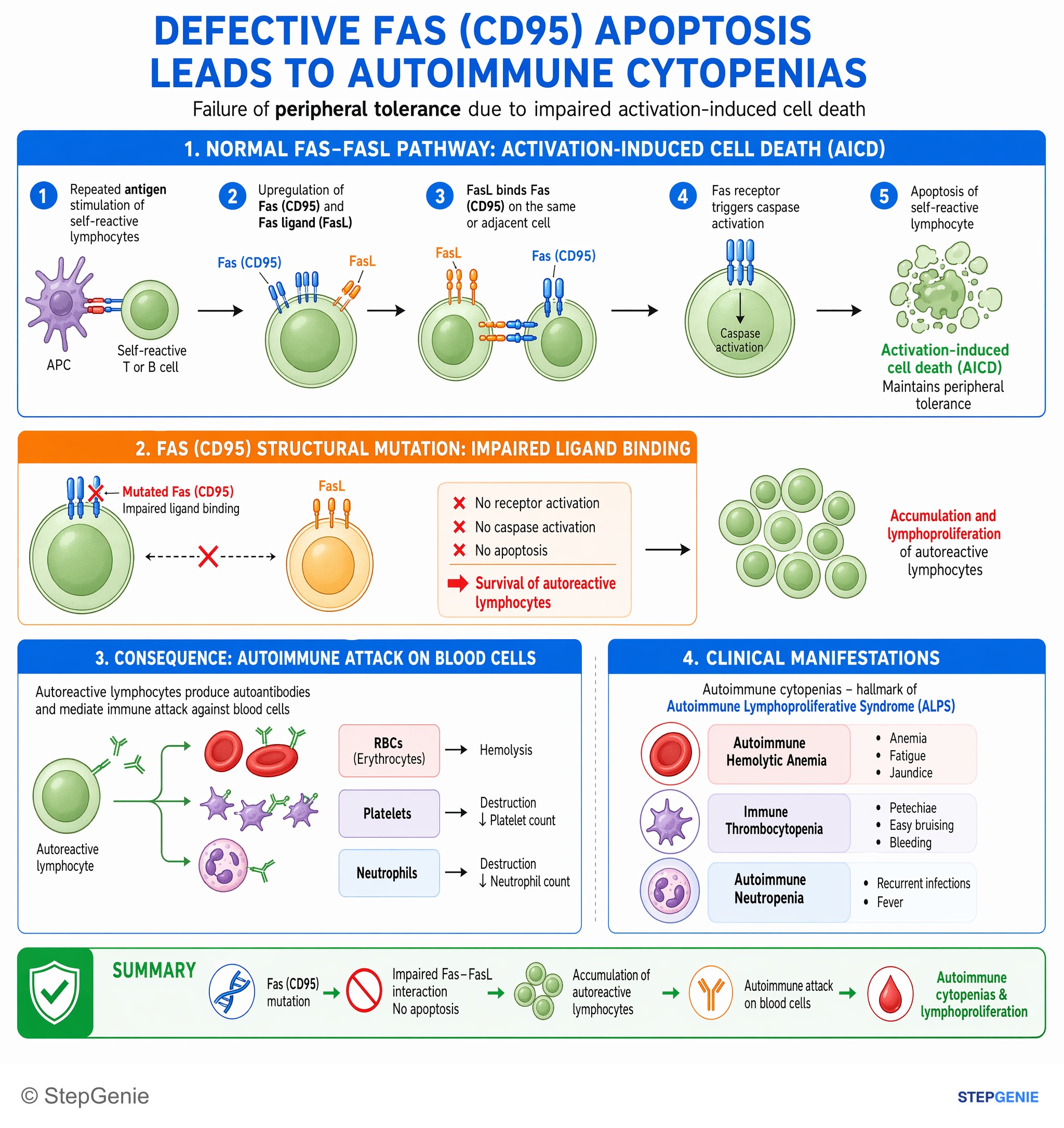
Defective Fas (CD95) apoptosis in autoreactive lymphocytes → failure of peripheral tolerance → autoimmune cytopenias and…
USMLE Step 1Frequently Asked Questions
How do you tell apoptosis from necrosis on a Step 1 question stem?
Apoptosis is regulated, ATP-dependent cell death with cell shrinkage, intact membranes, apoptotic bodies, and minimal inflammation, while necrosis is unregulated injury-related cell death with membrane rupture, cell swelling, enzyme leakage, and prominent inflammation. On stems, apoptosis fits embryogenesis, immune regulation, hepatitis, or chemotherapy effects, whereas necrosis fits ischemia, toxins, infection, trauma, MI, stroke, abscess, pancreatitis, or vasculitis.
Why does apoptosis usually cause little or no inflammation?
Apoptosis causes minimal inflammation because intracellular contents are not spilled into surrounding tissue. The dying cell breaks into membrane-bound apoptotic bodies that expose eat-me signals such as phosphatidylserine, allowing rapid phagocytosis. Since the membrane stays intact instead of rupturing, the process avoids the enzyme leakage and tissue irritation that typically trigger inflammation in necrosis.
What are the intrinsic and extrinsic apoptosis pathways you need to know for Step 1?
The intrinsic pathway is triggered by internal stress such as DNA damage, growth factor withdrawal, or ER stress and works through Bax and Bak, cytochrome c release, Apaf-1, and initiator caspase-9. The extrinsic pathway is triggered by external death signals through Fas or TNF receptors, recruits adaptor proteins like FADD, and activates initiator caspase-8. Both pathways converge on executioner caspases 3, 6, and 7.
Why does necrosis lead to inflammation and elevated serum enzymes?
Necrosis leads to inflammation and serum enzyme elevation because severe injury disrupts the cell membrane, causing intracellular contents to leak out. This leakage provokes inflammation and releases measurable enzymes into the blood. Common examples are troponin and CK-MB in myocardial infarction, lipase in pancreatitis, and AST and ALT in hepatocellular injury. ATP depletion, calcium influx, reactive oxygen species, and lysosomal enzyme leakage contribute to the damage.
What is coagulative necrosis, and where do you see it?
Coagulative necrosis is a necrosis pattern in which protein denaturation predominates over enzymatic digestion, so the basic tissue architecture is preserved. It classically occurs in ischemic infarcts of most solid organs, especially the heart, kidney, and spleen, but not the brain. Histology shows preserved tissue outlines or ghost cells, increased eosinophilia, and loss of nuclei through karyolysis.