Inflammation questions love to hide in plain sight: the vignette looks like “just an infection,” but the answer hinges on timing, cell types, and what the tissue is actually doing. If you train yourself to interpret every answer choice as a mini-pathway (not just a word), you’ll start picking up easy points—especially on acute vs chronic inflammation.
Tag: Pathology > General Pathology
The Clinical Vignette (Q-Bank Style)
A 54-year-old man with poorly controlled type 2 diabetes presents with a nonhealing ulcer on the plantar surface of his foot for 3 months. He reports minimal pain. Exam shows a 2-cm ulcer with a pale base and scant serous drainage. He is afebrile. Debridement is performed. Histology from the wound base shows numerous small blood vessels, proliferating fibroblasts, and a mixed inflammatory infiltrate with macrophages and lymphocytes.
Which of the following is the most likely predominant process occurring in this wound?
A. Neutrophil chemotaxis mediated by IL-8 and complement C5a
B. Granulation tissue formation driven by VEGF and TGF-β
C. Histamine-mediated vasodilation from mast cell degranulation
D. Formation of fibrin-rich exudate due to increased vascular permeability
E. Caseating granuloma formation mediated by Th1 cells and IFN-γ
Step 1: Identify the Pattern (Acute vs Chronic)
This is chronic inflammation + impaired wound healing, not acute:
- Time course: 3 months (chronic by definition)
- Cell types: macrophages + lymphocytes (chronic), not neutrophils (acute)
- Tissue response: angiogenesis + fibroblasts = repair response (granulation tissue)
- Clinical clue: diabetes → poor perfusion/neuropathy → chronic nonhealing ulcer
Correct Answer: B. Granulation tissue formation driven by VEGF and TGF-β
Why B is correct
Granulation tissue is the classic histologic hallmark of healing wounds and chronic ulcers:
- New capillaries (angiogenesis) → “numerous small blood vessels”
- Fibroblast proliferation → early collagen deposition (type III first)
- Loose ECM + inflammatory cells → often macrophage-predominant
Cytokines you’re expected to know
- VEGF: angiogenesis (new vessels)
- TGF-β: stimulates fibroblasts, collagen deposition, scar formation; anti-inflammatory effects too
- PDGF: fibroblast migration/proliferation, smooth muscle proliferation
- FGF: angiogenesis + fibroblasts
USMLE punchline:
Chronic inflammation = macrophages + lymphocytes + tissue destruction + repair (fibrosis/angiogenesis). Granulation tissue is the “repair” half of that equation.
Now the Real Game: Why Each Distractor Is Wrong (and When It’s Right)
A. Neutrophil chemotaxis mediated by IL-8 and complement C5a
Why it’s wrong here:
Neutrophils dominate acute inflammation, especially early (hours to a couple days). This wound is 3 months old and shows macrophages/lymphocytes, not neutrophils.
When A would be right:
- Acute bacterial infection (e.g., appendicitis, cellulitis)
- Early inflammation after MI (first 1–3 days)
- Classic chemotaxis mediators:
- C5a
- IL-8
- LTB4
- Bacterial N-formylmethionine peptides
High-yield memory hook:
Chemotaxis = C5a, IL-8, LTB4, bacteria.
C. Histamine-mediated vasodilation from mast cell degranulation
Why it’s wrong here:
Histamine is an immediate, early-phase mediator (seconds to minutes) causing:
- Arteriolar vasodilation
- Increased venular permeability (via endothelial contraction)
But a 3-month ulcer with angiogenesis and fibroblasts is way past the histamine window.
When C would be right:
- Urticaria (hives), anaphylaxis
- Early acute inflammation after trauma
- Type I hypersensitivity reactions
High-yield detail:
Histamine release triggers: IgE cross-linking, C3a/C5a (anaphylatoxins), physical injury.
D. Formation of fibrin-rich exudate due to increased vascular permeability
Why it’s wrong here:
A fibrinous exudate is classically acute and occurs with significant vascular leak—often on serosal surfaces.
This vignette points to granulation tissue (angiogenesis + fibroblasts) and chronic inflammatory cells, not an exudative acute process.
When D would be right:
- Fibrinous pericarditis (“bread and butter”) after MI
- Fibrinous pleuritis or peritonitis
- Severe acute inflammation where fibrinogen leaks and polymerizes to fibrin
Exudate vs transudate (fast differentiation):
| Feature | Exudate | Transudate |
|---|---|---|
| Mechanism | ↑ vascular permeability (inflammation) | ↑ hydrostatic pressure or ↓ oncotic pressure |
| Protein content | High | Low |
| Cells | Many inflammatory cells | Few |
| Examples | Pneumonia, abscess, pericarditis | CHF, cirrhosis, nephrotic syndrome |
E. Caseating granuloma formation mediated by Th1 cells and IFN-γ
Why it’s wrong here:
Granulomas are a special subtype of chronic inflammation, but the histology described is granulation tissue (healing), not granulomatous inflammation.
Granulomas = organized collections of epithelioid macrophages and giant cells, typically formed when the immune system can’t eradicate a pathogen or foreign material.
When E would be right:
- TB (caseating granulomas)
- Histoplasma, some fungal infections
- Sarcoidosis (noncaseating)
- Foreign body granulomas (suture, talc)
High-yield cytokine axis:
- Th1 → IFN-γ activates macrophages
- TNF-α helps maintain granulomas (and explains why anti–TNF-α increases TB reactivation risk)
Acute vs Chronic Inflammation: The Board-Friendly Framework
Acute inflammation (minutes → days)
Goal: deliver leukocytes + plasma proteins rapidly.
Key features
- Neutrophils (early dominant)
- Vascular changes: vasodilation + increased permeability
- Exudate and edema are common
Classic mediators
- Histamine, prostaglandins, leukotrienes
- Complement (especially C5a)
- Cytokines (IL-1, TNF-α), chemokines (IL-8)
Chronic inflammation (weeks → months)
Goal: contain and repair—often with ongoing injury.
Key features
- Macrophages, lymphocytes, plasma cells
- Tissue destruction
- Repair with angiogenesis and fibrosis
Big idea: chronic inflammation isn’t just “longer acute inflammation”—it’s a different cellular program.
Granulation Tissue vs Granuloma (Common USMLE Trap)
| Feature | Granulation tissue | Granuloma |
|---|---|---|
| Purpose | Wound healing/repair | Contain hard-to-eradicate agent |
| Main cells | Fibroblasts + new vessels + macrophages | Epithelioid macrophages + giant cells + T cells |
| Key mediators | VEGF, TGF-β, PDGF | IFN-γ, TNF-α |
| Typical setting | Healing wound, chronic ulcer | TB, sarcoid, foreign body |
If you see new capillaries + fibroblasts, think granulation tissue every time.
High-Yield Diabetes Tie-In (Why This Wound Won’t Heal)
Diabetes impairs wound healing through multiple mechanisms:
- Microvascular disease → reduced perfusion and oxygen delivery
- Neuropathy → unnoticed trauma → repeated injury
- Immune dysfunction → impaired neutrophil chemotaxis and phagocytosis
- Relative hypoxia → less efficient collagen synthesis and remodeling
USMLE connection: wound healing is oxygen dependent (hydroxylation of proline/lysine in collagen synthesis).
Rapid-Fire Takeaways (What to Remember on Test Day)
- Neutrophils = acute; macrophages/lymphocytes = chronic.
- Granulation tissue = angiogenesis (VEGF) + fibroblasts/collagen (TGF-β).
- Histamine is early and fast—think immediate vascular changes.
- Fibrinous exudate = acute, leaky vessels, often serosal surfaces.
- Granulomas = Th1/IFN-γ–driven macrophage activation; TB is the classic.
Visual explanations from the StepGenie QBank
Infographics StepGenie drew to explain questions in its own QBank, matched to this article's topic. Each caption is the takeaway of the question it explains.
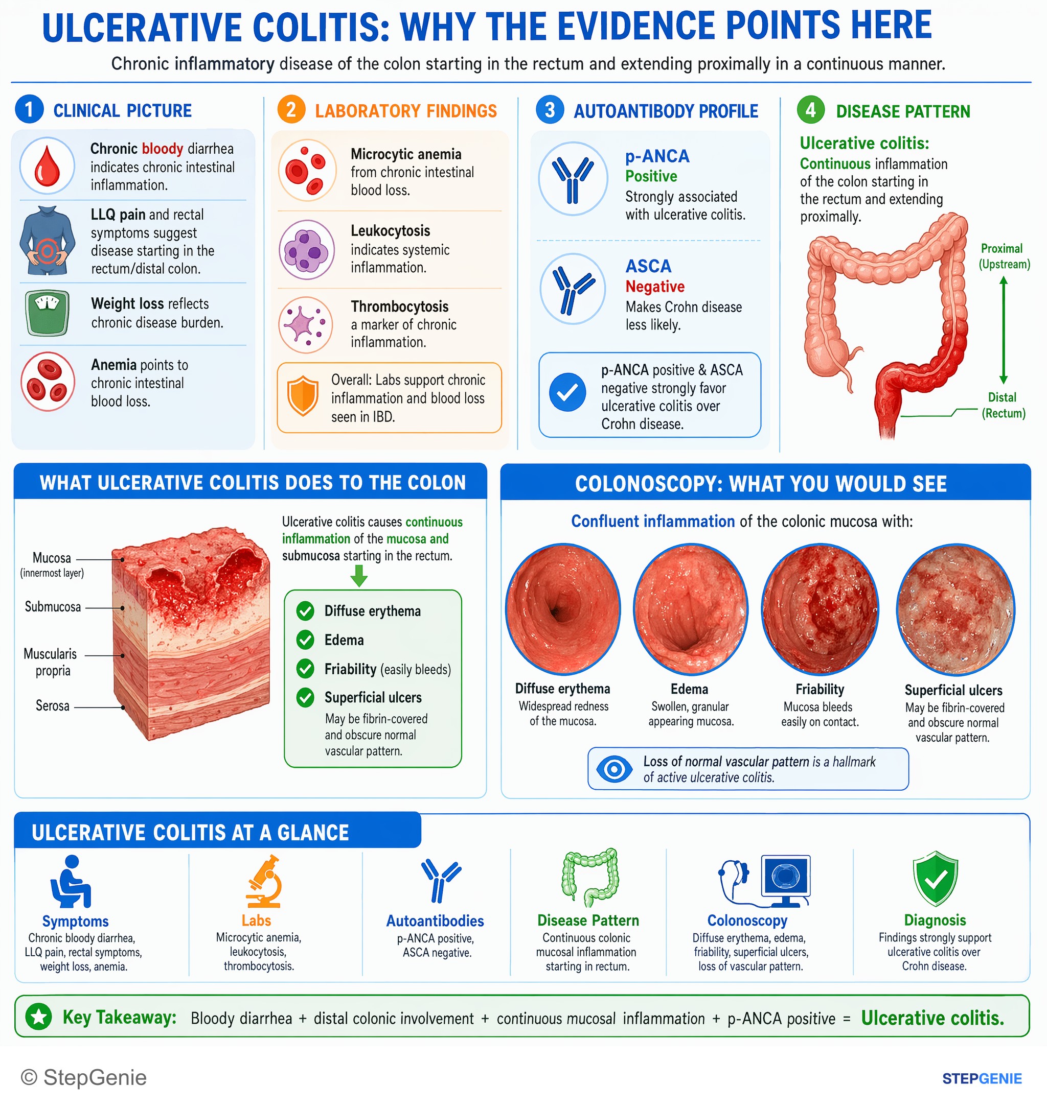
Ulcerative colitis: continuous colonic mucosal inflammation, bloody diarrhea, weight loss, positive p-ANCA; colonoscopy shows diffuse…
USMLE Step 1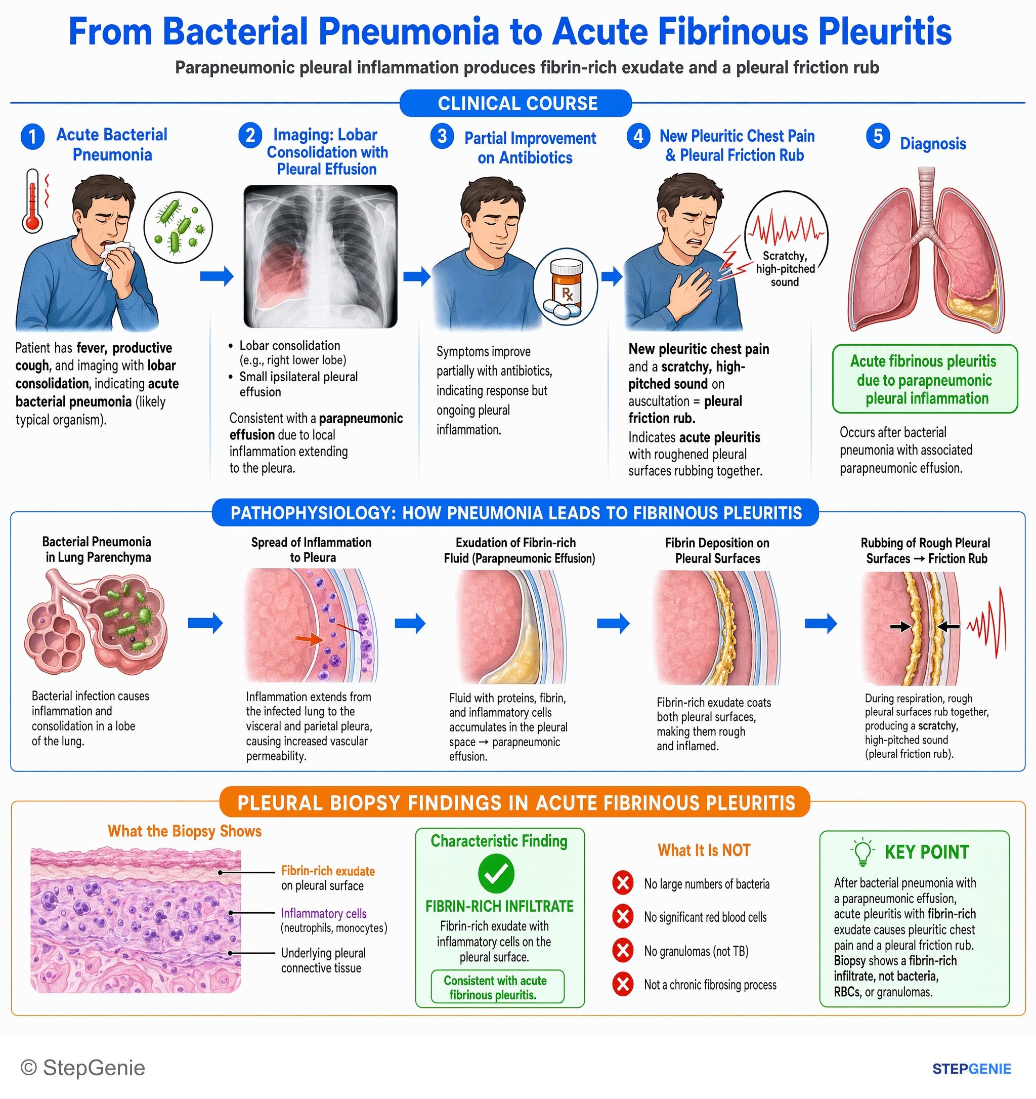
Acute fibrinous pleuritis from parapneumonic pleural inflammation shows a fibrin-rich exudate on the pleural surface after bacterial…
USMLE Step 1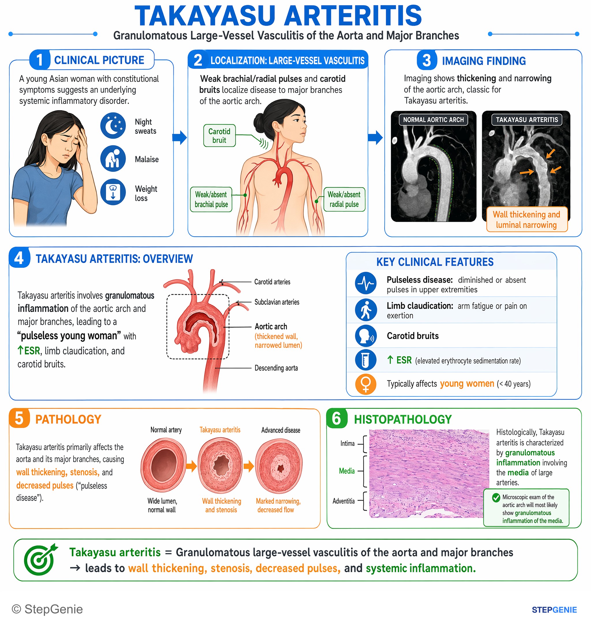
Takayasu arteritis: granulomatous inflammation of aortic arch and major branches → pulseless young woman, ↑ESR, limb claudication, carotid…
USMLE Step 1Frequently Asked Questions
How do you tell chronic inflammation from acute inflammation in a wound question?
Chronic inflammation is identified by a long time course, macrophages and lymphocytes, and evidence of repair such as angiogenesis and fibroblast proliferation. Acute inflammation is more likely when the process is minutes to days old and dominated by neutrophils, vasodilation, increased permeability, edema, and exudate. In a nonhealing wound present for months, chronic inflammation with impaired healing is the better fit.
What histology points to granulation tissue in a chronic ulcer?
Granulation tissue is suggested by numerous small blood vessels, proliferating fibroblasts, loose extracellular matrix, and inflammatory cells that are often macrophage-predominant. In a chronic ulcer, this pattern reflects the repair side of chronic inflammation. The combination of angiogenesis and fibroblast proliferation is especially important because it indicates wound healing activity rather than a purely acute exudative process.
Why is VEGF and TGF-beta the best answer for a nonhealing diabetic foot ulcer with small vessels and fibroblasts?
VEGF and TGF-β fit because they drive granulation tissue formation, which is the predominant process in a chronic healing wound. VEGF promotes angiogenesis, explaining the numerous small blood vessels, while TGF-β stimulates fibroblasts, collagen deposition, and scar formation. In a months-old diabetic ulcer with macrophages, lymphocytes, angiogenesis, and fibroblasts, this repair response is more consistent than acute inflammatory pathways.
Why are IL-8 and C5a wrong in a 3-month wound ulcer question?
IL-8 and C5a are wrong because they are classic mediators of neutrophil chemotaxis in acute inflammation, especially early in the process. A 3-month ulcer is chronic by definition and the histology shows macrophages and lymphocytes rather than neutrophils. Those findings point away from early acute inflammation and toward chronic inflammation with ongoing repair and granulation tissue formation.
How do you tell granulation tissue from a granuloma on pathology?
Granulation tissue and granulomas are both linked to chronic inflammation, but they look different and represent different processes. Granulation tissue is a healing response with new capillaries, proliferating fibroblasts, loose extracellular matrix, and inflammatory cells. A granuloma is an organized collection of epithelioid macrophages and giant cells, often forming when the immune system cannot eradicate a pathogen or foreign material.