High-yield pathology images for the USMLE
StepGenie explanation infographics. Free to view, no account needed.
What do StepGenie's pathology images cover?
Each pathology image is a StepGenie infographic drawn for one of its own practice questions. It connects the tissue-level change the question describes to the mechanism that produced it, so the histology or the biopsy finding stops being a label and becomes a step in a process.
Pathology is a discipline rather than an organ, so this set draws from everywhere. The largest groups are hepatic disorders, inflammatory dermatoses and bullous disease, glomerular and nephrotic-nephritic syndromes, intestinal and colorectal disease, white cell disorders and the general principles of oncology.
The images most worth your time are the ones that put a stain or a microscopic pattern next to the mechanism it reports. A membranous versus membranoproliferative panel does more for a glomerular question than either description alone, because the disagreement between them is exactly what the question is testing. The neoplasia panels work the other way round: they start at the mutation or the exposure and follow it forward to the tissue change.
Since these images are filed by the question's discipline, they overlap heavily with the organ-system pages. If you are revising renal pathology specifically, the nephrology gallery will be the denser page.
Cell injury and inflammation panels are the general-principles core of this set. They are the images most likely to help on a question that names no organ at all, and they are worth working through early in a Step 1 block rather than saving for the end.
Topics in this set
Labels are the question-bank topics these images are filed under. Step 1 and Step 2 file some of the same material under different names, so a few labels here merge both.
Pathology explanation images
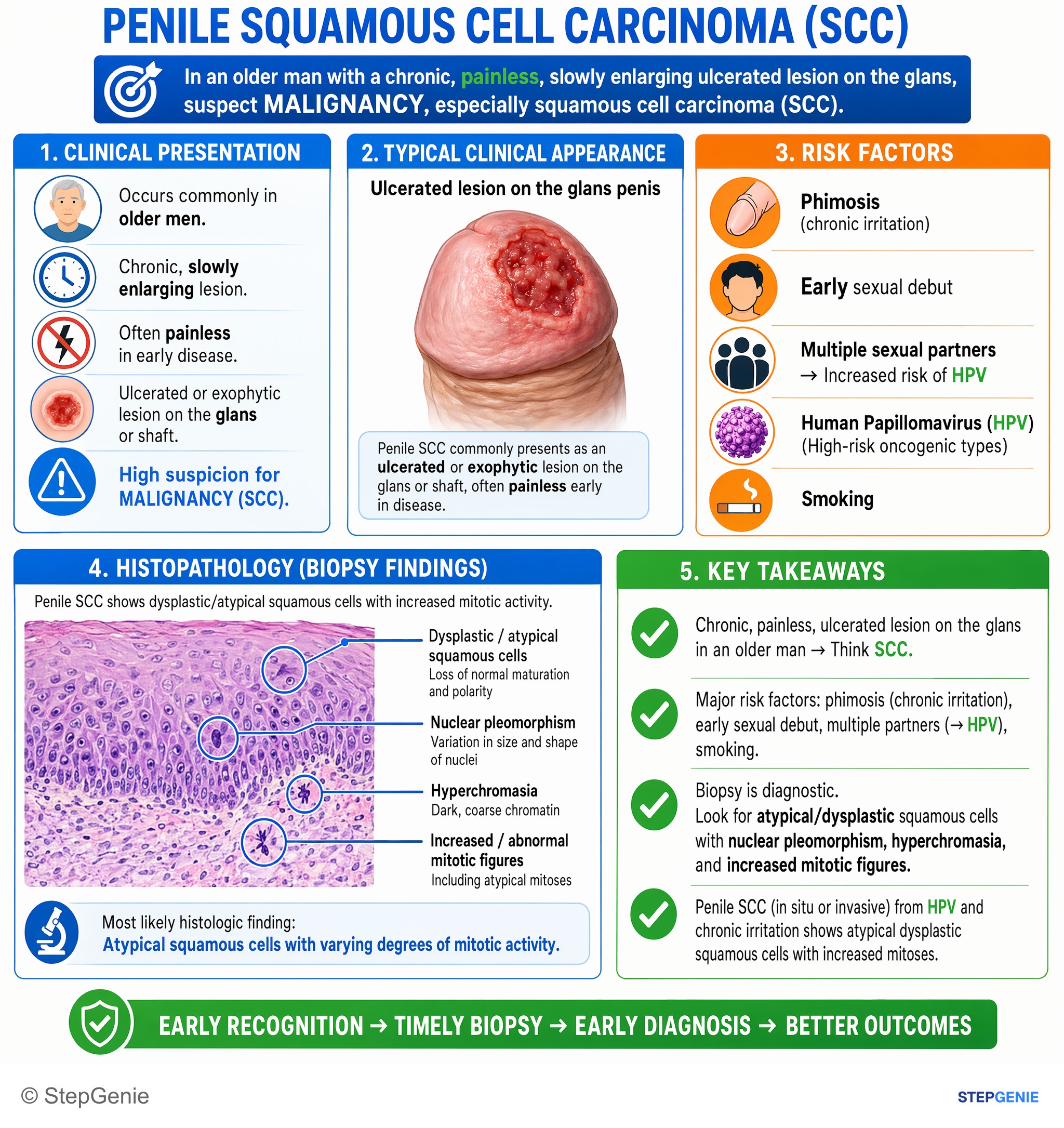
Penile squamous cell carcinoma in situ/invasive from HPV and chronic irritation shows atypical dysplastic squamous cells with increased…
USMLE Step 1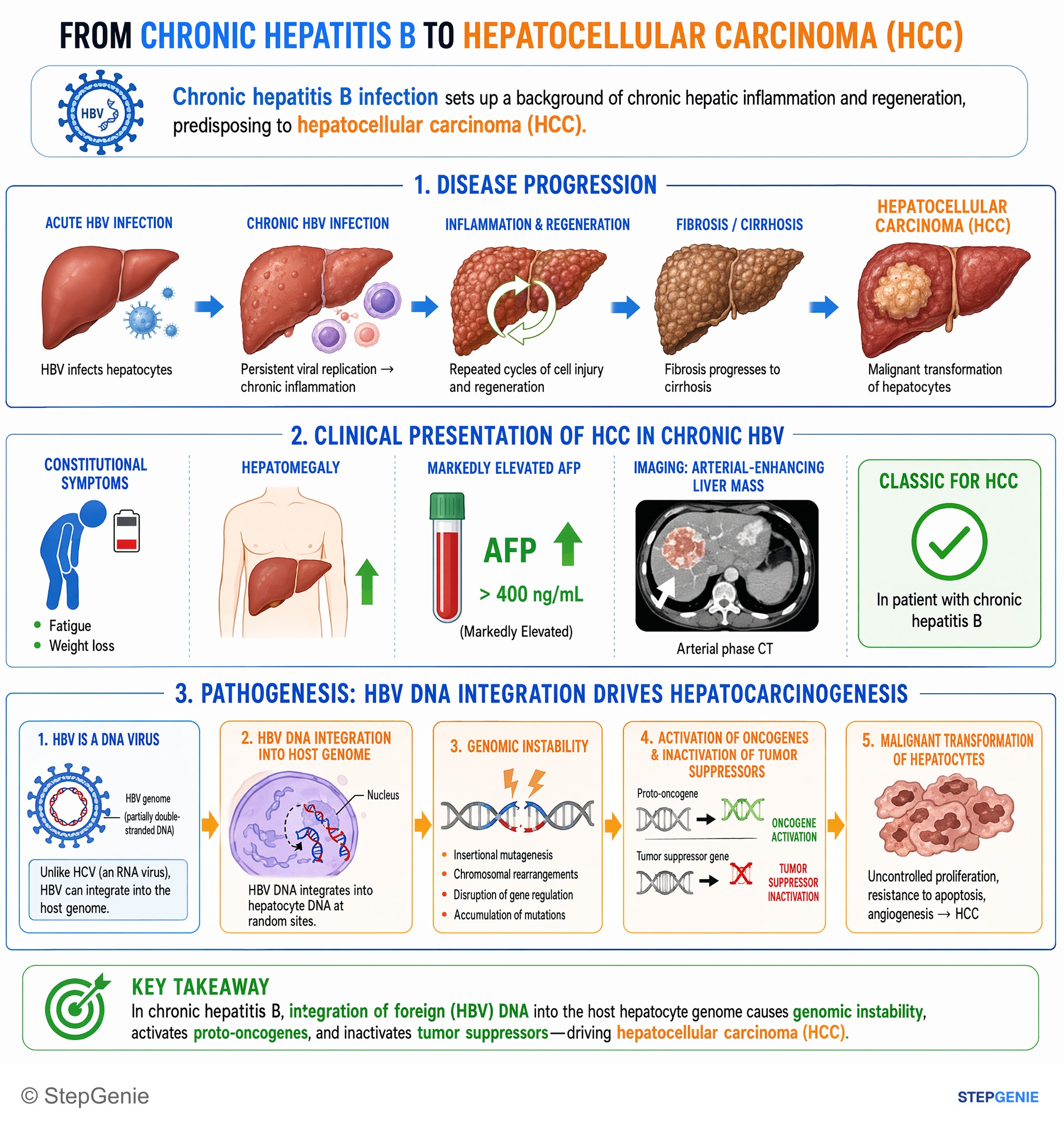
Hepatocellular carcinoma in chronic hepatitis B arises via HBV DNA integration causing genomic instability and oncogene activation in…
USMLE Step 1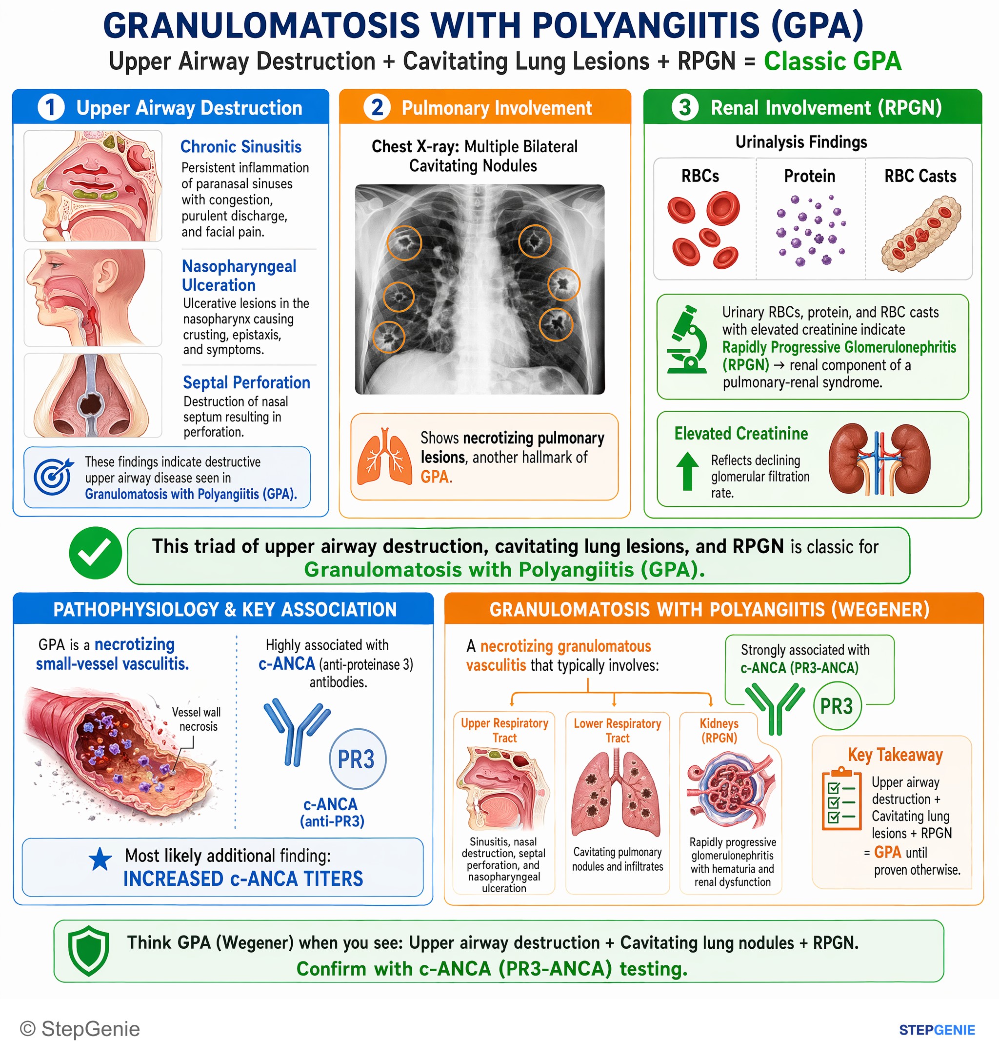
Granulomatosis with polyangiitis (Wegener): necrotizing granulomatous vasculitis of upper/lower respiratory tract plus RPGN; strongly…
USMLE Step 1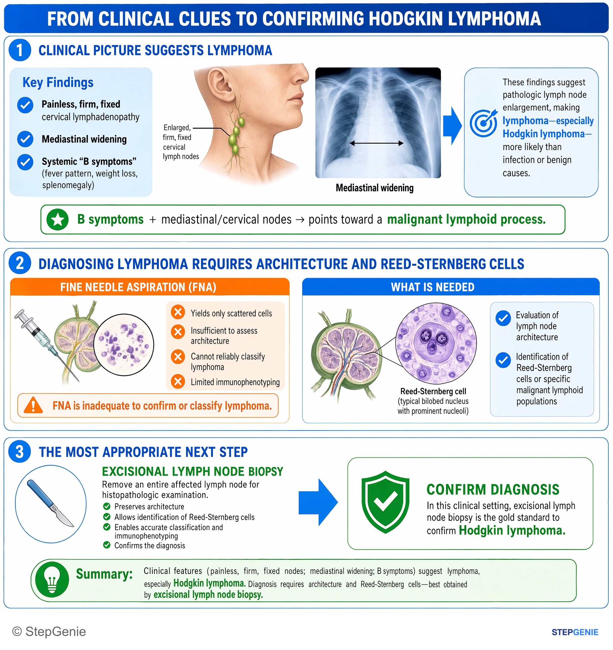
Hodgkin lymphoma suspected from B symptoms and mediastinal/cervical nodes; confirm with excisional lymph node biopsy for architecture and…
USMLE Step 1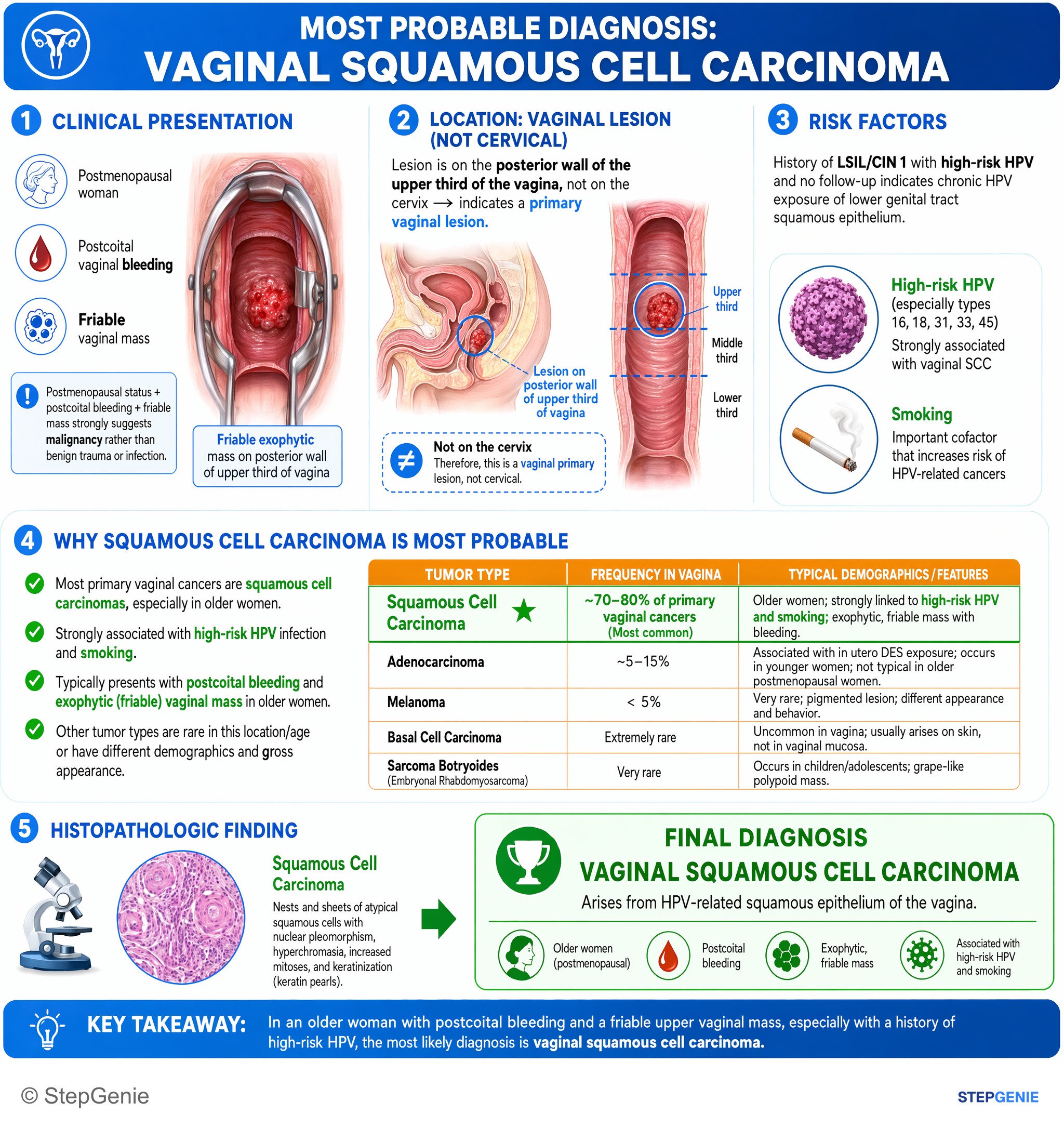
Vaginal squamous cell carcinoma arises from HPV-related squamous epithelium; presents with postcoital bleeding and exophytic vaginal mass…
USMLE Step 1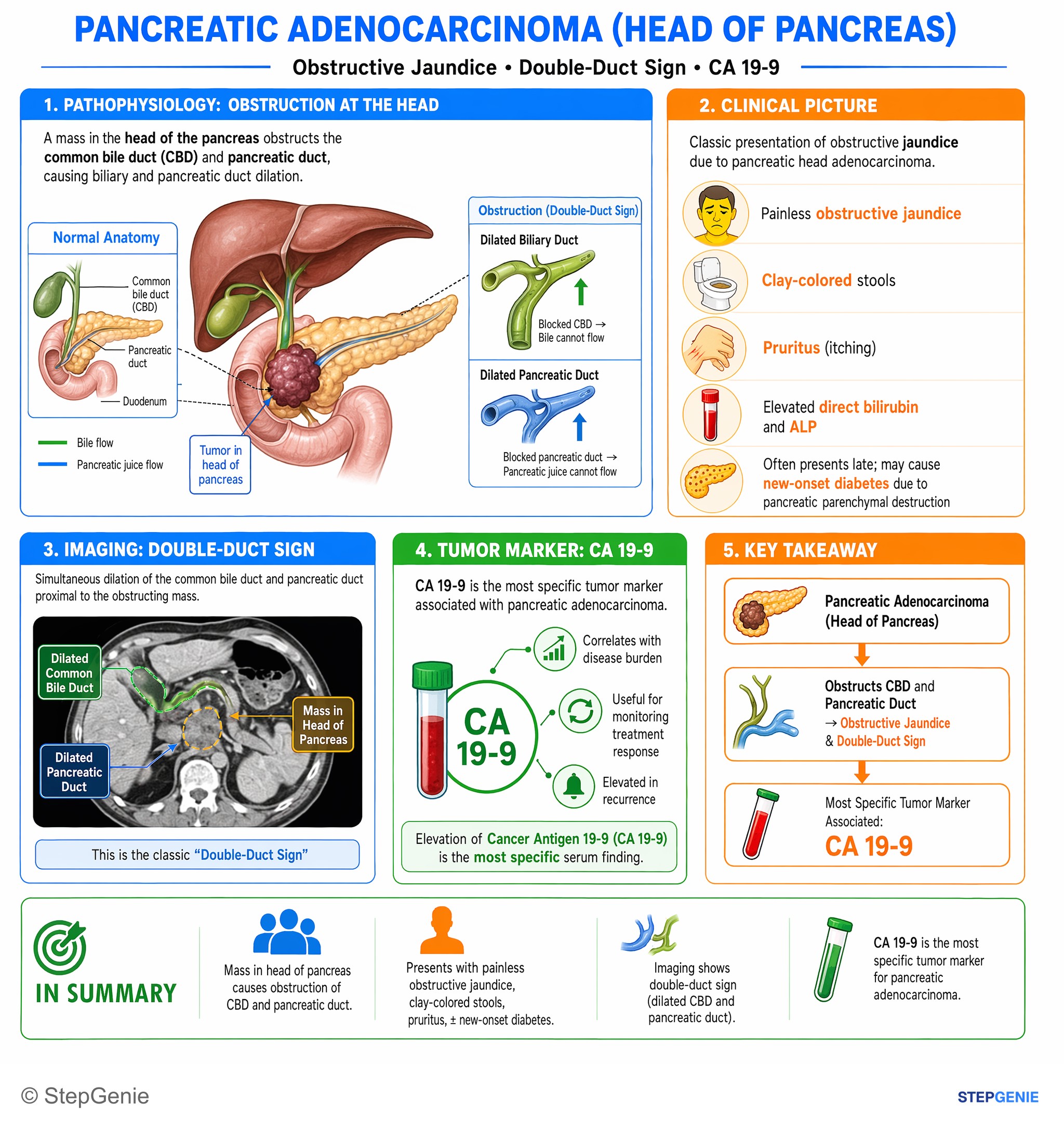
Pancreatic adenocarcinoma (head of pancreas) → obstructive jaundice, double-duct sign; most specific associated tumor marker is CA 19-9.
USMLE Step 1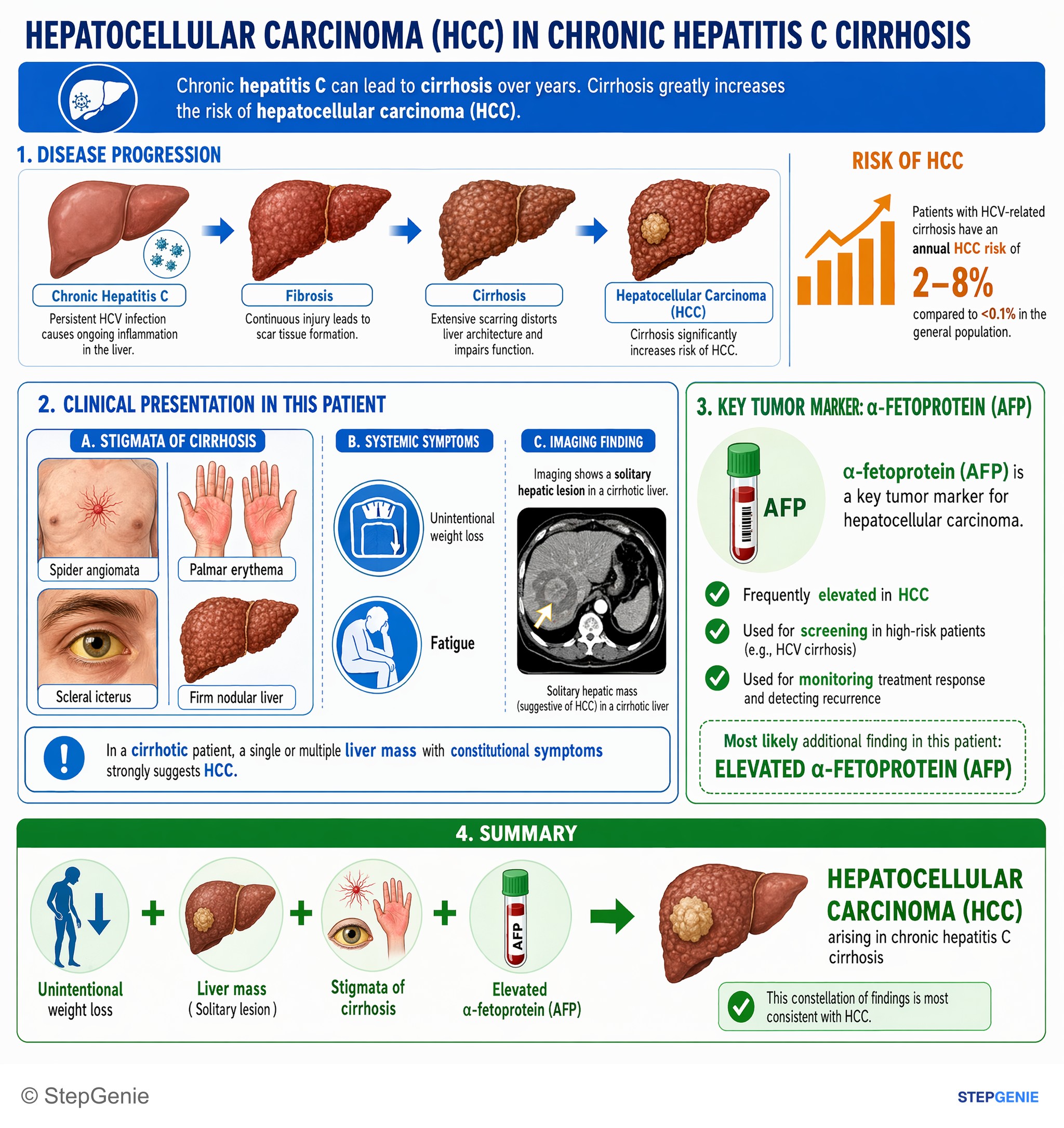
Hepatocellular carcinoma arising in chronic hepatitis C cirrhosis presents with weight loss, liver mass, stigmata of cirrhosis, and…
USMLE Step 1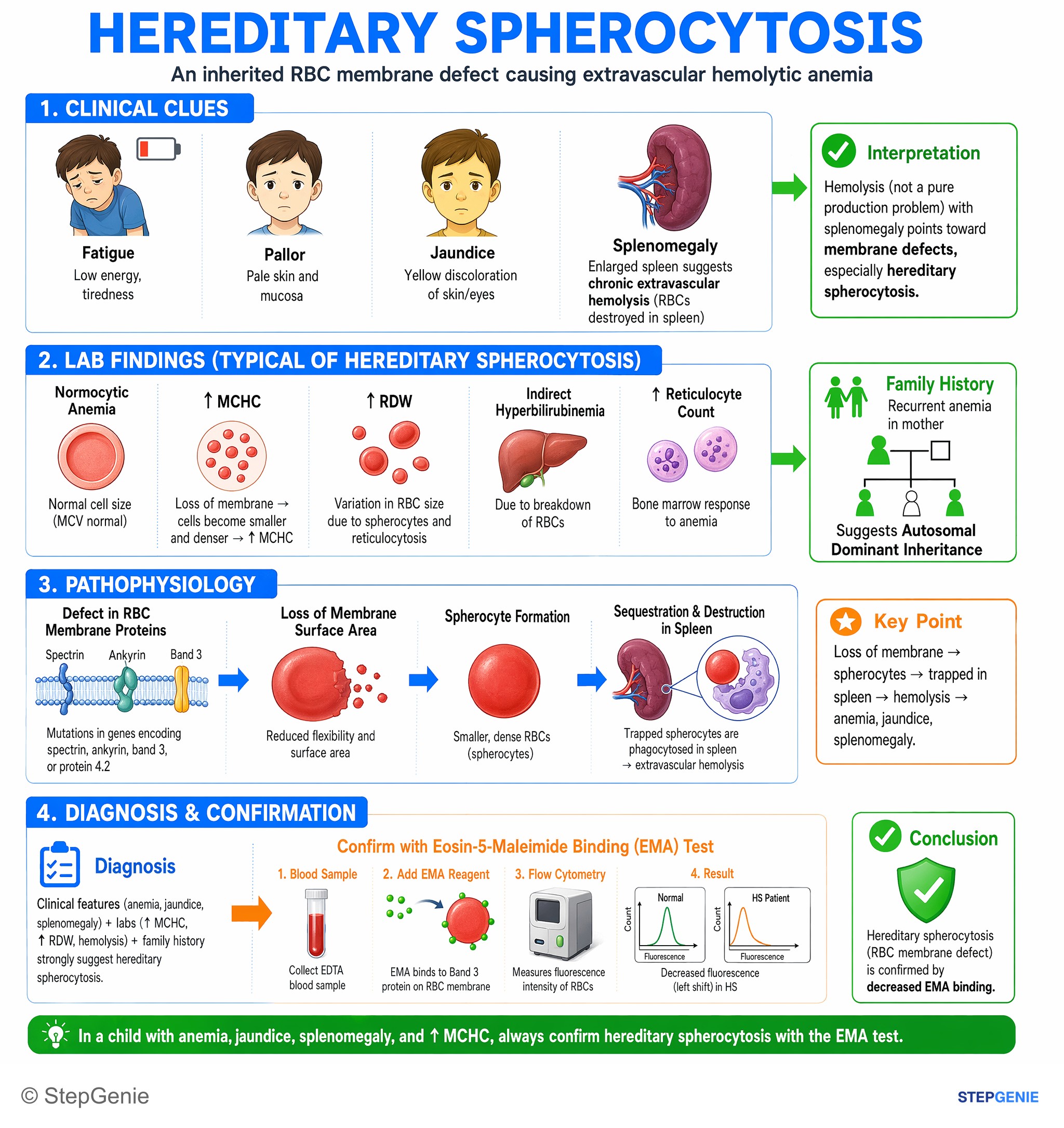
Hereditary spherocytosis in a child with anemia, jaundice, splenomegaly, ↑MCHC → confirm with eosin-5-maleimide binding (EMA) test for…
USMLE Step 1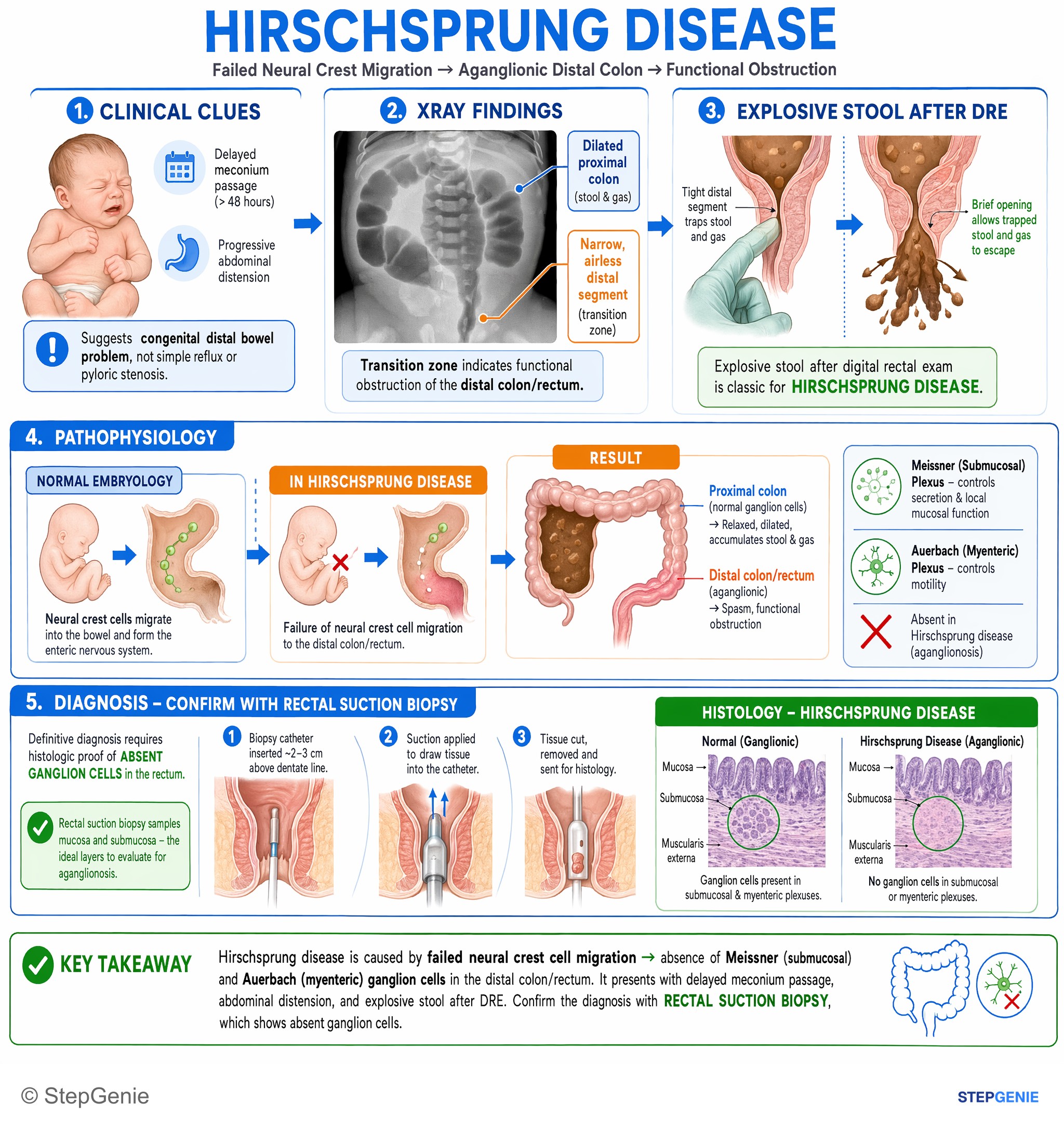
Hirschsprung disease from failed neural crest migration → aganglionic distal colon; confirm with rectal suction biopsy showing absent…
USMLE Step 1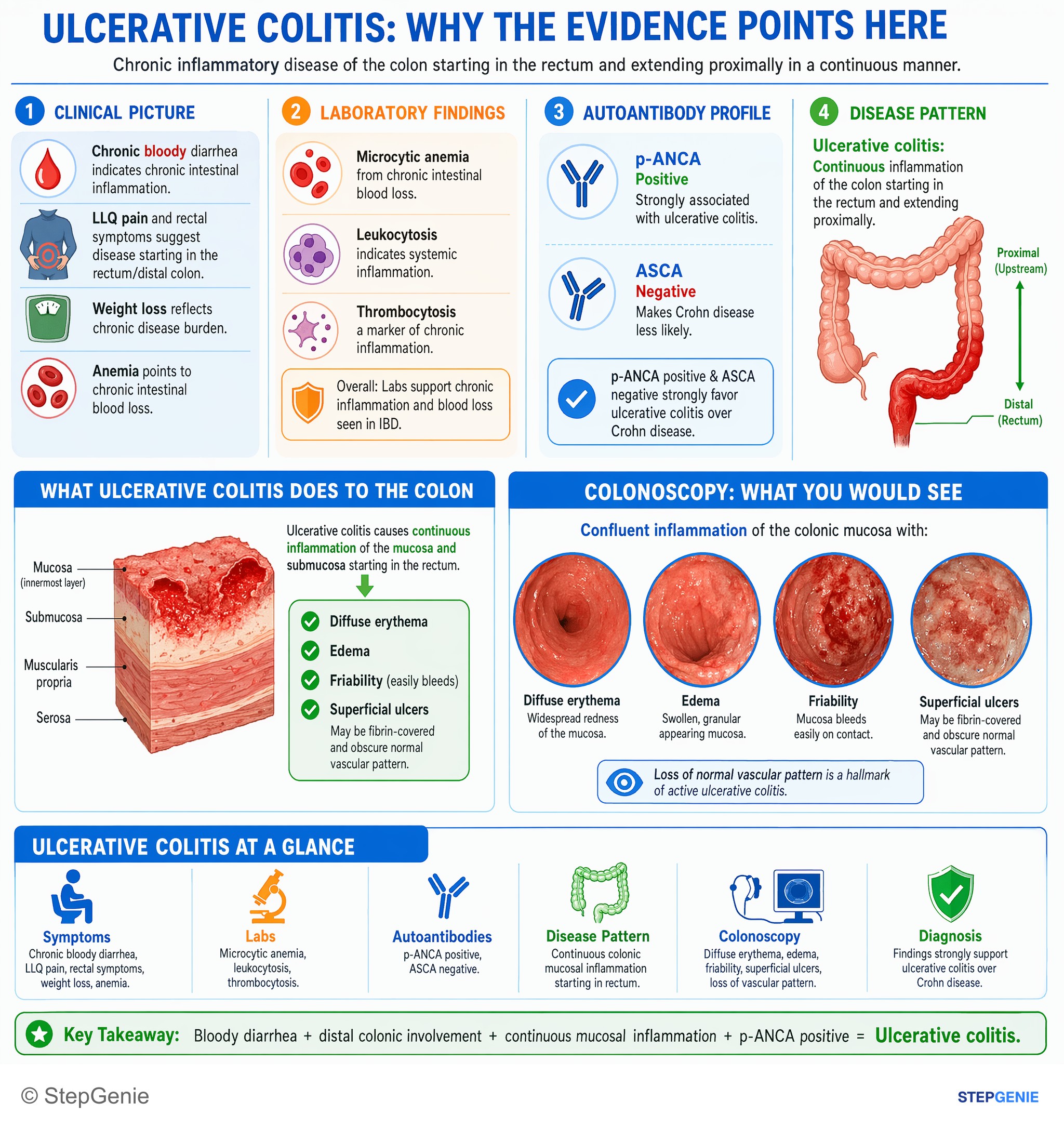
Ulcerative colitis: continuous colonic mucosal inflammation, bloody diarrhea, weight loss, positive p-ANCA; colonoscopy shows diffuse…
USMLE Step 1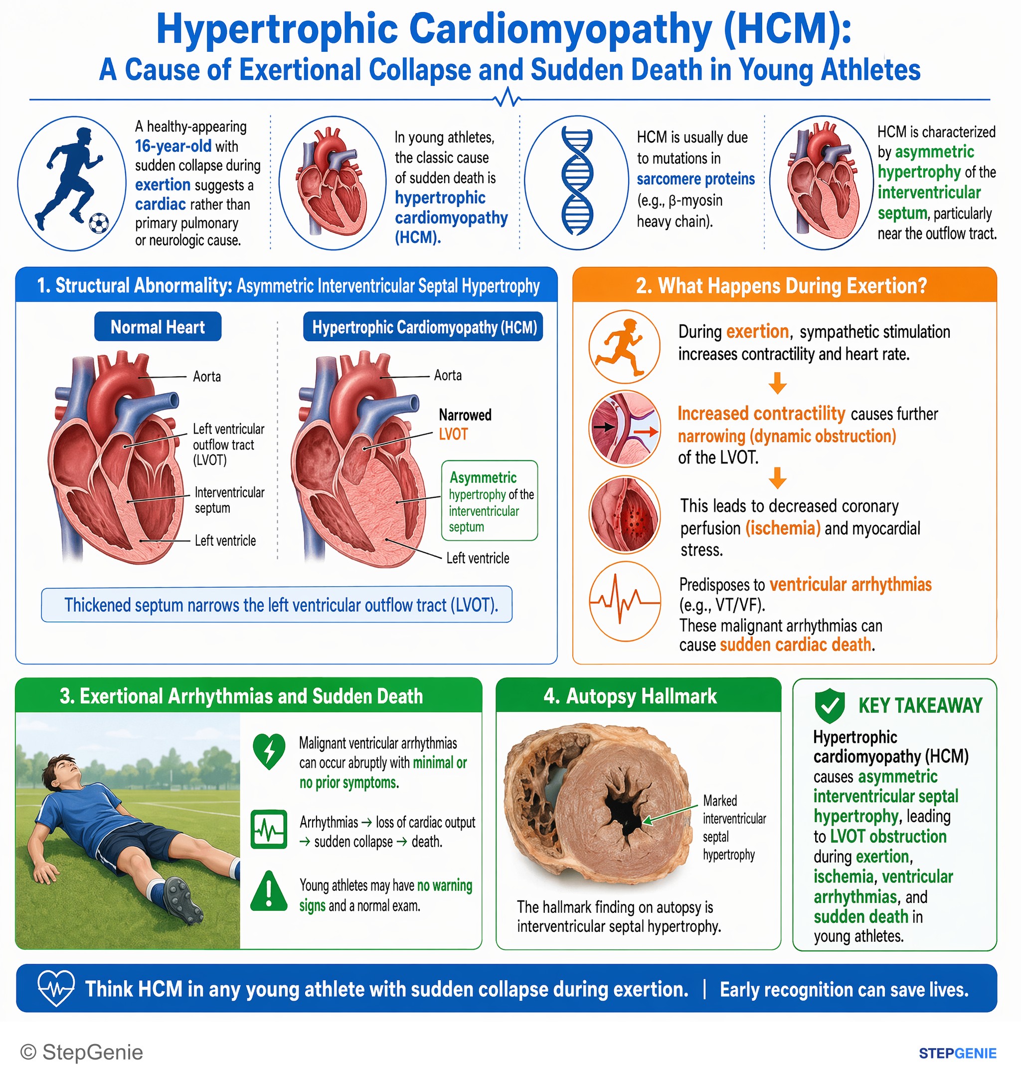
Hypertrophic cardiomyopathy with asymmetric interventricular septal hypertrophy causes exertional arrhythmias and sudden death in young…
USMLE Step 1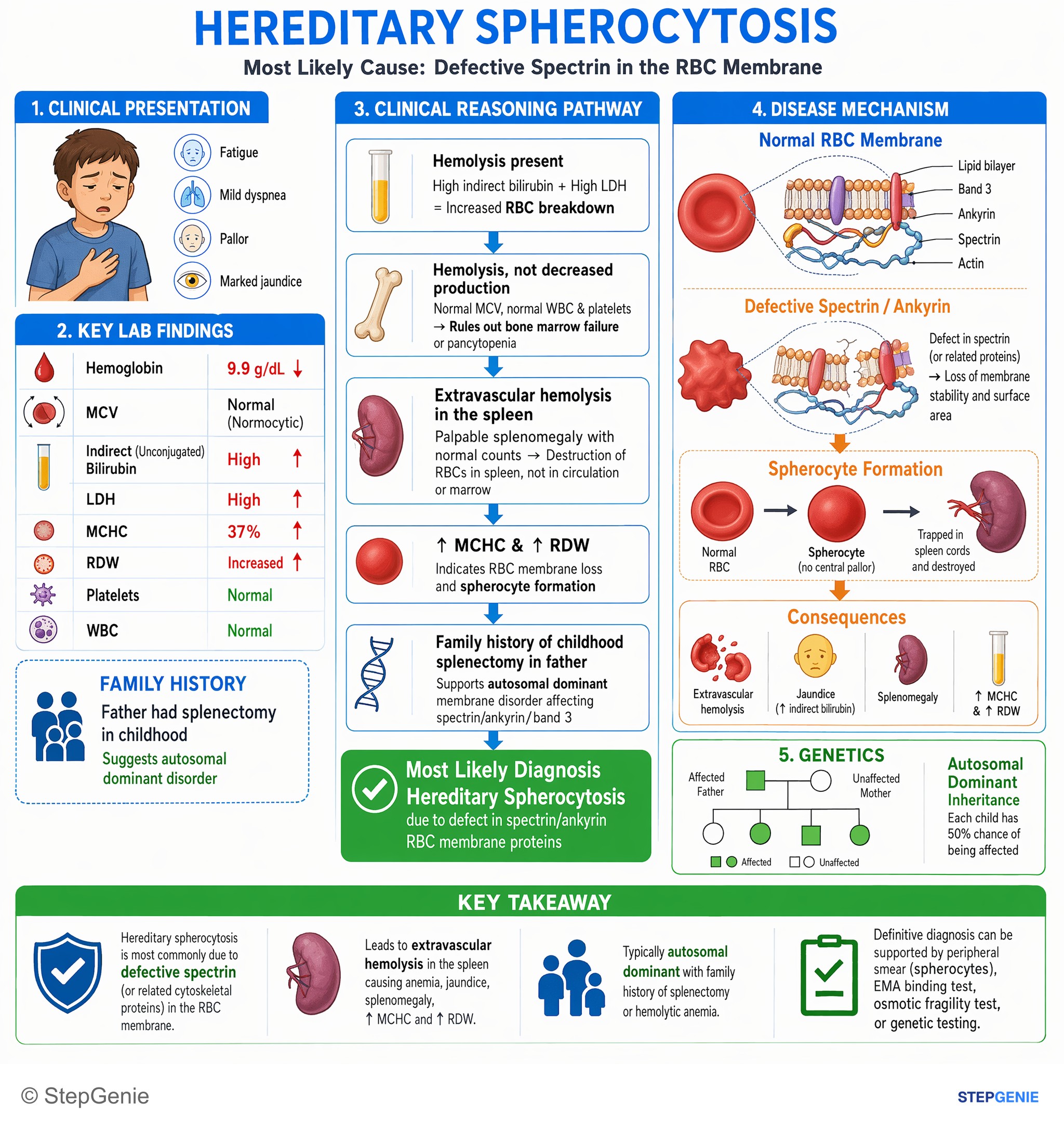
Hereditary spherocytosis due to spectrin/ankyrin defect → extravascular hemolysis, splenomegaly, ↑MCHC, jaundice; often autosomal dominant…
USMLE Step 1329 publishable pathology infographics in the library — all of them from Step 1 questions, counted on October 29, 2026. This page shows 48 of them. An image is listed when its question is active, has a full structured explanation, passed verification and carries no unresolved report.
This page caps out at 48 images. 281 more pathology infographics sit in the in-app image library, where they are searchable and filterable by subject, system and topic.
Open the image libraryPractice the questions behind these images
Each image explains one question. The pathology topic page has the written explanations and the question sets, and the preview lets you answer three questions without an account.
Other image sets
Study pathology with the image attached
In the app, each of these sits at the end of the question it explains, alongside the step-by-step reasoning and a panel for every option you did not pick. Starting is free.
Start practicing free