Psoriatic arthritis (PsA) is one of those “Step-friendly” diseases because it sits right at the intersection of derm, rheum, immunology, and radiology. If you can recognize the pattern—inflammatory arthritis + psoriasis features + classic imaging—you’ll pick up easy points on Step 1 and be set up well for Step 2 clinical reasoning.
Where Psoriatic Arthritis Fits (Big Picture)
Psoriatic arthritis is a seronegative spondyloarthropathy associated with psoriasis, characterized by inflammatory joint disease that can involve:
- Peripheral joints (often asymmetric)
- Entheses (enthesitis)
- Axial skeleton (sacroiliitis, spondylitis)
High-yield classification:
PsA is in the seronegative spondyloarthropathies group (typically RF-negative, often associated with HLA-B27).
Definition (Step-Ready)
Psoriatic arthritis = a chronic inflammatory arthritis associated with psoriasis, featuring joint inflammation, enthesitis, and sometimes axial involvement, with characteristic deformities like dactylitis and arthritis mutilans in severe disease.
Pathophysiology (What’s Actually Happening)
Core immune drivers
PsA is immune-mediated with prominent T-cell activation and cytokine signaling, especially:
- TNF-α
- IL-17
- IL-23
These promote synovial inflammation and—importantly—enthesis-centered inflammation (a hallmark concept for the spondyloarthropathies).
Why entheses matter (HY mechanism)
The enthesis is where tendons/ligaments insert into bone. In PsA:
- Inflammation at entheses → pain at tendon insertions (e.g., Achilles)
- Can lead to bone erosions plus new bone formation (this combo helps explain some classic imaging patterns)
Genetics (what to remember)
- HLA-B27 association is strongest when there’s axial involvement
- Other HLA associations exist, but for Step purposes: think HLA-B27 = spondyloarthropathy family
Clinical Presentation (Recognize the Pattern Fast)
Joint pattern (classic Step clues)
PsA arthritis is often:
- Inflammatory (morning stiffness, improves with movement)
- Often asymmetric
- Frequently involves DIP joints (key differentiator from RA)
Common phenotypes
- Asymmetric oligoarthritis (most common)
- DIP-predominant arthritis
- Symmetric polyarthritis (can mimic RA but usually RF−, plus psoriasis clues)
- Arthritis mutilans (severe, deforming)
- Axial disease (sacroiliitis/spondylitis)
Signature physical exam findings (very HY)
- Dactylitis (“sausage digits”)
- Diffuse swelling of an entire finger/toe due to tenosynovitis + joint inflammation
- Enthesitis
- Achilles tendon insertion pain is classic
- Nail changes (big Step giveaway)
- Nail pitting
- Onycholysis
- Psoriasis clues
- Extensor surface plaques (silvery scale)
- Scalp involvement
- Intergluteal cleft plaques (often missed in vignettes but high-yield)
Extra-articular associations
- Uveitis can occur (shared with other spondyloarthropathies)
Diagnosis (Clinical + Targeted Labs/Imaging)
Key point
PsA is primarily a clinical diagnosis supported by labs and imaging.
Labs (what Step expects you to say)
- RF negative (seronegative)
- Anti-CCP usually negative (helps differentiate from RA)
- ESR/CRP often elevated (inflammatory)
- Consider HLA-B27 testing if axial symptoms
Quick comparison table (PsA vs RA vs OA)
| Feature | Psoriatic arthritis | Rheumatoid arthritis | Osteoarthritis |
|---|---|---|---|
| Inflammation | Yes | Yes | No/low-grade |
| RF / anti-CCP | Usually negative | Often positive | Negative |
| Joint distribution | Often asymmetric, DIP common | Symmetric, MCP/PIP, spares DIP | DIP/PIP, weight-bearing; bony enlargement |
| Morning stiffness | Yes (>1 hr) | Yes (>1 hr) | Brief (<30 min) |
| Key clues | Psoriasis, nail pitting, dactylitis, enthesitis | Nodules, deformities, erosions | Heberden/Bouchard nodes |
Imaging (high-yield radiology buzzwords)
Classic X-ray findings:
- “Pencil-in-cup” deformity
- Due to erosions + bone remodeling
- Joint space narrowing + erosions (inflammatory)
- New bone formation can be seen
- Possible sacroiliitis if axial involvement
Clinical pearl: RA is famous for marginal erosions and uniform joint space narrowing; PsA can show erosions too, but DIP involvement + pencil-in-cup + psoriasis/nails should push you to PsA.
Treatment (Step 1 Mechanism + Step 2 Practical)
Treatment depends on severity and axial vs peripheral disease.
Mild disease (peripheral, few joints)
- NSAIDs (symptomatic relief)
Moderate to severe peripheral disease
- DMARDs (to prevent progression)
- Methotrexate
- Sulfasalazine
- Leflunomide
Severe disease and/or axial disease, enthesitis, dactylitis
- Biologics (high-yield mechanism targets)
- TNF-α inhibitors: etanercept, infliximab, adalimumab
- IL-17 inhibitors: secukinumab, ixekizumab
- IL-12/23 inhibitor: ustekinumab
- IL-23 inhibitors (more psoriasis-focused but relevant): guselkumab, etc.
HY testable adverse effects (biologics)
- TNF-α inhibitors → reactivation of TB + serious infections
- Screening: TB testing before starting
- Risk of opportunistic infections overall with biologics
Glucocorticoids?
Systemic steroids aren’t a first-line long-term solution for PsA; on Step exams, the bigger steroid warning is in psoriasis, where systemic steroid withdrawal can trigger severe flares (including pustular psoriasis). In joint disease, localized injections may be used in real life, but don’t overreach on Step unless asked.
High-Yield Associations & “Vignette Triggers”
When to think PsA immediately
- Patient with psoriasis + joint pain + DIP involvement
- Sausage digits (dactylitis)
- Nail pitting or onycholysis
- RF negative inflammatory arthritis
- X-ray: pencil-in-cup
“Seronegative spondyloarthropathy” family cues (Step pattern recognition)
PsA shares features with:
- Ankylosing spondylitis
- Reactive arthritis
- IBD-associated arthritis
Shared themes:
- RF negative
- Enthesitis
- Possible uveitis
- HLA-B27 association (especially axial involvement)
First Aid Cross-References (What to Tie It To)
Use these First Aid “anchor points” when reviewing (section names may vary slightly by edition, but these are consistent concepts):
- MSK/Rheumatology: Seronegative spondyloarthropathies
- Psoriatic arthritis listed with RF− and HLA-B27 association
- Dermatology: Psoriasis
- Classic plaques + nail pitting
- Association with PsA
- Immunology/Pharm: TNF-α inhibitors
- Mechanism + TB reactivation risk
- Radiology/Ortho concepts
- Pencil-in-cup deformity as a distinguishing imaging clue
Rapid Review (Last-Minute Step Checklist)
Psoriatic arthritis =
- Seronegative inflammatory arthritis associated with psoriasis
- DIP involvement, often asymmetric
- Dactylitis, enthesitis, nail pitting
- Imaging: pencil-in-cup
- Tx: NSAIDs → DMARDs → biologics (TNF-α, IL-17, IL-23/12-23)
- Biologics adverse effect: TB reactivation, serious infections
Visual explanations from the StepGenie QBank
Infographics StepGenie drew to explain questions in its own QBank, matched to this article's topic. Each caption is the takeaway of the question it explains.
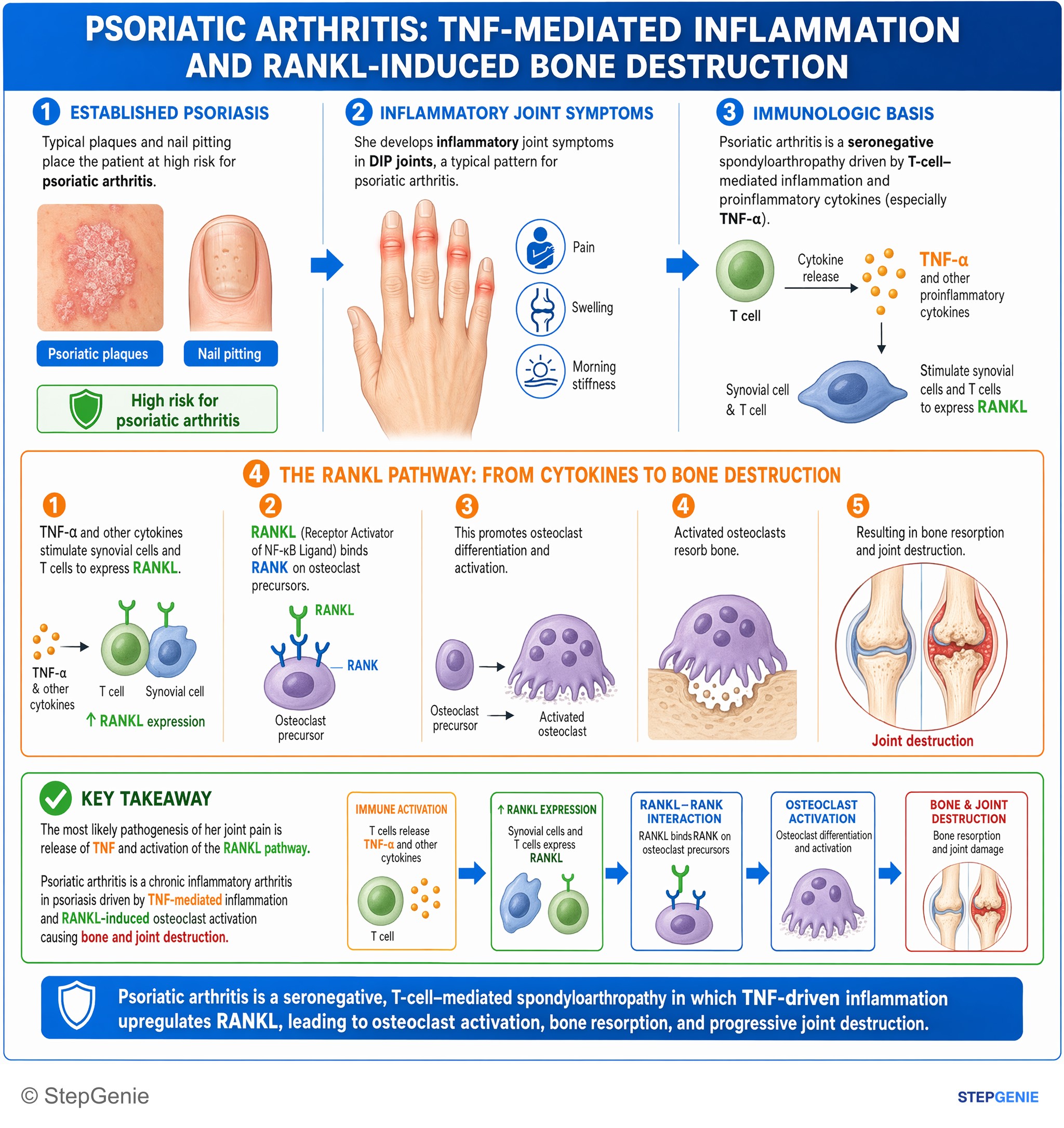
Psoriatic arthritis: chronic inflammatory arthritis in psoriasis driven by TNF-mediated inflammation and RANKL-induced osteoclast…
USMLE Step 1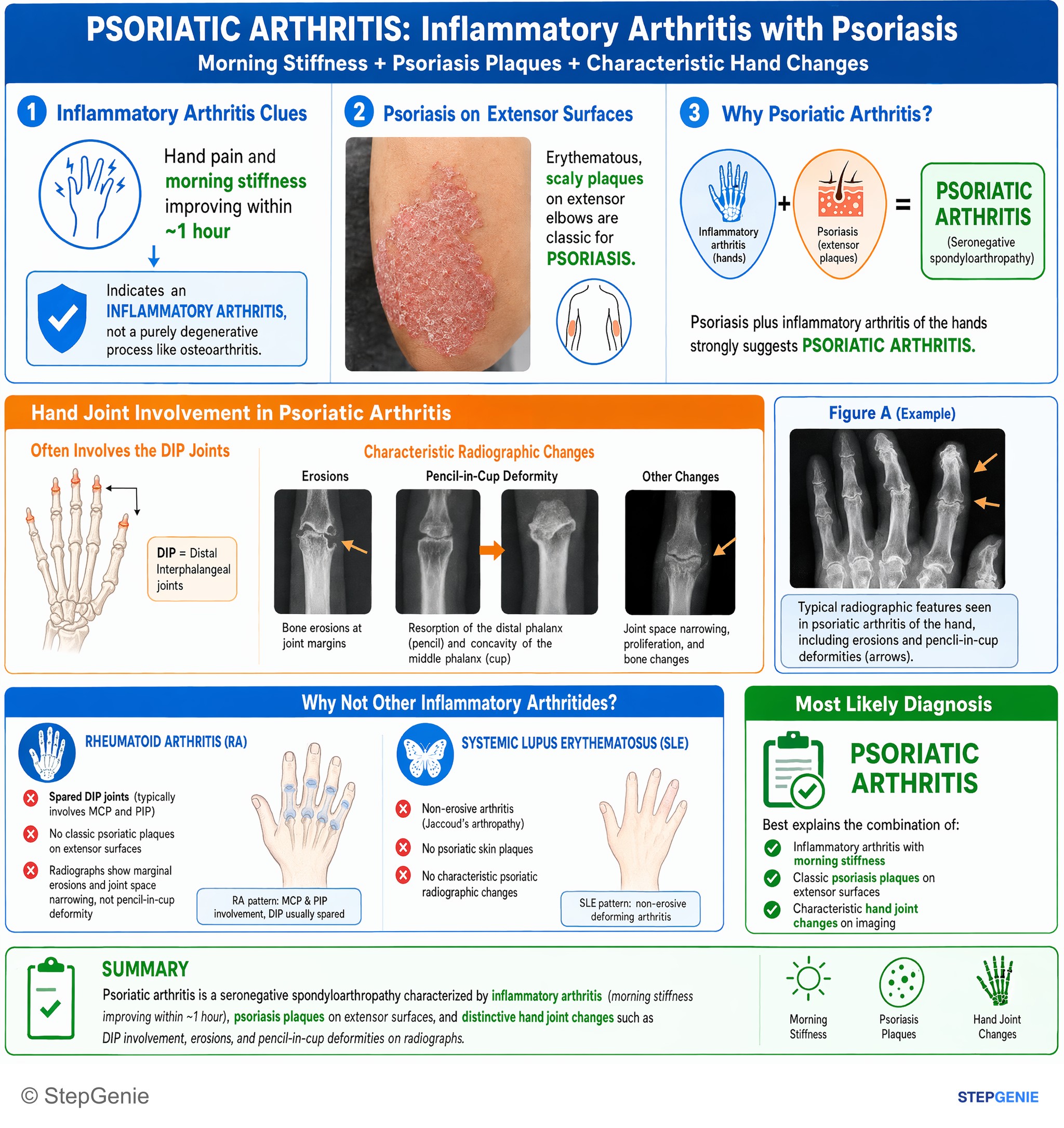
Psoriatic arthritis: inflammatory arthritis with morning stiffness plus classic psoriasis plaques on extensor surfaces and characteristic…
USMLE Step 2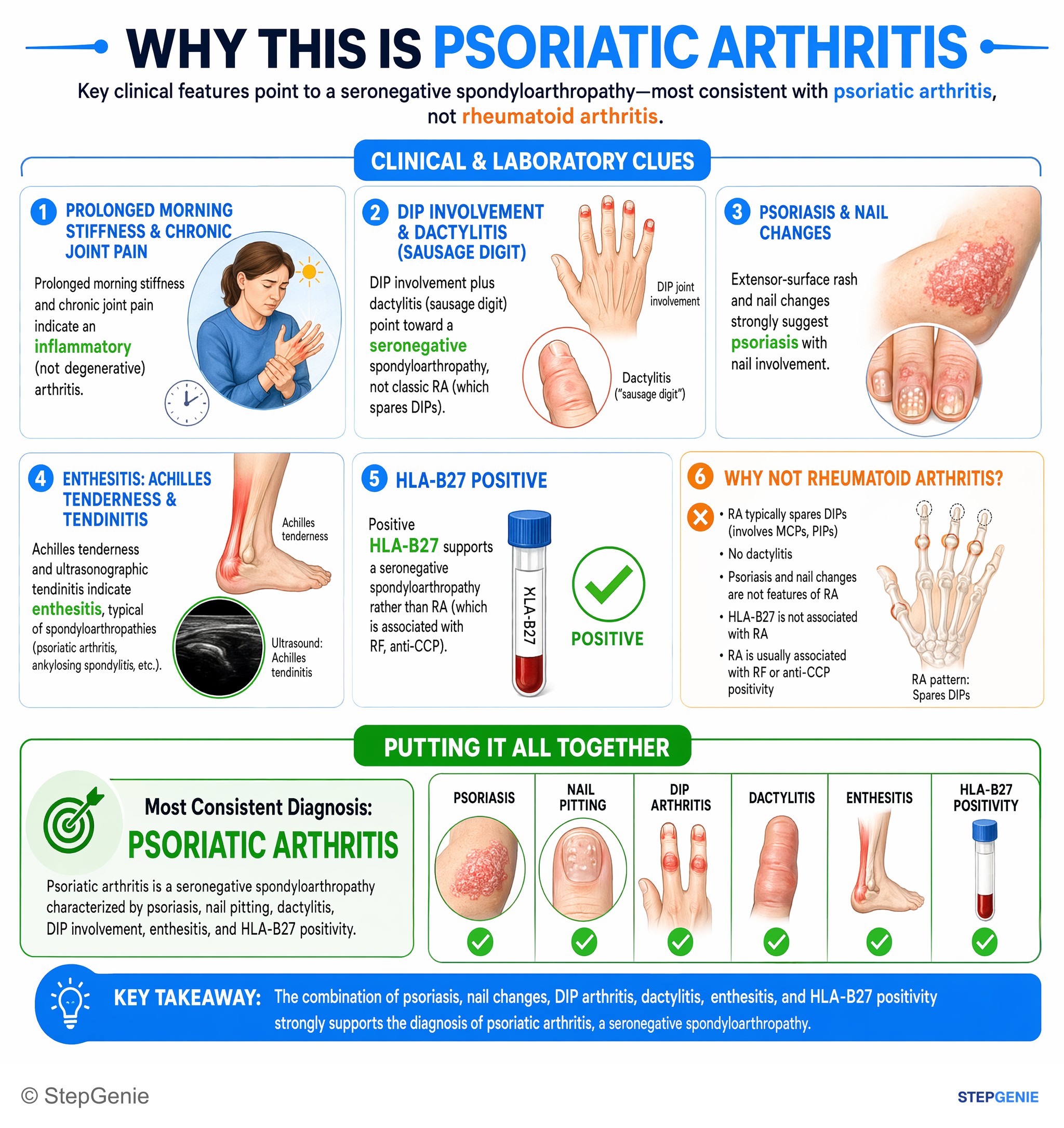
Psoriatic arthritis: seronegative spondyloarthropathy with psoriasis, nail pitting, dactylitis, DIP involvement, enthesitis, and HLA-B27…
USMLE Step 2Frequently Asked Questions
How do you tell psoriatic arthritis from rheumatoid arthritis on a Step question?
Psoriatic arthritis is suggested by inflammatory arthritis with psoriasis clues, DIP joint involvement, and seronegative labs. It is often asymmetric, commonly involves the DIP joints, and may show nail pitting, onycholysis, dactylitis, or enthesitis. Rheumatoid arthritis more often causes symmetric MCP and PIP disease with positive RF or anti-CCP and typically spares the DIP joints.
Why does psoriatic arthritis cause enthesitis and sausage digits?
Psoriatic arthritis causes enthesitis because it is an immune-mediated inflammatory disease centered in part on the enthesis, where tendons and ligaments insert into bone. Cytokines such as TNF-α, IL-17, and IL-23 drive this process. Dactylitis, or sausage digits, reflects diffuse swelling of an entire finger or toe from tenosynovitis plus joint inflammation.
What are the classic X-ray findings in psoriatic arthritis?
The classic X-ray finding in psoriatic arthritis is a pencil-in-cup deformity. This comes from erosions plus bone remodeling, and imaging can also show joint space narrowing, erosions, and new bone formation. If there is axial involvement, sacroiliitis may be present. DIP involvement together with pencil-in-cup changes and psoriasis or nail findings strongly supports psoriatic arthritis.
What is the first-line treatment for psoriatic arthritis, and when do you use biologics?
First-line treatment depends on severity: mild peripheral disease is treated with NSAIDs, while moderate to severe peripheral disease is treated with DMARDs such as methotrexate, sulfasalazine, or leflunomide. Biologics are used for severe disease and for axial disease, enthesitis, or dactylitis. High-yield targets include TNF-α, IL-17, IL-12/23, and IL-23 pathways.
What lab pattern supports psoriatic arthritis?
Psoriatic arthritis is usually supported by a seronegative inflammatory pattern. RF is typically negative, anti-CCP is usually negative, and ESR or CRP are often elevated. HLA-B27 is an important association, especially when there is axial involvement. The diagnosis is primarily clinical, but these laboratory findings help support it and distinguish it from rheumatoid arthritis.