Cavernous sinus thrombosis (CST) is a rare, usually septic clot in the cavernous sinus that spreads from an infection of the central face, the sphenoid or ethmoid sinuses, or the teeth; Staphylococcus aureus causes about 70% of cases. A progressively severe, usually one-sided retro-orbital headache comes first, often with high fever; then ophthalmoplegia that typically starts with a CN VI (abducens) palsy, plus proptosis, chemosis and eyelid edema that often spread to the other eye, and numbness in the V1/V2 territory. Contrast-enhanced MR or CT venography is the most sensitive test. Treatment is immediate IV antibiotics (an antistaphylococcal drug plus a third-generation cephalosporin), sinus drainage when needed and heparin if there is no contraindication; mortality is still about 10-15%.
The vignette
A 28-year-old man comes to the emergency department with 4 days of worsening left-sided headache behind the eye and fevers. Ten days ago he had nasal congestion and facial pressure that he treated with over-the-counter decongestants. Temperature is 39.4°C (102.9°F). The left eyelids are swollen, the left eye is proptotic with marked chemosis, and he reports double vision. At rest the left eye is turned slightly inward, and it cannot abduct past the midline; there is mild left ptosis. Light touch sensation is reduced over the left forehead and cheek. Fundoscopy shows papilledema. Over the past 12 hours his right upper eyelid has started to swell.
Which of the following is the most likely diagnosis?
A. Orbital cellulitis
B. Cavernous sinus thrombosis
C. Subarachnoid hemorrhage
D. Carotid artery dissection
E. Tolosa-Hunt syndrome
Answer: B. Cavernous sinus thrombosis. Four clues lock it in:
- An infectious source next door: recent sinusitis. Sphenoid or ethmoid sinusitis is the source in more than half of cases.
- Orbital venous congestion: proptosis, chemosis, eyelid edema and papilledema, because the ophthalmic veins drain into a clotted sinus.
- A nerve pattern only the cavernous sinus explains: CN VI palsy (eye turned in, no abduction), ptosis (CN III or the carotid sympathetic fibers) and V1/V2 numbness (forehead and cheek).
- Spread to the other eye: the two cavernous sinuses communicate, so signs often become bilateral.
Cavernous sinus anatomy: the wall versus the inside
| Where | Structure | What you see when it is hit |
|---|---|---|
| Lateral wall (top) | CN III | Ptosis, eye down and out, dilated or unequal pupil |
| Lateral wall | CN IV | Vertical diplopia, trouble looking down and in |
| Lateral wall | V1 (ophthalmic) | Numb forehead and cornea, weak corneal reflex |
| Lateral wall (lowest) | V2 (maxillary) | Numb cheek and upper lip |
| Inside the sinus | CN VI | Eye turned in, cannot abduct, horizontal diplopia worse looking toward that side |
| Inside the sinus | Internal carotid artery with its sympathetic plexus | Horner syndrome (ptosis, miosis) |
Why CN VI usually goes first: CN III, IV, V1 and V2 sit inside the dural lateral wall, but CN VI runs through the middle of the sinus next to the internal carotid artery, surrounded by venous blood, so clot and inflammation reach it early. Merck describes ophthalmoplegia that is "typically the 6th cranial nerve in the initial stage," which may come with a Horner syndrome, because the carotid's sympathetic fibers run right beside the nerve. An abducens palsy plus a Horner syndrome on the same side therefore points to the cavernous sinus. For where these nerves start in the brainstem, see cranial nerve nuclei.
How infection reaches the sinus
- The facial "danger triangle": blood from the nose, lips and inner corner of the eye normally leaves through the facial vein, but it can also run back through the ophthalmic veins into the cavernous sinus, or through the pterygoid plexus and emissary veins. These veins are classically described as valveless, and the dural sinuses have no valves, so infected clot can travel backward. Classic stem: a squeezed nasal furuncle (boil).
- Paranasal sinuses: sphenoid or ethmoid sinusitis accounts for more than 50% of cases in Merck's figures (an older StatPearls review ranks nasal furuncles first).
- Teeth: with a dental source, anaerobes become more common.
- Organisms: S. aureus (about 70%), then streptococci; compare its toxins and MRSA patterns in the S. aureus comparison table. In poorly controlled diabetes (especially ketoacidosis) or immunosuppression, rhinocerebral mucormycosis can also invade the cavernous sinus.
- Why both eyes: intercavernous sinuses in front of and behind the pituitary stalk connect the left and right sinuses.
Clinical course: the usual order
- Headache or facial pain, progressively severe, usually one-sided, behind the eye and in the forehead. High fever is common.
- Eye and nerve signs: ophthalmoplegia (CN VI first), proptosis, eyelid edema and chemosis; reduced facial sensation from trigeminal involvement; anisocoria or a dilated pupil (CN III); papilledema; vision loss.
- The other eye: signs often become bilateral.
- Spread to the brain: confusion, decreased consciousness, seizures or focal deficits.
Complications are meningoencephalitis, brain abscess, stroke, blindness and pituitary insufficiency.
Diagnosis
- Image immediately. MRI is better than CT, but contrast-enhanced MR venography or CT venography is more sensitive than either, so venography is the study that shows the clot.
- Blood cultures and a lumbar puncture are useful adjuncts.
- Think of it early. CST is often missed because it is rare. Compared with orbital cellulitis, the features that point to CST are cranial nerve dysfunction, involvement of both eyes and mental status changes (Merck).
Treatment
| Step | What to do | Detail |
|---|---|---|
| Empiric IV antibiotics | Nafcillin or oxacillin plus a third-generation cephalosporin | Use vancomycin instead of nafcillin or oxacillin where MRSA is prevalent; add metronidazole for a dental source; IV therapy usually runs 3-4 weeks, or 6-7 weeks depending on the clinical picture (StatPearls) |
| Source control | Surgical drainage of sphenoid sinusitis | Especially if there is no clinical response to antibiotics within 24 hours |
| Anticoagulation | Unfractionated or low-molecular-weight heparin if no contraindication | Merck recommends it and states it reduces morbidity and mortality; StatPearls notes the support is retrospective (heparin may reduce mortality in carefully selected cases), so the decision is individualized |
| Glucocorticoids | Possible secondary treatment (eg, dexamethasone) | Merck lists it for cranial nerve dysfunction, as an add-on to antibiotics; evidence is thin, and in acute cerebral venous thrombosis without parenchymal lesions corticosteroids were not useful and were harmful (StatPearls) |
Even with treatment, mortality is about 10-15%, and about one-third of survivors have serious sequelae such as ophthalmoplegia or blindness. Heparin mechanisms and monitoring are reviewed in the anticoagulants guide.
Why every other answer choice is wrong
A. Orbital cellulitis
Why it tempts: fever, swollen lids, proptosis, chemosis, pain with eye movement and reduced motility, usually spreading from ethmoid sinusitis.
Why it is wrong: orbital cellulitis is infection behind the orbital septum, confined to the orbit. It does not explain forehead and cheek numbness (V1/V2) or swelling that has started in the other eye. It can, however, progress to CST, which Merck lists among its complications.
C. Subarachnoid hemorrhage
Why it tempts: severe headache, and a posterior communicating (PComm) artery aneurysm can compress CN III.
Why it is wrong: SAH headache peaks within seconds ("the worst headache of my life"), often with loss of consciousness and, within 24 hours, meningismus. This headache built over days with fever from the start, and SAH does not cause proptosis, chemosis or V1/V2 numbness. Fever can appear in the first 5-10 days after SAH, so the time course, not the fever, rules it out. A PComm aneurysm causes a CN III palsy with a dilated pupil, not a CN VI-plus-trigeminal picture. More in hemorrhagic stroke (ICH, SAH).
D. Carotid artery dissection
Why it tempts: the internal carotid runs through the sinus, and dissection causes headache and Horner syndrome.
Why it is wrong: carotid dissection presents with neck pain or headache, an ipsilateral Horner syndrome (ptosis, miosis) and sometimes TIA or ischemic stroke. It does not explain fever, a sinus infection, orbital venous congestion or combined CN III, CN VI and V1/V2 deficits.
E. Tolosa-Hunt syndrome
Why it tempts: same territory, same complaint: one-sided orbital pain with palsies of CN III, IV and/or VI.
Why it is wrong: Tolosa-Hunt is idiopathic granulomatous inflammation of the cavernous sinus, superior orbital fissure or orbit, demonstrated by MRI or biopsy, and its pain and palsies resolve with adequate corticosteroid treatment (ICHD-3). It is a diagnosis of exclusion: tumors, vasculitis, basal meningitis, sarcoidosis and diabetes have to be ruled out. The classic stem has no fever and no infectious source; this patient has both, plus spread to the other eye.
Look-alikes at a glance
| Condition | Clues that separate it | Confirm with |
|---|---|---|
| Cavernous sinus thrombosis | Facial, sinus or dental infection; high fever; CN VI first; V1/V2 numbness; spread to the other eye | Contrast MR or CT venography |
| Orbital cellulitis | Ethmoid sinusitis; pain with eye movement; proptosis; reduced acuity; one orbit | CT or MRI of the orbits |
| Carotid-cavernous fistula | Direct (carotid to sinus, high-flow) fistulas mostly follow head trauma such as a skull base fracture, or a ruptured cavernous carotid aneurysm: classic triad of pulsatile proptosis, orbital bruit and chemosis, plus corkscrew conjunctival vessels. Indirect (dural, low-flow) fistulas arise from dural branches, and some close on their own | Digital subtraction angiography (gold standard); endovascular embolization |
| Tolosa-Hunt syndrome | Painful ophthalmoplegia without fever or infection | MRI or biopsy showing granulomatous inflammation; response to corticosteroids |
| Rhinocerebral mucormycosis | Ketoacidosis or immunosuppression; necrotic lesions of the nasal mucosa or palate | Biopsy: broad nonseptate hyphae with right-angle branching; liposomal amphotericin B plus debridement |
| SAH from a PComm aneurysm | Thunderclap headache; CN III palsy with a dilated pupil | Noncontrast CT, then LP if imaging is not diagnostic |
Key takeaways
- CST is a septic clot that follows a central facial (nasal furuncle), sphenoid or ethmoid sinus, or dental infection; S. aureus causes about 70%.
- Lateral wall: CN III, IV, V1, V2. Inside the sinus: CN VI and the internal carotid artery with its sympathetic fibers.
- CN VI palsy comes first, sometimes with Horner syndrome; then ptosis, a dilated pupil, V1/V2 numbness and spread to the other eye.
- Contrast MR or CT venography is the most sensitive test; add blood cultures and a lumbar puncture.
- Treat now: nafcillin or oxacillin (vancomycin where MRSA is common) plus a third-generation cephalosporin, metronidazole for dental sources, drainage of sphenoid sinusitis, and heparin if there is no contraindication.
- Orbital cellulitis vs CST: cranial nerve dysfunction, both eyes and altered mental status point to CST.
- Mortality is about 10-15%, and a third of survivors have serious sequelae.
Sources
- Cavernous Sinus Thrombosis — Merck Manual Professional Edition (2026)
- Preseptal and Orbital Cellulitis — Merck Manual Professional Edition (2026)
- Neuroanatomy, Cavernous Sinus — StatPearls, NCBI Bookshelf (2023)
- Cavernous Sinus Syndromes — StatPearls, NCBI Bookshelf (2023)
- Sixth Cranial Nerve (Abducens) Palsy — Merck Manual Professional Edition (2025)
- Third Cranial Nerve (Oculomotor) Disorders — Merck Manual Professional Edition (2025)
- Carotid Cavernous Fistula — EyeWiki, American Academy of Ophthalmology (2026)
- 13.8 Tolosa-Hunt Syndrome — International Classification of Headache Disorders, 3rd edition (ICHD-3)
- Mucormycosis — Merck Manual Professional Edition (2026)
- Subarachnoid Hemorrhage — Merck Manual Professional Edition (2026)
- Horner Syndrome — Merck Manual Professional Edition (2025)
- Approach to the Patient With Headache — Merck Manual Professional Edition (2025)
Visual explanations from the StepGenie QBank
Infographics StepGenie drew to explain questions in its own QBank, matched to this article's topic. Each caption is the takeaway of the question it explains.
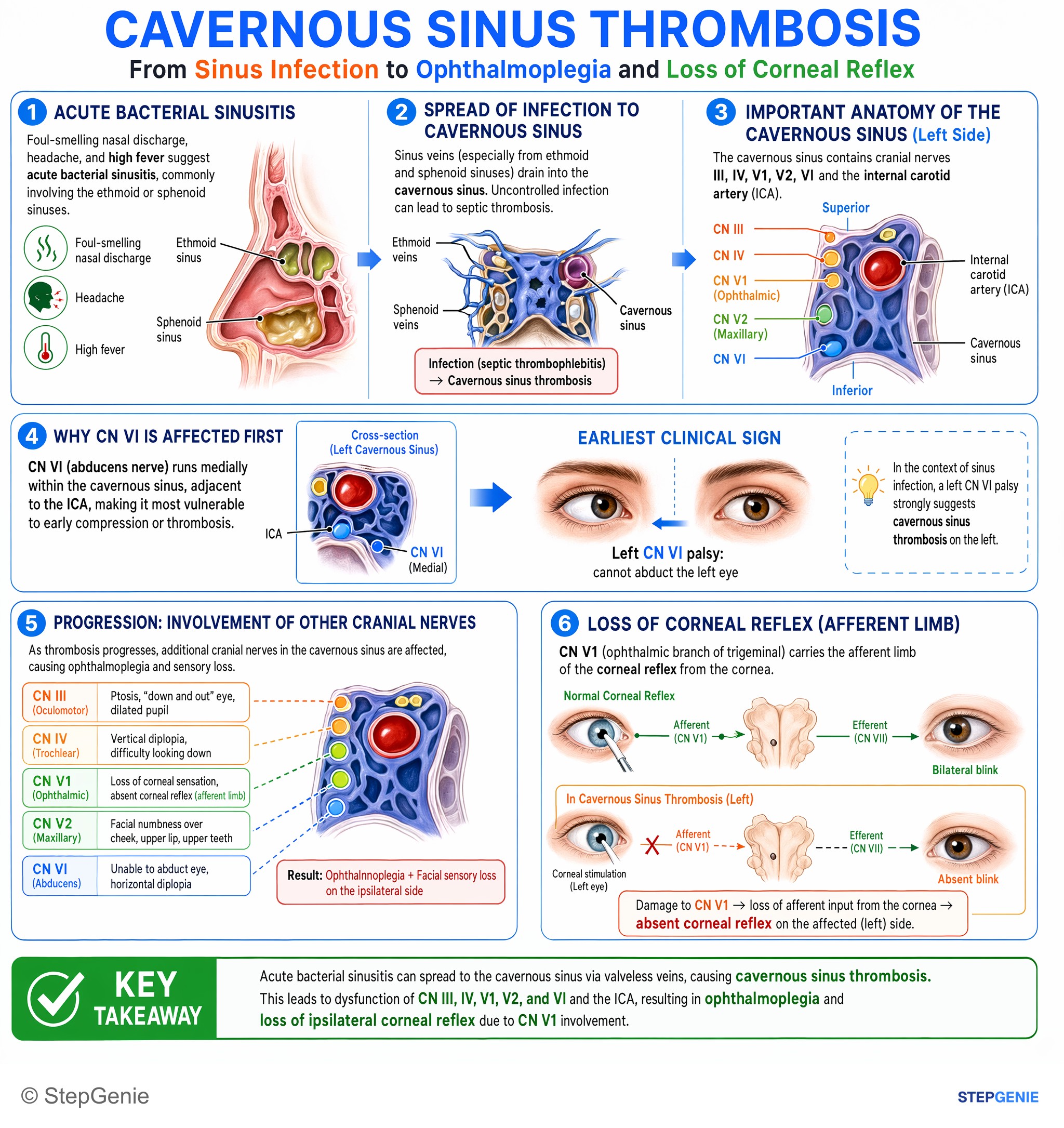
Cavernous sinus thrombosis from sinus infection → CN III, IV, V1/V2, VI dysfunction → ophthalmoplegia and loss of ipsilateral corneal…
USMLE Step 2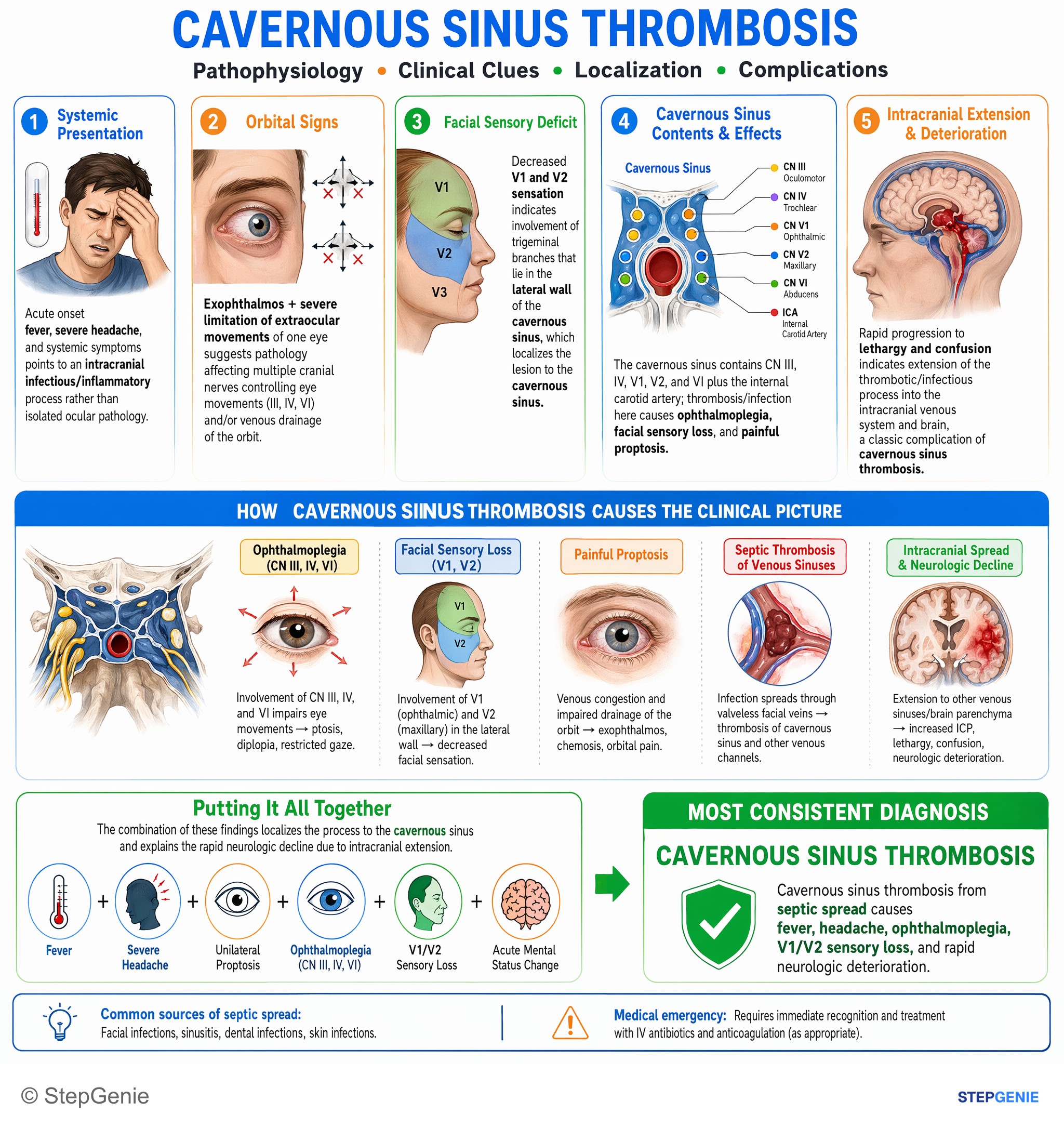
Cavernous sinus thrombosis from septic spread causes fever, headache, ophthalmoplegia, V1/V2 sensory loss, and rapid neurologic…
USMLE Step 2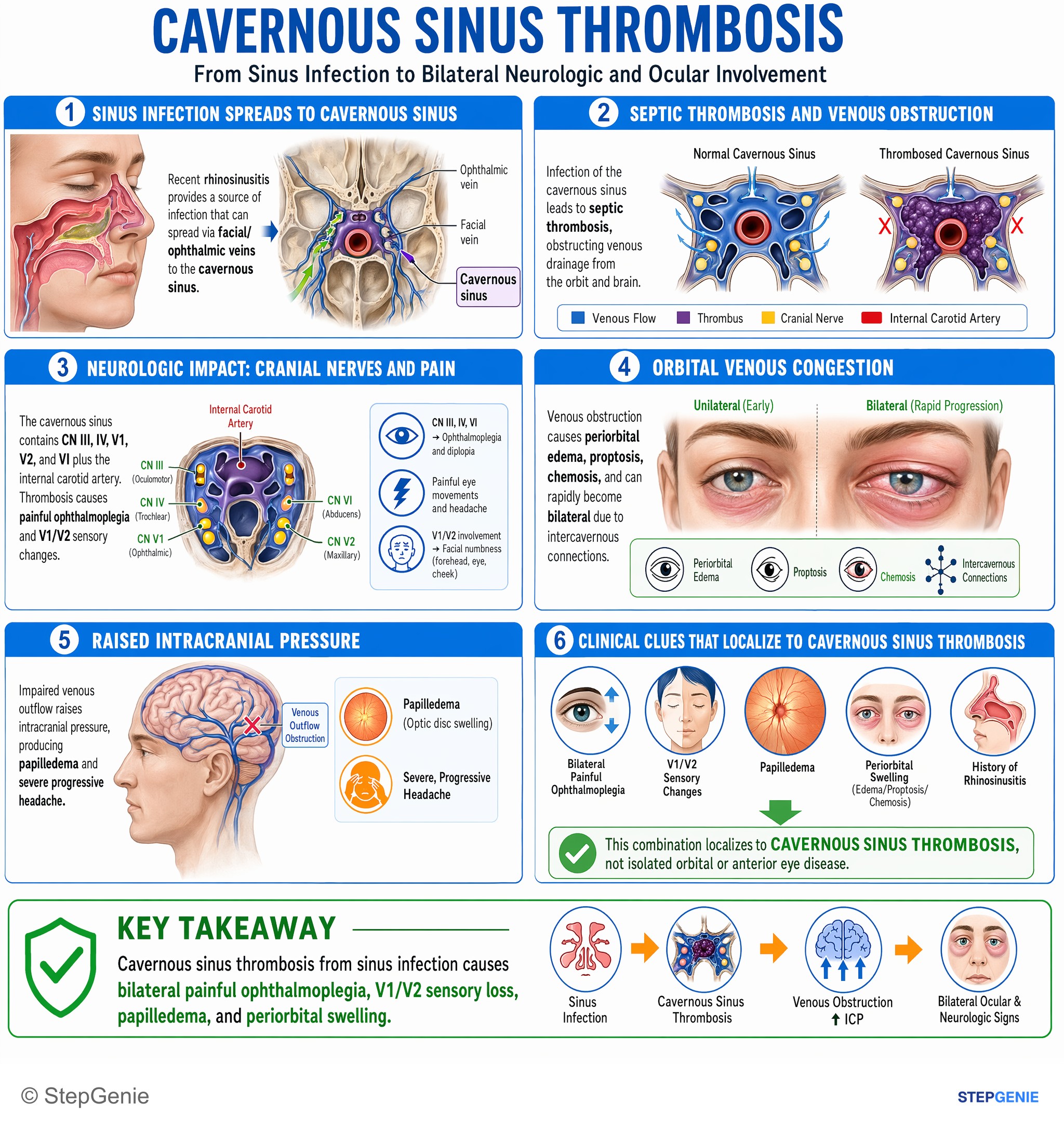
Cavernous sinus thrombosis from sinus infection causes bilateral painful ophthalmoplegia, V1/V2 sensory loss, papilledema, and periorbital…
USMLE Step 2Frequently Asked Questions
Which cranial nerve is affected first in cavernous sinus thrombosis?
The abducens nerve (CN VI) is usually affected first. CN III, CN IV, V1 and V2 run in the lateral wall of the cavernous sinus, but CN VI travels through the middle of the sinus beside the internal carotid artery, surrounded by venous blood, so clot and inflammation reach it early. The result is an eye that turns inward and cannot abduct, with horizontal double vision. Because the carotid's sympathetic fibers run alongside, the early CN VI palsy may come with a Horner syndrome.
What passes through the cavernous sinus?
The lateral wall of the cavernous sinus carries, from top to bottom, the oculomotor nerve (CN III), the trochlear nerve (CN IV), and the ophthalmic (V1) and maxillary (V2) divisions of the trigeminal nerve. Running through the sinus itself are the internal carotid artery, wrapped in its sympathetic plexus, and the abducens nerve (CN VI). Damage produces ophthalmoplegia, ptosis, a dilated pupil, numbness of the forehead and cheek, and sometimes a Horner syndrome.
Why is the danger triangle of the face linked to cavernous sinus thrombosis?
Blood from the nose, lips and inner corner of the eye usually drains through the facial vein, but it can also flow back through the ophthalmic veins, or through the pterygoid plexus and emissary veins, into the cavernous sinus. Because these veins are classically described as valveless, an infection such as a squeezed nasal boil can seed the sinus with bacteria, most often Staphylococcus aureus. Sphenoid or ethmoid sinusitis and dental infections are the other main sources.
How do you tell cavernous sinus thrombosis from orbital cellulitis?
Both cause fever, eyelid swelling, proptosis, chemosis and painful, limited eye movements. Orbital cellulitis is an infection behind the orbital septum, usually spreading from ethmoid sinusitis, and it stays in the orbit. Cavernous sinus thrombosis adds cranial nerve dysfunction, such as numbness over the forehead and cheek, involvement of the other eye and changes in mental status. Contrast-enhanced MR or CT venography shows the clot. Orbital cellulitis can itself spread to the cavernous sinus.
What is the treatment for cavernous sinus thrombosis?
IV antibiotics start immediately: nafcillin or oxacillin plus a third-generation cephalosporin, with vancomycin instead where MRSA is common and metronidazole added if a dental infection is the source. IV therapy usually lasts 3 to 4 weeks, sometimes 6 to 7. Sphenoid sinusitis is drained surgically, especially if there is no response within 24 hours. Merck recommends unfractionated or low-molecular-weight heparin when there is no contraindication, although the supporting data are retrospective, and glucocorticoids may be added for cranial nerve dysfunction. Even with treatment, mortality is about 10 to 15 percent.
What is the best imaging test for cavernous sinus thrombosis?
Contrast-enhanced MR venography or CT venography is the most sensitive test, more sensitive than standard CT or MRI. If venography is not available, MRI is better than CT, but imaging should never be delayed in a patient with fever, orbital swelling and new cranial nerve palsies. Blood cultures and a lumbar puncture are added to identify the organism and to look for spread to the meninges, since meningoencephalitis and brain abscess are among the complications.