You’re cruising through a neuro Q-bank and hit a classic: fever + focal deficits + ring-enhancing lesion. Easy, right? Except the test writers know you’ll jump to “brain tumor” or “toxoplasmosis” unless you slow down and interrogate the details. This post breaks down a brain abscess vignette the way Step wants you to think: pick the best answer, then prove every other choice is wrong.
Tag: Neurology > CNS Tumors & Infections
The Clinical Vignette (Q-bank style)
A 45-year-old man presents with 4 days of worsening headache, fever, and new-onset left arm weakness. He had chronic otitis media for months and recently stopped antibiotics early. Exam shows T = 38.9°C, mild nuchal discomfort, and left upper extremity pronator drift. Labs: mild leukocytosis. MRI brain with contrast shows a solitary ring-enhancing lesion in the right temporal lobe with surrounding vasogenic edema and restricted diffusion in the center.
Question: What is the most likely diagnosis?
The Correct Answer: Brain Abscess
Why it fits
A brain abscess is a localized intracerebral infection that classically presents with:
- Headache
- Fever (may be absent, but when present it’s a big clue)
- Focal neurologic deficits
- ± Seizures
- Imaging: ring-enhancing lesion with vasogenic edema
- Key imaging discriminator: restricted diffusion on DWI (purulent material)
This vignette also hands you a major risk factor: chronic otitis media → contiguous spread to the temporal lobe (or cerebellum).
High-yield microbiology (think source → bug)
| Source / Risk factor | Common organisms | Typical location |
|---|---|---|
| Otitis media / mastoiditis / sinusitis | Streptococci (esp. viridans), anaerobes (Bacteroides, Peptostreptococcus) | Temporal lobe, cerebellum |
| Dental infection | Viridans strep, anaerobes | Frontal lobe |
| Hematogenous spread (endocarditis, lung abscess) | Staph aureus, strep, sometimes Gram-negatives | Often at gray-white junction, can be multiple |
| Immunocompromised | Nocardia, fungi, Toxo (separate entity) | Variable |
Path & imaging pearls Step loves
- Ring-enhancement reflects a capsule with neovascularization and leaky BBB.
- Restricted diffusion strongly favors abscess over most tumors because pus limits water movement.
- Increased ICP signs can appear (vomiting, papilledema), especially with larger lesions/edema.
- Lumbar puncture is often contraindicated if mass effect is suspected (risk of herniation).
Treatment framework (Step-level)
- Empiric antibiotics (tailor to source)
- Common empiric: ceftriaxone (or cefotaxime) + metronidazole
- Add vancomycin if trauma/neurosurgery or concern for MRSA
- Drainage often needed (especially if large, multiloculated, or poor response)
- Manage edema/ICP (e.g., dexamethasone only if significant mass effect—can impair capsule formation and antibiotic penetration, so not routine)
Why Every Other Answer Choice Is Wrong (and how they trick you)
Below are high-yield distractors that commonly appear alongside “brain abscess.”
Distractor 1: Glioblastoma (GBM)
Why it’s tempting
- Ring-enhancing lesion + edema + focal deficits = tumor vibe.
Why it’s wrong here
- Time course: GBM typically evolves over weeks to months, not 4 febrile days.
- Systemic signs: fever and leukocytosis push you toward infection.
- Imaging clue: GBM often shows necrotic ring enhancement, but restricted diffusion is more characteristic of abscess.
- Typical features (Step 1 recall):
- Older adults
- Hemorrhage/necrosis
- “Butterfly” crossing corpus callosum is classic (not required, but a clue when present)
Rule of thumb: Acute febrile neurologic deficits + diffusion restriction → think abscess first.
Distractor 2: Toxoplasma encephalitis
Why it’s tempting
- Ring-enhancing brain lesions are toxo-famous.
Why it’s wrong here
- Host matters: toxoplasmosis is classically in AIDS (CD4 < 100) or other severe immunosuppression.
- Lesion pattern: typically multiple ring-enhancing lesions, often in basal ganglia.
- Clinical context: may have subacute confusion, seizures; fever can occur—but you’d expect immunocompromised clues.
High-yield comparator
| Feature | Brain abscess | Toxoplasma |
|---|---|---|
| Patient | Any, often contiguous infection risk | Immunocompromised (esp. AIDS) |
| Number | Often single (can be multiple) | Often multiple |
| Location | Temporal/frontal, gray-white junction | Basal ganglia common |
| DWI restriction | Yes (classic) | Variable/less classic |
Distractor 3: Primary CNS lymphoma
Why it’s tempting
- Enhancing mass lesions in immunocompromised patients.
Why it’s wrong here
- Again, host: classically EBV-associated in AIDS/transplant patients.
- Imaging: tends to be homogeneously enhancing (ring-enhancing can occur in AIDS but is less classic).
- Systemic infection source (otitis media) + diffusion restriction + acute fever favors abscess.
Step pearl: If the stem screams “AIDS,” you should be toggling between toxo vs CNS lymphoma; if it screams “ear/sinus infection,” toggle toward abscess.
Distractor 4: Neurocysticercosis
Why it’s tempting
- Multiple lesions, seizures, sometimes ring enhancement.
Why it’s wrong here
- Usually presents with seizures as the dominant symptom; fever is not a classic driver.
- Epidemiology: exposure/travel to endemic areas; ingestion of Taenia solium eggs.
- Imaging often shows multiple cystic lesions in different stages; may see scolex (“dot”).
- DWI restriction is not the go-to clue.
High-yield: Neurocysticercosis is the most common cause of adult-onset seizures worldwide.
Distractor 5: Tuberculoma (CNS TB)
Why it’s tempting
- Ring-enhancing lesion + systemic symptoms can overlap.
Why it’s wrong here
- TB tends to be subacute/chronic with weight loss/night sweats, risk factors, and sometimes basilar meningitis findings.
- Imaging may show ring enhancement, but DWI restriction and a clear contiguous ear infection story lean abscess.
- TB often involves basilar cisterns and can cause hydrocephalus (meningitis angle).
Distractor 6: Subdural empyema
Why it’s tempting
- Also infectious, can follow sinusitis/otitis, can cause fever + neuro deficits.
Why it’s wrong here
- It’s extra-axial, not an intra-parenchymal round lesion.
- Imaging: crescent-shaped collection along the subdural space (often with midline shift), not a focal intraparenchymal ring-enhancing mass.
High-yield urgency: Subdural empyema is a neurosurgical emergency—rapid deterioration, seizures, signs of increased ICP.
The “Step Strategy”: How to Win Ring-Enhancing Lesions
1) Start with the timeline
- Hours–days: abscess, hemorrhage, acute infarct (not ring enhancing early)
- Weeks–months: tumors, TB/fungal, demyelination (tumefactive), slow infections
2) Check the immune status
- Immunocompromised → toxo, CNS lymphoma, fungal, nocardia
- Immunocompetent + ear/sinus/dental source → abscess
3) Use DWI like a cheat code
- Restricted diffusion in a ring-enhancing lesion = abscess until proven otherwise
4) Don’t LP a mass
If imaging suggests mass effect/increased ICP:
- Avoid lumbar puncture (herniation risk)
- Treat and image-guided diagnose (aspiration/culture) instead
Rapid-Fire High-Yield Facts (for Step 1 & 2)
- Brain abscess triad (often incomplete): fever, headache, focal neuro deficits
- Otitis media/mastoiditis → temporal lobe or cerebellar abscess
- Restricted diffusion helps distinguish abscess from necrotic tumor
- Common empiric therapy: ceftriaxone + metronidazole (± vancomycin depending on risk)
- Seizures are common—consider prophylaxis in many clinical settings
- Steroids only if significant mass effect (can reduce antibiotic penetration/capsule formation)
Visual explanations from the StepGenie QBank
Infographics StepGenie drew to explain questions in its own QBank, matched to this article's topic. Each caption is the takeaway of the question it explains.
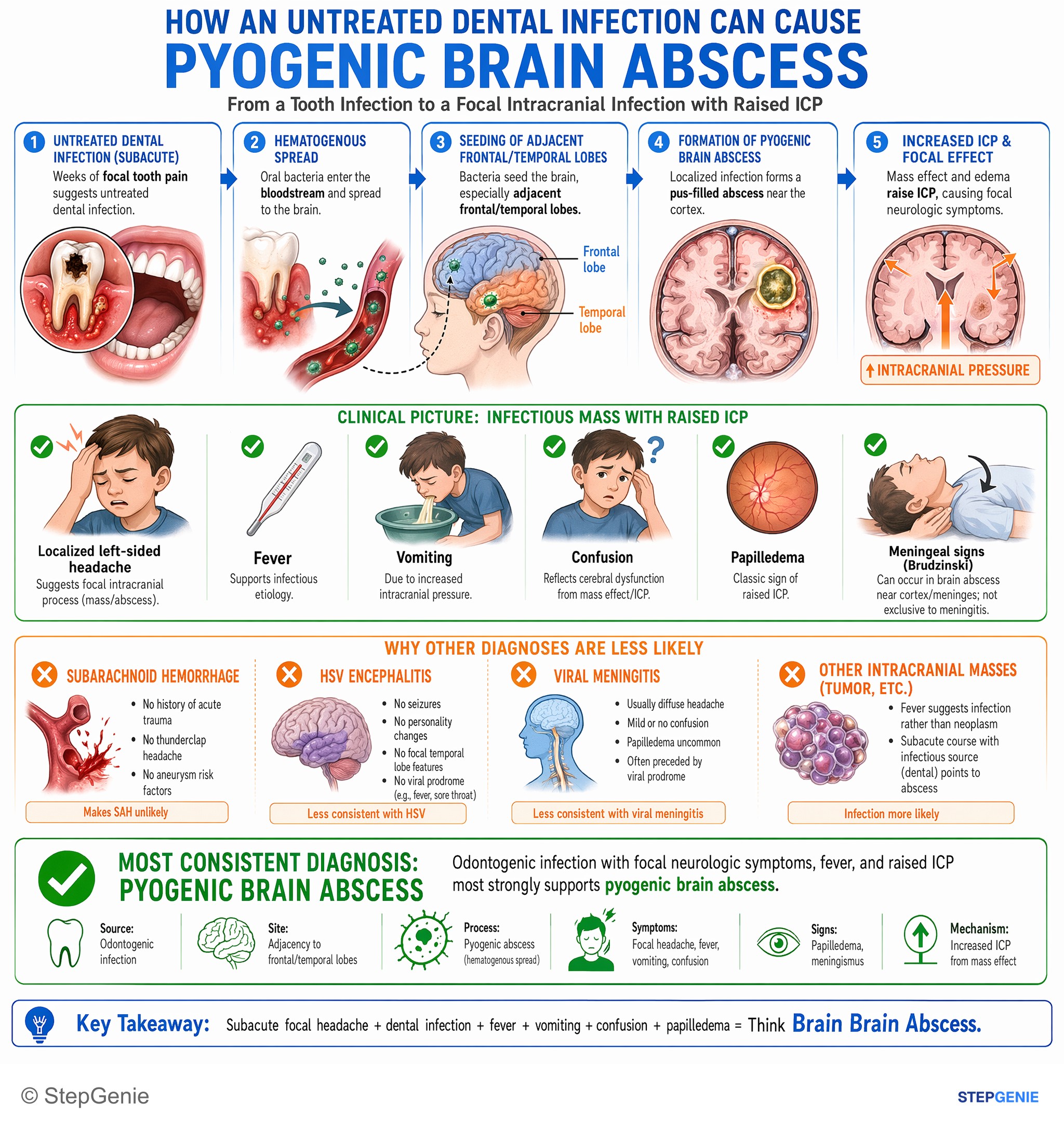
Pyogenic brain abscess from odontogenic infection causes focal headache, fever, vomiting, confusion, papilledema, and meningismus due to…
USMLE Step 2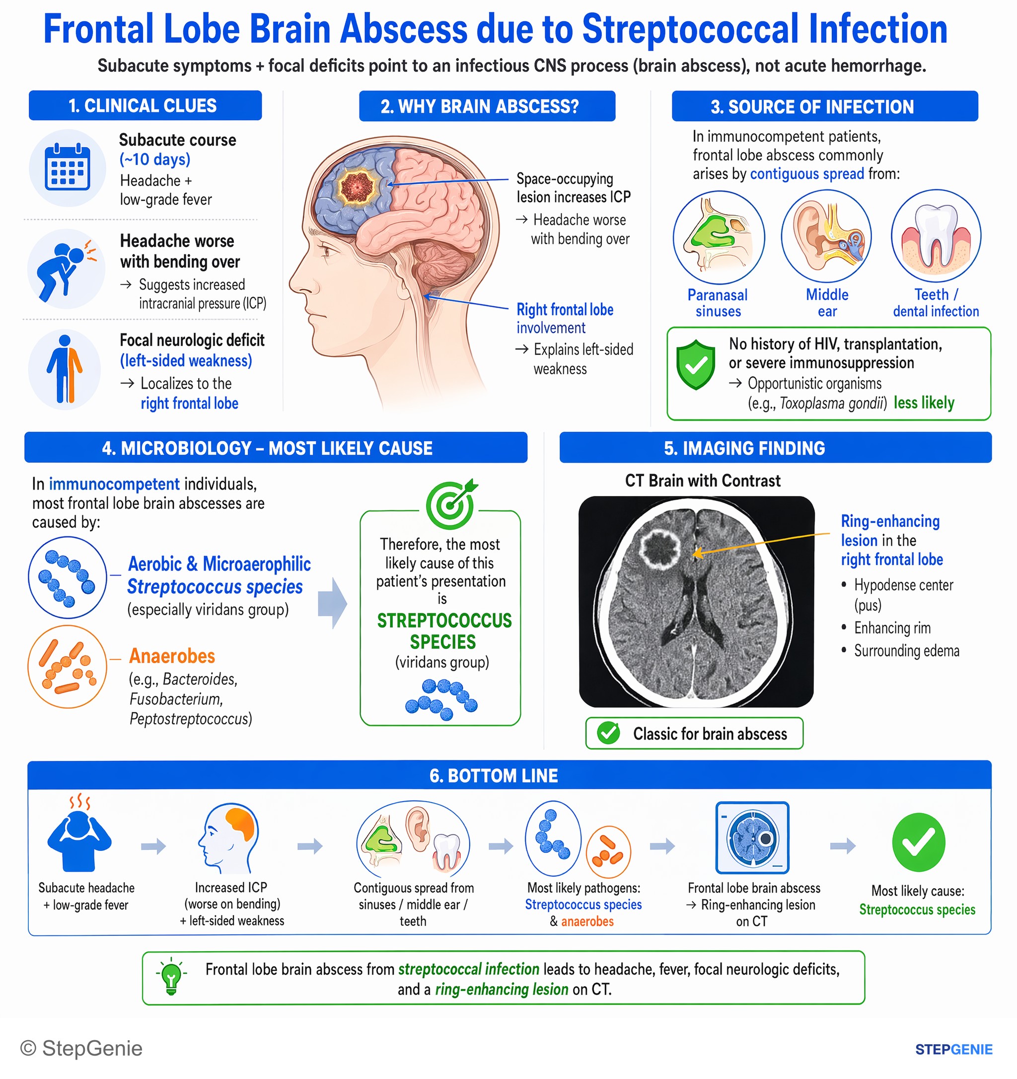
Frontal lobe brain abscess from streptococcal infection → headache, fever, focal neurologic deficits, ring-enhancing lesion on CT.
USMLE Step 2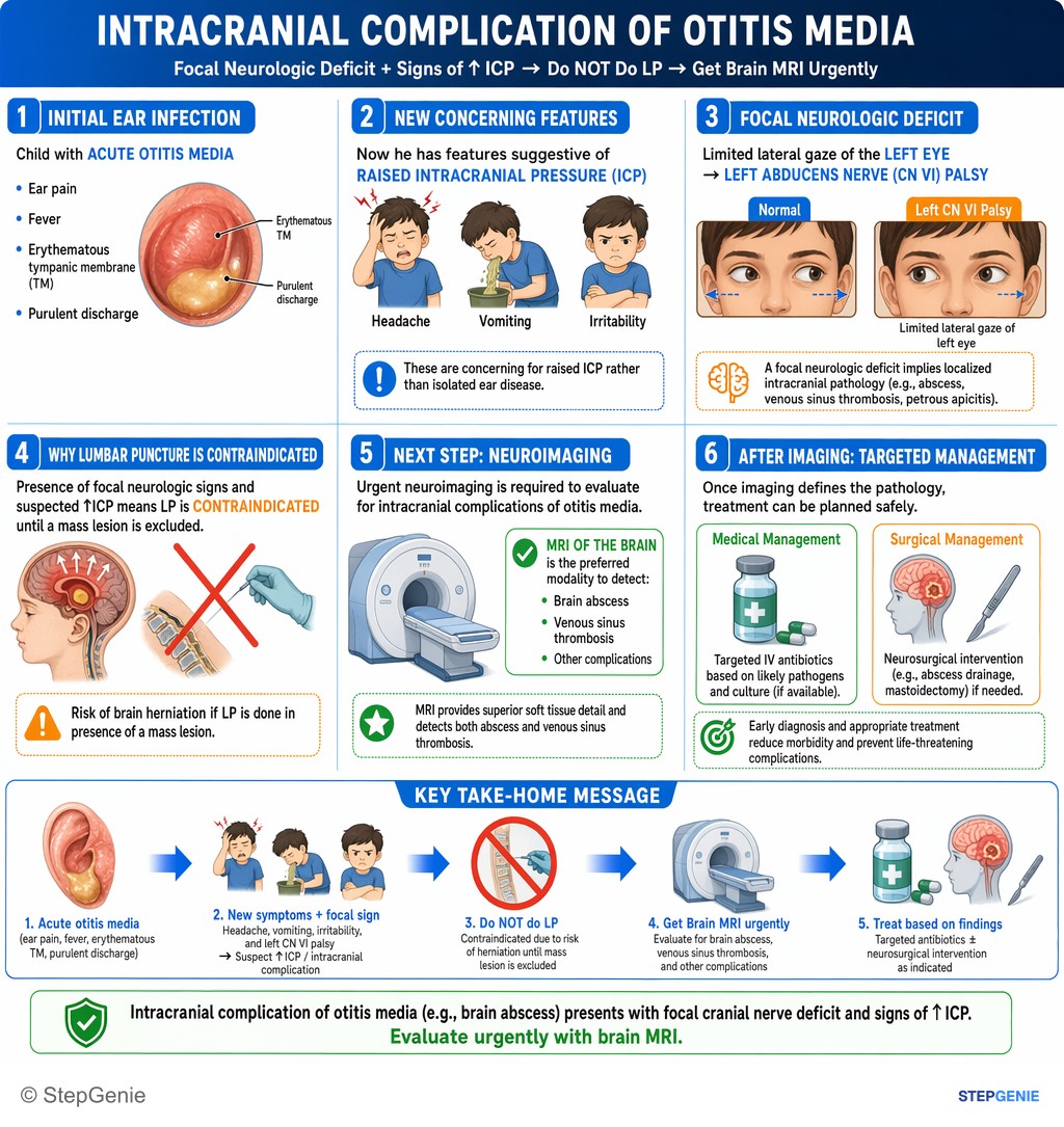
Intracranial complication of otitis media (e.g., brain abscess) presents with focal cranial nerve deficit and signs of ↑ICP; evaluate…
USMLE Step 2Frequently Asked Questions
How do you tell a brain abscess from a brain tumor on imaging?
Restricted diffusion in the center strongly favors a brain abscess over most tumors. Both can show a ring-enhancing lesion with surrounding vasogenic edema, but abscesses contain purulent material that limits water movement on diffusion-weighted imaging. An acute febrile presentation with leukocytosis also supports infection, whereas glioblastoma more often evolves over weeks to months.
Why does chronic otitis media cause a brain abscess in the temporal lobe?
Chronic otitis media can lead to a brain abscess by contiguous spread of infection, and the temporal lobe is a classic location. In this setting, common organisms include streptococci, especially viridans streptococci, and anaerobes such as Bacteroides and Peptostreptococcus. A temporal lobe ring-enhancing lesion with fever and focal deficits fits that pattern well.
What is the first-line treatment for a brain abscess?
Initial treatment is empiric antibiotics tailored to the suspected source, commonly ceftriaxone or cefotaxime plus metronidazole. Vancomycin is added if there is trauma, neurosurgery, or concern for MRSA. Drainage is often needed, especially for a large lesion, multiloculated abscess, or poor response, and edema or elevated intracranial pressure may also need management.
How do you tell a brain abscess from toxoplasmosis?
A brain abscess is more likely when there is a contiguous infection source such as otitis media, a solitary lesion, and classic diffusion restriction. Toxoplasmosis is classically seen in severely immunocompromised patients, especially those with AIDS, and it often causes multiple ring-enhancing lesions in the basal ganglia. Host context and lesion pattern are the key clues.
Why is lumbar puncture avoided in suspected brain abscess?
Lumbar puncture is often contraindicated when a brain abscess has possible mass effect because of the risk of herniation. Brain abscesses can cause surrounding edema and increased intracranial pressure, especially when lesions are larger. In that setting, imaging findings such as a ring-enhancing lesion with vasogenic edema are safer and more informative than spinal fluid sampling.