Neoplasia questions on Step exams love to punish “pattern recognition without reasoning.” The stem may scream benign, but the answer choices try to bait you into confusing dysplasia, carcinoma in situ, invasion, and metastasis—because those distinctions determine prognosis, staging, and management. Let’s do a Q-bank–style breakdown where every distractor is teaching you something.
Clinical Vignette (Q-bank style)
A 52-year-old woman notices a slowly enlarging, painless lump in her right breast. She has no weight loss, fevers, or night sweats. Physical exam reveals a 2-cm, mobile, well-circumscribed mass. Mammography shows a round, sharply demarcated lesion without calcifications. Core needle biopsy demonstrates a biphasic lesion composed of proliferating fibrous stroma and benign ductal epithelium. The lesion is encapsulated and shows no invasion into surrounding adipose tissue.
Which of the following features most strongly supports that this lesion is benign?
A. High nuclear-to-cytoplasmic ratio
B. Ability to metastasize via lymphatics
C. Invasion through basement membrane into adjacent stroma
D. Well-differentiated cells resembling the tissue of origin
E. Tumor necrosis with “comedo” pattern
Step-Style Answer
✅ Correct Answer: D. Well-differentiated cells resembling the tissue of origin
A core hallmark of benign tumors is differentiation: the tumor cells look and behave like the normal tissue they came from. In this vignette, the mass is also well-circumscribed, mobile, encapsulated, and noninvasive, all classic benign features (think fibroadenoma in the breast).
High-yield pearl:
- Benign ≠ harmless, but benign typically implies:
- No invasion
- No metastasis
- Better differentiation
- Slower growth
- Often encapsulated and well-circumscribed
Why Each Distractor Is Wrong (and What It’s Testing)
A. High nuclear-to-cytoplasmic ratio
This is a classic feature of malignancy (anaplasia). A high N:C ratio reflects increased nuclear size and hyperchromasia due to increased DNA content and dysregulated growth.
High-yield malignancy cytology features:
- High N:C ratio
- Hyperchromatic, pleomorphic nuclei
- Prominent nucleoli
- Atypical mitoses
- Loss of polarity
- Tumor giant cells
Takeaway: If you see high N:C ratio, you should be thinking malignant, not benign.
B. Ability to metastasize via lymphatics
Metastasis is definitively malignant (with rare exceptions like benign “metastasizing leiomyoma,” which is a zebras-only concept and not how Step expects you to reason).
Step rule:
- Benign tumors do not metastasize.
- Metastasis = malignant until proven otherwise.
Extra high-yield detail:
- Carcinomas (epithelial) tend to spread via lymphatics.
- Sarcomas (mesenchymal) tend to spread via hematogenous routes.
- But both can do either.
C. Invasion through basement membrane into adjacent stroma
This is the defining step from in situ disease to invasive cancer.
- Carcinoma in situ: full-thickness dysplasia without basement membrane invasion
- Invasive carcinoma: breaks through the basement membrane into stroma → access to lymphatics/blood → metastasis potential
High-yield staging anchor:
- In many epithelial cancers, invasion (not just cytologic atypia) is what makes it stage-worthy and dangerous.
Takeaway: Basement membrane invasion is a malignant feature.
E. Tumor necrosis with “comedo” pattern
“Comedo necrosis” is associated with high-grade ductal carcinoma in situ (DCIS) of the breast: malignant cytology and necrotic debris in ducts.
Why necrosis suggests malignancy:
- Rapid growth can outstrip blood supply → ischemic tumor necrosis
- Often seen in aggressive, poorly differentiated tumors
Takeaway: Necrosis—especially comedo-type—pushes you toward malignant/pre-malignant, not benign.
The Big Picture: Benign vs Malignant (What Step Wants You to Say)
Core clinical and pathologic differences
| Feature | Benign | Malignant |
|---|---|---|
| Differentiation | Well-differentiated | Variable, often poorly differentiated/anaplastic |
| Growth rate | Slow, progressive | Faster; may be erratic |
| Borders | Well-circumscribed, often encapsulated | Infiltrative, poorly circumscribed |
| Invasion | Absent | Present (breaks basement membrane) |
| Metastasis | Absent | Possible/likely (key defining feature) |
| Mitotic figures | Few, normal | Many, may be atypical |
Memory hook:
- Benign = “B” for “Bounded” (well-circumscribed)
- Malignant = “M” for “Moves” (invades/metastasizes)
Rapid-Fire High-Yield Clarifications (Common Trap Concepts)
Dysplasia vs metaplasia vs neoplasia
- Metaplasia: reversible change from one differentiated cell type to another (adaptive)
- Example: smoking → squamous metaplasia in bronchi
- Dysplasia: disordered growth, atypia; can be reversible; premalignant in many settings
- Features: pleomorphism, hyperchromasia, increased mitoses, loss of polarity
- Neoplasia: autonomous clonal proliferation (benign or malignant)
Carcinoma in situ (CIS)
- Full-thickness dysplasia without invasion
- Still cannot metastasize because basement membrane is intact
- Clinically serious because it’s one step away from invasive cancer
“Benign” tumors can still cause harm
Even benign lesions can be dangerous due to:
- Mass effect (e.g., meningioma compressing brain tissue)
- Hormone production (e.g., pituitary adenoma)
- Obstruction (e.g., colonic polyp causing intussusception)
- Location matters as much as histology
How to Nail These Questions on Test Day
A Step-optimized approach to the stem
- Look for invasion/metastasis language first
- If present → malignant
- Check differentiation and borders
- Well-differentiated + well-circumscribed → benign leaning
- Identify the “single best discriminator”
- Metastasis and invasion beat everything
- Don’t overvalue cytology alone
- Atypia matters, but invasion is the pivot point for carcinoma
One-Line Wrap-Up
When Step asks “benign vs malignant,” the highest-yield discriminator is invasion/metastasis, but the best benign-supporting feature in many answer sets is good differentiation—and every distractor is basically a different way of saying “malignant.”
Visual explanations from the StepGenie QBank
Infographics StepGenie drew to explain questions in its own QBank, matched to this article's topic. Each caption is the takeaway of the question it explains.
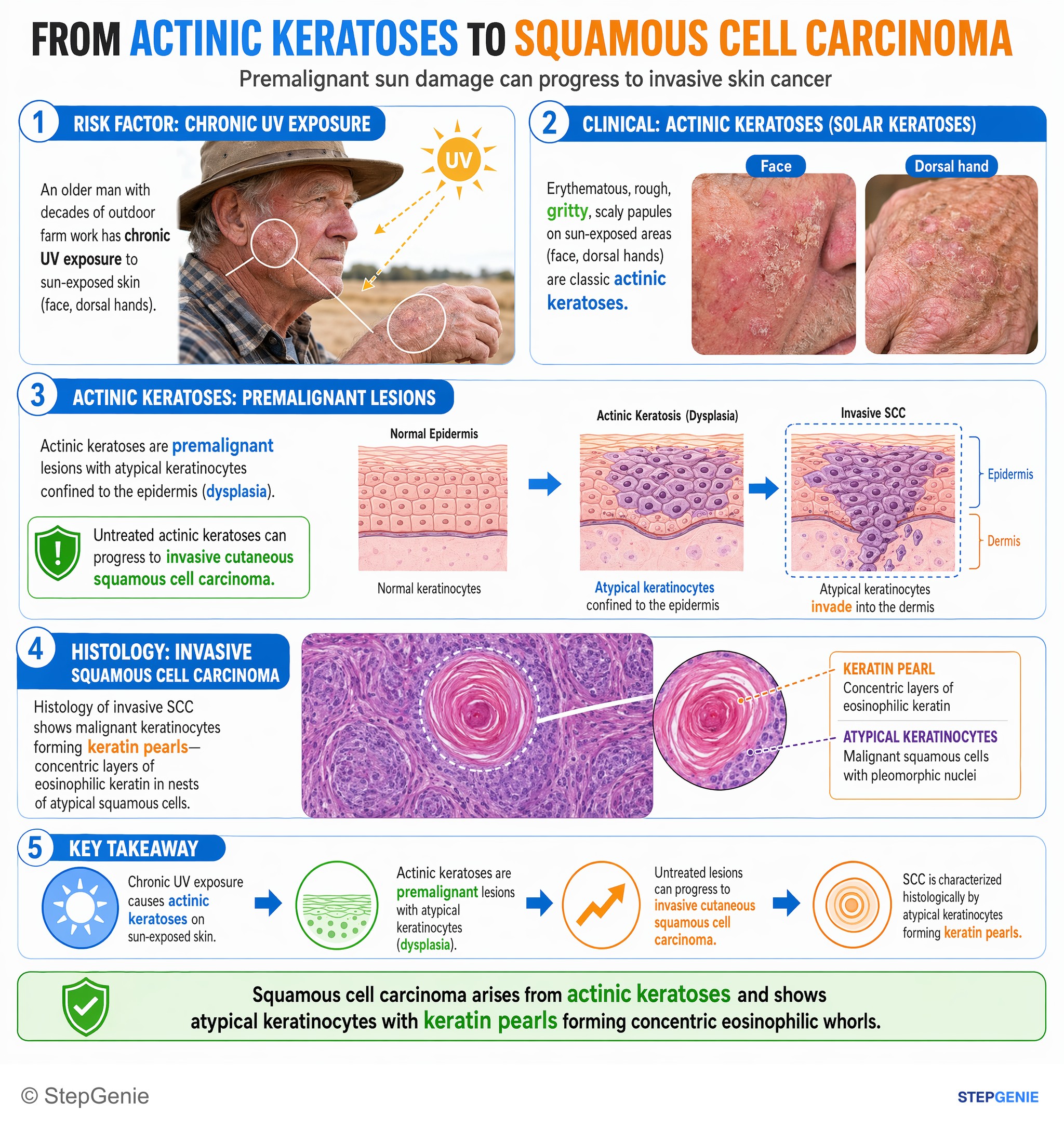
Squamous cell carcinoma arises from actinic keratoses and shows atypical keratinocytes with keratin pearls forming concentric eosinophilic…
USMLE Step 1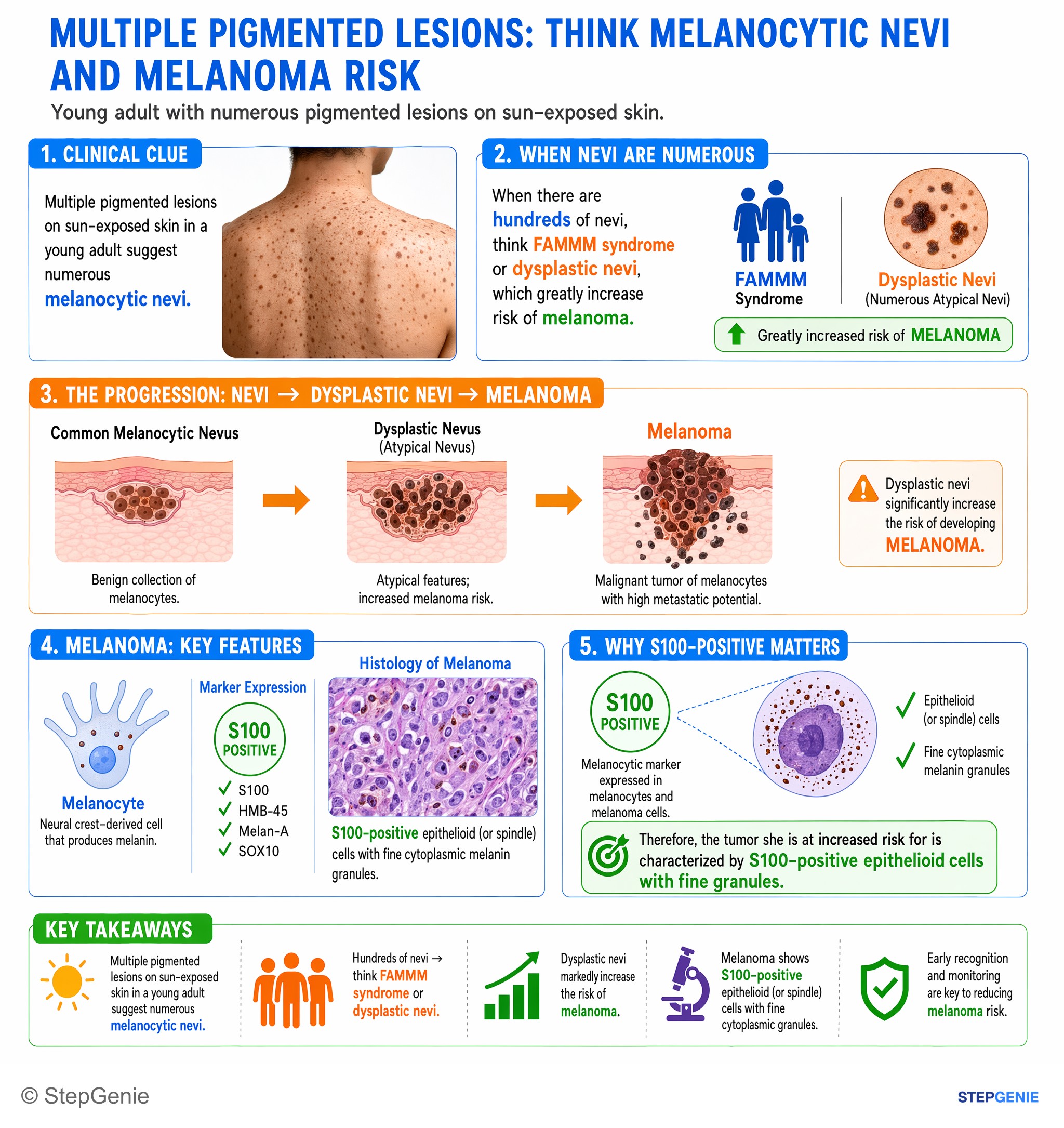
Dysplastic nevi (numerous atypical nevi) increase risk of melanoma, a tumor of S100-positive melanocytes with epithelioid cells and fine…
USMLE Step 1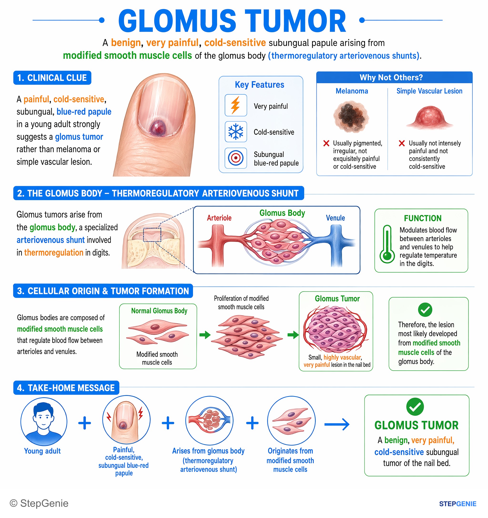
Glomus tumor: benign, very painful, cold-sensitive subungual papule arising from modified smooth muscle cells of the glomus body…
USMLE Step 1Frequently Asked Questions
What feature most strongly supports that a tumor is benign on pathology?
Well-differentiated cells resembling the tissue of origin most strongly support a benign tumor. Benign neoplasms typically preserve differentiation, meaning the cells still look and behave like normal tissue. They are also commonly well-circumscribed, mobile, encapsulated, and noninvasive, with slower growth and no metastatic potential.
Why does invasion through the basement membrane make a tumor malignant?
Invasion through the basement membrane makes a tumor malignant because it marks the transition from in situ disease to invasive cancer. Once tumor cells break into the adjacent stroma, they gain access to lymphatics and blood vessels, which creates metastatic potential. On exams, invasion is a key feature that changes prognosis, staging, and clinical significance.
Can carcinoma in situ metastasize if the cells look malignant?
No, carcinoma in situ cannot metastasize because the basement membrane remains intact. It consists of full-thickness dysplasia without invasion, so the abnormal cells have not yet entered the stroma or gained access to lymphatic or hematogenous routes. It is still clinically serious because it is one step away from invasive carcinoma.
How do you tell a benign tumor from a malignant one in a question stem?
The fastest way is to look for invasion and metastasis first, because their presence supports malignancy. Benign tumors are usually well-differentiated, slow-growing, well-circumscribed, often encapsulated, and lack invasion or metastasis. Malignant tumors are more likely to be infiltrative, poorly circumscribed, variable in differentiation, and show increased or atypical mitoses.
Why is a high nuclear-to-cytoplasmic ratio a clue for malignancy?
A high nuclear-to-cytoplasmic ratio is a classic cytologic feature of malignancy because it reflects enlarged, hyperchromatic nuclei and dysregulated growth. It is part of the broader picture of anaplasia, which can also include pleomorphism, prominent nucleoli, atypical mitoses, loss of polarity, and tumor giant cells. These findings point away from a benign lesion.