Breast cancer questions on UWorld/AMBOSS/NBME are rarely just “pick the diagnosis.” They’re usually a test of risk factors, screening, pathology, receptor status, lymphatic spread, and management—and the distractors are deliberately chosen to probe the exact things you’re shaky on. Let’s do a classic clinical vignette, then break down why the right answer is right and why every wrong answer is tempting—but wrong.
Clinical Vignette (Q-bank style)
A 52-year-old woman comes to the clinic for evaluation of a breast lump she noticed 3 weeks ago. She has no fever or recent trauma. Menarche was at age 11. She had her first child at age 34 and breastfed for 2 months. She drinks 2–3 glasses of wine nightly. Her mother was diagnosed with breast cancer at age 45. Exam shows a firm, irregular, non-tender mass in the upper outer quadrant of the left breast with skin dimpling. No nipple discharge. A diagnostic mammogram shows a spiculated mass with microcalcifications. Core needle biopsy shows malignant ductal epithelial cells in cords and nests surrounded by dense fibrous (desmoplastic) stroma.
Question: Which of the following is the most likely diagnosis?
A. Fibroadenoma
B. Intraductal papilloma
C. Invasive ductal carcinoma
D. Invasive lobular carcinoma
E. Phyllodes tumor
F. Fat necrosis
The Correct Answer: C. Invasive Ductal Carcinoma (IDC)
Why this is IDC
This vignette screams invasive ductal carcinoma:
- Firm, irregular mass + skin dimpling
- Dimpling is from traction on Cooper ligaments due to desmoplastic reaction (fibrosis) around invasive tumor.
- Mammogram: spiculated mass (classic for malignancy) + microcalcifications
- Microcalcifications are strongly associated with ductal carcinoma in situ (DCIS), and IDC often coexists with DCIS.
- Histology: malignant ductal cells + desmoplastic stroma
- That dense fibrous response is a hallmark clue for IDC.
High-yield IDC facts (Step 1 + Step 2)
- Most common breast cancer overall.
- Metastasis pattern: first to axillary nodes (especially for upper outer quadrant tumors).
- Sentinel node biopsy is used for staging.
- Receptor status matters (treatment + prognosis):
- ER/PR+ → endocrine therapy (e.g., tamoxifen, aromatase inhibitor)
- HER2+ → trastuzumab/pertuzumab (cardiotoxicity risk)
- Triple-negative → more aggressive; chemo mainstay
- Risk factors to recognize quickly:
- Increased lifetime estrogen exposure: early menarche, late menopause, nulliparity, first pregnancy after 30
- Alcohol
- Family history/BRCA (mother <50 is a red flag)
- Prior chest radiation (e.g., Hodgkin therapy)
Why Each Distractor Is Wrong (and How to Recognize It)
A. Fibroadenoma
Why it tempts you: “Breast lump in a woman” is often benign in younger patients.
Why it’s wrong here:
- Fibroadenoma is typically:
- Women <30
- Well-circumscribed, mobile, rubbery (“breast mouse”)
- Histology: proliferation of stroma that compresses ducts (benign)
- This vignette instead has:
- Irregular, fixed, skin dimpling
- Spiculations on mammogram (malignancy)
- Invasive malignant ductal cells, not benign stroma/duct architecture
High-yield pearl: A mobile, well-defined mass is reassuring; spiculated + fixed + dimpling is not.
B. Intraductal Papilloma
Why it tempts you: Common benign lesion that shows up in Q-banks.
Why it’s wrong here:
- Intraductal papilloma classically presents with:
- Unilateral bloody (or serous) nipple discharge
- Subareolar mass
- Benign papillary fronds in a lactiferous duct
- This patient has:
- No nipple discharge
- A peripheral upper outer quadrant mass
- Imaging and biopsy consistent with invasive malignancy
High-yield pearl: If the stem says bloody nipple discharge + subareolar, your brain should immediately pull up papilloma (benign) vs Paget disease (eczema-like nipple/areola changes).
D. Invasive Lobular Carcinoma (ILC)
Why it tempts you: Also common, also invasive, and often tested alongside IDC.
Why it’s wrong here (based on provided histology/imaging clues):
- ILC classically has:
- Loss of E-cadherin (cells can’t stick together)
- Single-file infiltrating pattern
- More likely multifocal and bilateral
- Often less conspicuous on mammogram (can be subtle)
- This vignette has:
- Cords and nests + desmoplastic stroma → more in line with IDC
- Spiculated mass with microcalcifications → more typical of ductal pathology
High-yield pearl:
- ILC = “single-file” + E-cadherin negative
- IDC = desmoplasia + often spiculated mass, can be associated with calcifications via DCIS
E. Phyllodes Tumor
Why it tempts you: Another “breast mass” entity with a distinctive name.
Why it’s wrong here:
- Phyllodes tumor features:
- Leaf-like architecture (phyllodes = “leaf”)
- Stromal hypercellularity (can be benign or malignant)
- Often large, fast-growing, well-circumscribed
- This vignette describes:
- Spiculated, irregular lesion
- Malignant ductal epithelial invasion with desmoplasia (not a stromal-driven tumor)
High-yield pearl: Phyllodes is more like a “fibroadenoma on steroids” (larger, leaf-like); it’s not your typical spiculated carcinoma presentation.
F. Fat Necrosis
Why it tempts you: Can mimic breast cancer on exam and imaging—very testable.
Why it’s wrong here:
- Fat necrosis is classically:
- After trauma, surgery, or radiation
- Can cause a firm mass and even skin retraction
- Histology: necrotic adipocytes, foamy macrophages, giant cells, calcifications
- This patient has:
- No trauma/procedure history
- Biopsy shows malignant ductal epithelial cells, not necrotic fat/inflammation
High-yield pearl: Fat necrosis is a classic false friend—it can look malignant clinically and radiographically, so biopsy is often needed.
Rapid Pattern Recognition Table (High Yield)
| Entity | Typical Age | Key Presentation | Imaging | Histology/Clue |
|---|---|---|---|---|
| Invasive ductal carcinoma | >50 | Firm, irregular mass; may cause dimpling | Spiculated mass; may have microcalcifications (via DCIS) | Desmoplastic stroma, malignant ducts |
| Invasive lobular carcinoma | >50 | Subtle thickening; multifocal/bilateral | Often subtle | Single-file cells, E-cadherin– |
| Fibroadenoma | <30 | Mobile, rubbery, well-circumscribed | Well-circumscribed | Benign stroma compressing ducts |
| Intraductal papilloma | 30–50 | Bloody nipple discharge, subareolar | Usually not spiculated | Papillary fronds in duct |
| Phyllodes tumor | 40–50 | Rapidly growing, large mass | Well-circumscribed | Leaf-like stromal tumor |
| Fat necrosis | Any | Firm mass after trauma/surgery | Can mimic cancer | Necrotic fat + macrophages/giant cells |
Step 2 Add-On: What’s the Next Best Step (Management Angle)
In a real NBME/Step 2 framing, the question often becomes: “What next?”
- New palpable mass in a woman >30 → diagnostic mammography (not screening) ± targeted ultrasound
- Suspicious imaging → core needle biopsy (preferred for tissue diagnosis)
- After confirmed invasive cancer → staging and receptor testing (ER/PR/HER2), then multidisciplinary treatment.
Screening vs diagnostic mammogram is a common trap:
- Screening = asymptomatic
- Diagnostic = symptom or abnormal screening result
Takeaway “Answer Choice Mindset”
When you review breast cancer questions, don’t just memorize “IDC is most common.” Train yourself to map each clue to a category:
- Spiculations + desmoplasia + fixed mass → invasive carcinoma (often ductal)
- Single-file + E-cadherin loss → lobular
- Bloody discharge + subareolar → papilloma
- Mobile, well-circumscribed in young woman → fibroadenoma
- Trauma history → fat necrosis
- Large, fast-growing, leaf-like → phyllodes
That’s how you turn “one question” into a repeatable framework.
Visual explanations from the StepGenie QBank
Infographics StepGenie drew to explain questions in its own QBank, matched to this article's topic. Each caption is the takeaway of the question it explains.
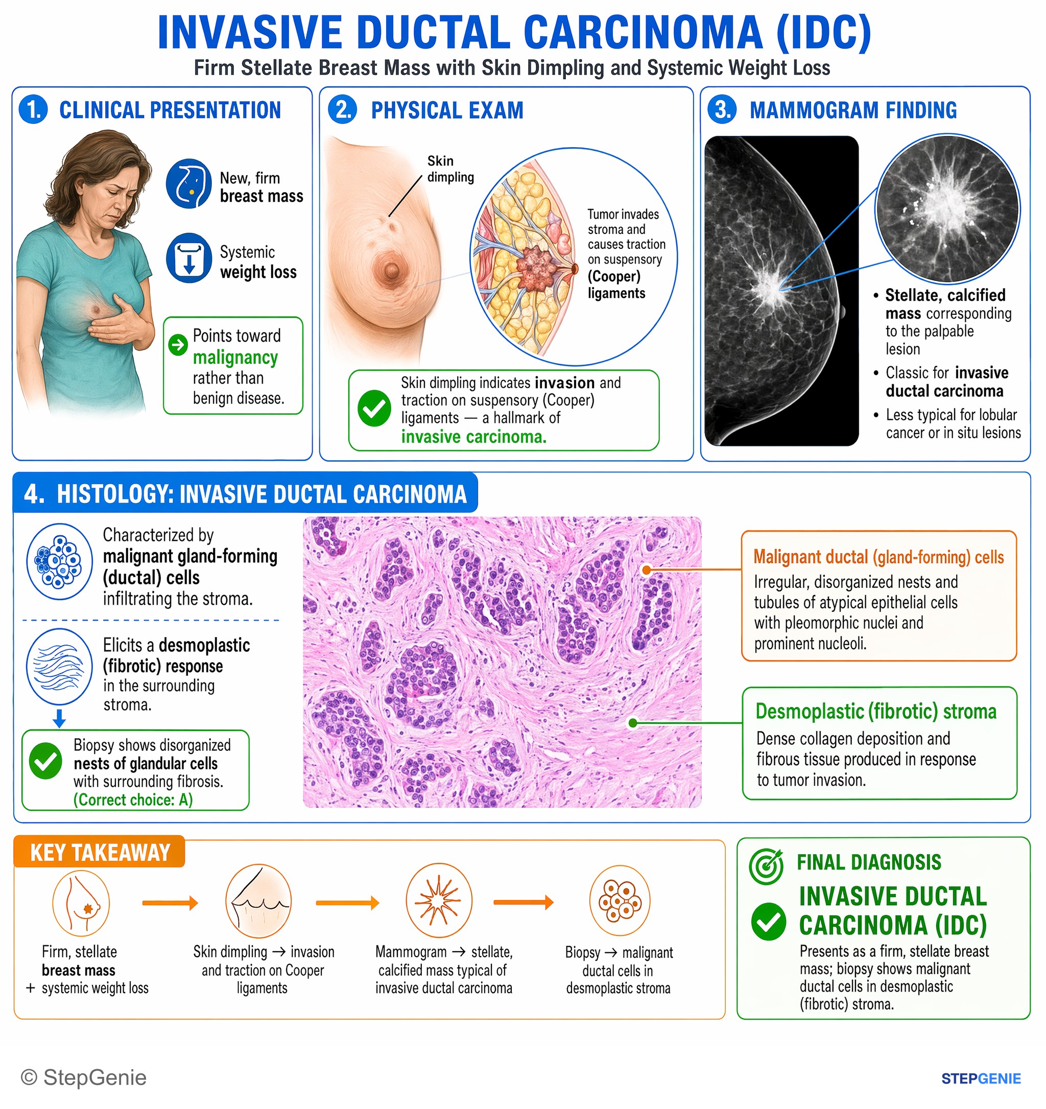
Invasive ductal carcinoma presents as a firm, stellate breast mass; biopsy shows malignant ductal cells in desmoplastic (fibrotic) stroma.
USMLE Step 1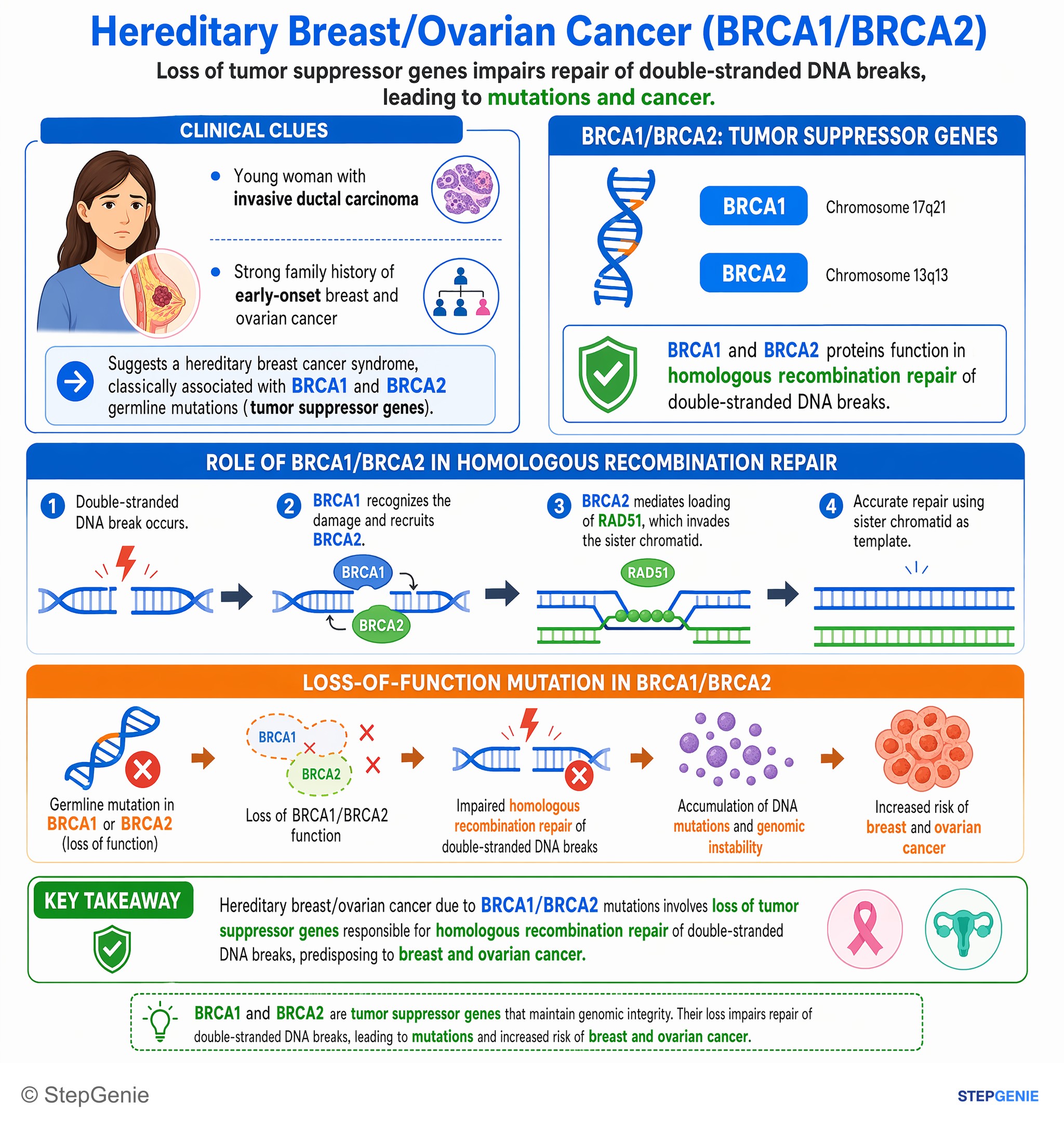
Hereditary breast cancer (BRCA1/BRCA2) involves loss of tumor suppressor genes responsible for homologous recombination repair of…
USMLE Step 1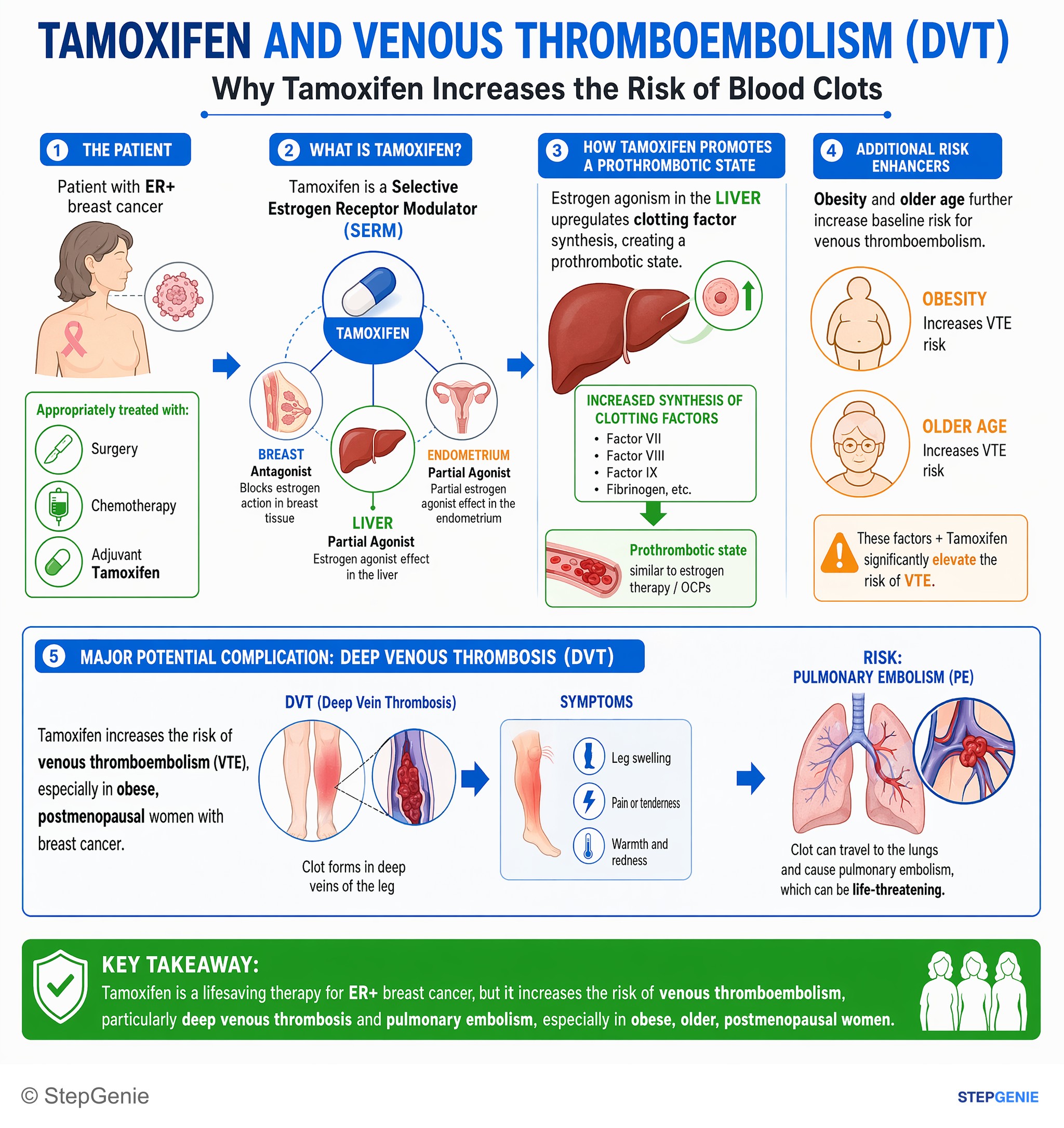
Tamoxifen is a selective estrogen receptor modulator that increases risk of venous thromboembolism, especially in obese, postmenopausal…
USMLE Step 2Frequently Asked Questions
What diagnosis fits a firm irregular breast mass with skin dimpling and microcalcifications?
The most likely diagnosis is invasive ductal carcinoma. A firm, irregular, non-tender mass with skin dimpling points to an invasive malignancy, and the dimpling occurs from traction on Cooper ligaments due to a desmoplastic fibrotic reaction. A spiculated mass on mammogram is classic for malignancy, and microcalcifications strongly suggest ductal pathology because invasive ductal carcinoma often coexists with ductal carcinoma in situ.
Why does invasive ductal carcinoma cause skin dimpling?
Invasive ductal carcinoma causes skin dimpling because the invasive tumor triggers a desmoplastic reaction that pulls on Cooper ligaments. This fibrotic response creates traction on the suspensory structures of the breast, producing the classic dimpling seen on exam. In the right clinical setting, skin dimpling paired with a firm irregular mass strongly favors an invasive breast carcinoma rather than a benign lesion.
How do you tell invasive ductal carcinoma from invasive lobular carcinoma?
Invasive ductal carcinoma is suggested by malignant ductal cells in cords and nests surrounded by dense desmoplastic stroma, often with a spiculated mass and microcalcifications on mammogram. Invasive lobular carcinoma instead classically shows loss of E-cadherin with a single-file infiltrating pattern, is more likely to be multifocal and bilateral, and can be less conspicuous on mammography.
How do you distinguish invasive ductal carcinoma from fibroadenoma?
Invasive ductal carcinoma is more likely when the mass is irregular, fixed, and associated with skin dimpling or spiculations on mammogram. Fibroadenoma is typically seen in women younger than 30 and presents as a well-circumscribed, mobile, rubbery mass. Histology also differs: fibroadenoma shows benign stromal proliferation compressing ducts, whereas invasive ductal carcinoma shows malignant ductal cells with desmoplastic stroma.
What breast cancer risk factors are being tested in this vignette?
The vignette highlights several classic risk factors for breast cancer: early menarche, first pregnancy after age 30, alcohol use, and a family history of breast cancer in a mother diagnosed before age 50. These features reflect increased lifetime estrogen exposure and possible hereditary risk. Prior chest radiation and BRCA-associated family history are also important clues to recognize quickly.