You’re cruising through a hemostasis question set when a “warfarin complication” vignette shows up—and suddenly every answer choice looks plausible. Protein C and Protein S deficiencies are classic USMLE traps because they sit at the intersection of thrombophilia, warfarin pharmacology, and coagulation cascade logic. The key to scoring reliably isn’t just knowing the right diagnosis—it’s knowing why the wrong ones are wrong.
Tag: Heme/Onc > Hemostasis & Coagulation
The Clinical Vignette (Q-bank style)
A 24-year-old woman presents with painful swelling of her left calf. Doppler ultrasound confirms a DVT. She’s started on warfarin without heparin bridging. Two days later she develops painful, well-demarcated, violaceous plaques on her thighs and abdomen that progress to necrosis. Labs show normal platelet count. She has no history of autoimmune disease.
What is the most likely underlying predisposing condition?
Correct Answer: Protein C deficiency (± Protein S deficiency)
Why this is the answer
This is the classic setup for warfarin-induced skin necrosis, caused by a transient hypercoagulable state right after starting warfarin.
Warfarin inhibits vitamin K–dependent factors:
- Procoagulants: II, VII, IX, X
- Anticoagulants: Protein C and Protein S
The kicker: Protein C has a short half-life (about ~8 hours), so it drops first. That means early warfarin therapy can temporarily remove anticoagulant protection before procoagulant factors have fallen enough—creating a window of increased thrombosis in skin microvasculature → painful necrotic plaques.
High-yield mechanisms
- Protein C, activated by thrombin bound to thrombomodulin, inactivates factors Va and VIIIa.
- Protein S is a cofactor for activated Protein C.
- Deficiency of either predisposes to venous thromboembolism (VTE).
- Warfarin can precipitate skin necrosis particularly in Protein C deficiency (also can occur in Protein S deficiency).
What you should say on Step exams
- Warfarin-induced skin necrosis = think Protein C deficiency + no heparin bridge
- Treat acutely: stop warfarin, give vitamin K, start heparin, consider protein C concentrate (or FFP if needed)
The “Why Every Answer Choice Matters” Breakdown (Systematic Distractors)
Below are the common distractors tested with this vignette—and how to eliminate them quickly.
1) Heparin-induced thrombocytopenia (HIT)
Why it’s tempting: Thrombosis + skin findings after anticoagulation.
Why it’s wrong here:
- HIT requires heparin exposure (usually 5–10 days after start; can be earlier with prior exposure).
- HIT classically features thrombocytopenia (often >50% drop), not normal platelets.
- Skin necrosis can occur in HIT, but it’s typically at injection sites and with platelet drop.
USMLE trigger for HIT:
- Heparin + platelet fall + thrombosis → anti-PF4 IgG, treat with argatroban/bivalirudin (not warfarin initially)
2) Antiphospholipid syndrome (APS)
Why it’s tempting: Young patient with thrombosis.
Why it’s wrong here:
- APS is associated with arterial and venous thromboses and pregnancy morbidity.
- You’d expect history of:
- Recurrent miscarriages
- Livedo reticularis
- Autoimmune context (often SLE)
- Labs: prolonged PTT that does not correct on mixing study (due to lupus anticoagulant), plus anticardiolipin/β2-glycoprotein antibodies.
- APS doesn’t specifically cause warfarin-induced skin necrosis as the signature complication in the first days.
USMLE trigger for APS:
- “Paradox”: prolonged PTT but hypercoagulable patient
3) Factor V Leiden mutation
Why it’s tempting: Most common inherited thrombophilia; causes DVTs.
Why it’s wrong here:
- Factor V Leiden = resistance to activated Protein C, leading to increased clotting.
- It predisposes to VTE, but it does not classically cause the early-warfarin skin necrosis vignette.
- Also, Factor V Leiden is a factor V mutation, not a vitamin K–dependent issue.
USMLE trigger for Factor V Leiden:
- Recurrent DVTs, often provoked by estrogen/OCPs/pregnancy
- “Activated protein C resistance” test abnormal
4) Prothrombin (G20210A) mutation
Why it’s tempting: Another inherited thrombophilia → DVT.
Why it’s wrong here:
- Causes increased prothrombin levels → VTE risk.
- Again, no special linkage to warfarin-induced skin necrosis.
USMLE trigger:
- Unprovoked VTE + family history; labs often otherwise normal
5) Disseminated intravascular coagulation (DIC)
Why it’s tempting: Skin necrosis, thrombosis possible.
Why it’s wrong here:
- DIC is a consumption coagulopathy with bleeding + thrombosis.
- Expect:
- Low platelets
- ↑ PT and ↑ PTT
- ↓ fibrinogen
- ↑ D-dimer
- Often triggered by sepsis, trauma, malignancy, obstetric catastrophe—not “day 2 of warfarin.”
USMLE trigger:
- Sick patient + diffuse bleeding/oozing + abnormal coag labs
6) Thrombotic thrombocytopenic purpura (TTP)
Why it’s tempting: Thrombosis in microvasculature can cause skin findings.
Why it’s wrong here:
- TTP is marked thrombocytopenia and hemolytic anemia—think:
- MAHA (schistocytes)
- Neuro symptoms
- Renal dysfunction
- Fever
- Coag tests (PT/PTT) are usually normal (unlike DIC), but platelets are low.
USMLE trigger:
- Pentad (or dyad): MAHA + thrombocytopenia, treat with plasmapheresis
7) Von Willebrand disease
Why it’s tempting: Common hemostasis disorder.
Why it’s wrong here:
- vWD causes mucocutaneous bleeding (epistaxis, menorrhagia), not thrombosis.
- Platelet function defect with possible ↑ PTT (due to decreased factor VIII stabilization).
USMLE trigger:
- Bleeding with normal platelet count but prolonged bleeding time; responds to desmopressin
Quick Table: Thrombophilia vs Bleeding Patterns (High Yield)
| Condition | Primary problem | Typical presentation | Platelets | PT/PTT |
|---|---|---|---|---|
| Protein C/S deficiency | ↓ anticoagulants → ↑ factors Va/VIIIa | VTE, warfarin skin necrosis | Normal | Normal |
| Factor V Leiden | APC resistance | VTE (common) | Normal | Normal |
| Prothrombin mutation | ↑ prothrombin | VTE | Normal | Normal |
| APS | Autoantibodies | Arterial/venous clots, miscarriages | Normal (often) | ↑ PTT (paradoxical) |
| HIT | Anti-PF4 antibodies | Thrombosis + thrombocytopenia | Low | Normal |
| DIC | Consumption | Bleeding + thrombosis, sick patient | Low | ↑ PT, ↑ PTT |
| TTP | ADAMTS13 deficiency | MAHA + thrombocytopenia + neuro/renal | Low | Normal |
Step-Style “Rules” You Can Apply in Seconds
Warfarin early complication rule
- Early (days 1–3) + skin necrosis → think Protein C deficiency (or Protein S)
- Prevention: bridge with heparin when starting warfarin for acute VTE
Bleeding vs clotting rule of thumb
- Mucosal bleeding → platelet/vWF problems
- Deep tissue bleeding (hemarthroses) → coag factor deficiencies (hemophilia)
- VTE in young person → inherited thrombophilia (Factor V Leiden, prothrombin, Protein C/S, antithrombin)
One More Level: The Mechanism You Should Be Ready to Say Out Loud
Warfarin lowers vitamin K–dependent factors, but:
- Protein C falls fastest → transient hypercoagulability
- Microthrombi form in skin → ischemia → hemorrhagic necrosis
- This is why heparin bridging matters: it covers the early hypercoagulable window until factor II (long half-life) decreases.
Rapid-Fire Practice: If the Stem Changes, Your Answer Changes
- Thrombosis + thrombocytopenia + recent heparin → HIT
- Unprovoked VTE + family history, no warfarin detail → Factor V Leiden / prothrombin mutation
- Clots + miscarriages + prolonged PTT → APS
- Bleeding + low fibrinogen + high D-dimer → DIC
- MAHA + neuro symptoms + low platelets → TTP
Visual explanations from the StepGenie QBank
Infographics StepGenie drew to explain questions in its own QBank, matched to this article's topic. Each caption is the takeaway of the question it explains.
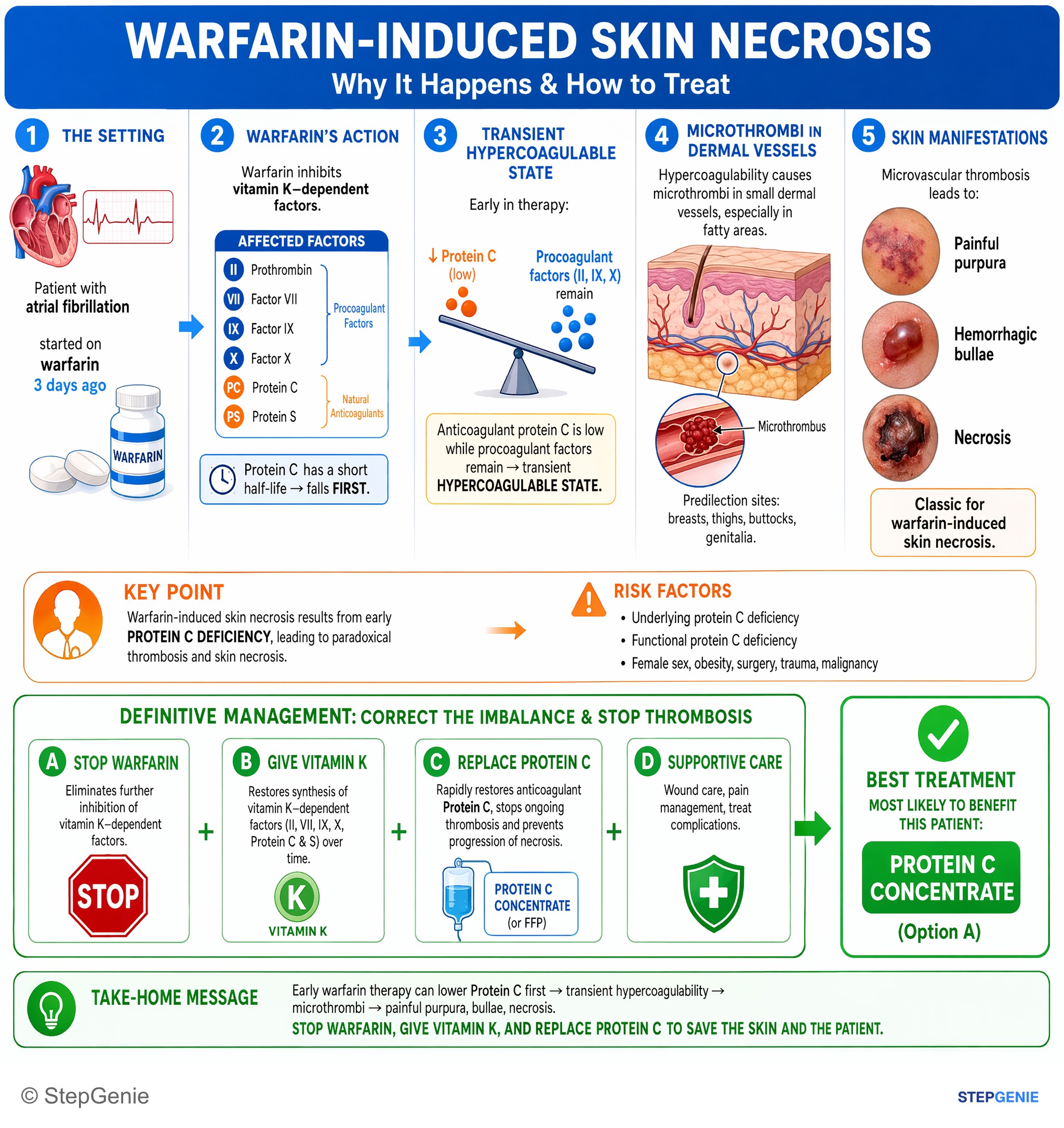
Warfarin-induced skin necrosis from early protein C deficiency → paradoxical thrombosis and skin necrosis; treat by replacing protein C…
USMLE Step 2Frequently Asked Questions
Why can starting warfarin without heparin bridging cause skin necrosis in protein C deficiency?
It happens because warfarin first lowers Protein C, creating a transient hypercoagulable state before the procoagulant factors fall enough. Protein C is a vitamin K–dependent anticoagulant with a short half-life, so its anticoagulant protection disappears early. In someone with Protein C deficiency, this can trigger thrombosis in the skin microvasculature, leading to painful violaceous plaques that progress to necrosis.
How do you tell warfarin-induced skin necrosis from heparin-induced thrombocytopenia?
Warfarin-induced skin necrosis is suggested by painful necrotic plaques shortly after starting warfarin, especially without heparin bridging, with a normal platelet count. HIT instead requires heparin exposure and classically causes thrombocytopenia, often with a major platelet drop. HIT can also cause skin necrosis, but it is typically at injection sites and occurs in the setting of platelet decline and anti-PF4 antibodies.
What is the mechanism of Protein C and Protein S in coagulation?
Protein C is an anticoagulant that, once activated by thrombin bound to thrombomodulin, inactivates factors Va and VIIIa. Protein S functions as a cofactor for activated Protein C. Because both help restrain clotting, deficiency of either predisposes to venous thromboembolism. This is also why early warfarin therapy can be dangerous in these patients, since warfarin lowers these vitamin K–dependent anticoagulants.
How do you distinguish Protein C deficiency from factor V Leiden in a DVT question?
Protein C deficiency is favored when a DVT patient develops early warfarin-induced skin necrosis, especially after starting warfarin without a heparin bridge. Factor V Leiden also causes venous thromboembolism, but its mechanism is resistance to activated Protein C rather than loss of Protein C itself. It is a factor V mutation and is not the classic cause of the early-warfarin skin necrosis vignette.
What is the acute treatment for warfarin-induced skin necrosis?
The acute treatment is to stop warfarin, give vitamin K, and start heparin. Protein C concentrate can also be considered, or fresh frozen plasma if needed. This approach addresses the transient hypercoagulable state caused by early warfarin therapy, which lowers Protein C before the vitamin K–dependent procoagulant factors have fallen enough to provide effective anticoagulation.