You’ve probably seen this pattern in a q-bank: an older adult with fatigue and weird neuro/bleeding symptoms, plus an “M spike” on serum protein studies. The trap is that a lot of lymphoid/plasma cell disorders share overlapping buzzwords—so the only way to reliably get these questions right is to force yourself to explain why each answer choice is wrong. Let’s do exactly that for Waldenström macroglobulinemia.
Tag: Heme/Onc > WBC Disorders & Lymphomas
The Clinical Vignette (Typical Q-Bank Style)
A 68-year-old man presents with progressive fatigue, headaches, and blurry vision. He reports intermittent epistaxis and easy bruising. Exam shows mild hepatosplenomegaly and scattered ecchymoses. Labs reveal normocytic anemia and elevated total protein. Peripheral smear shows rouleaux formation. Serum protein electrophoresis shows an M spike. Bone marrow biopsy demonstrates a lymphoplasmacytic infiltrate.
Question: What is the most likely diagnosis / key mechanism / expected finding?
Correct Answer: Waldenström Macroglobulinemia (Lymphoplasmacytic Lymphoma)
What it is
Waldenström macroglobulinemia (WM) is a lymphoplasmacytic lymphoma that secretes monoclonal IgM.
Why IgM matters (the “macroglobulinemia”)
IgM is a large pentamer → increases serum viscosity → causes hyperviscosity syndrome:
- Neurologic: headache, dizziness, confusion
- Visual: blurry vision, “sausage-link” retinal veins
- Bleeding: epistaxis, gingival bleeding (impaired platelet function from sludging/coating)
- Rouleaux can appear due to increased serum proteins (classically highlighted in plasma cell dyscrasias, but can show up with WM too)
Key distinguishing clinical clues
- Hyperviscosity is common in WM (high-yield differentiator)
- Hepatosplenomegaly/lymphadenopathy can occur (it’s a lymphoma)
- No punched-out lytic bone lesions (think multiple myeloma instead)
- No prominent Bence Jones renal failure as a classic feature (again, more myeloma)
Classic associations to know for USMLE
- MYD88 mutation (very common in WM; often tested as a “supports diagnosis” detail)
- Cold agglutinin hemolytic anemia can occur (IgM-mediated)
- Cryoglobulinemia can occur (IgM precipitates in cold)
Management pearl (often tested)
If the question hints at symptomatic hyperviscosity (vision changes, neuro symptoms, bleeding):
- Immediate: plasmapheresis (removes IgM from plasma quickly)
- Long-term disease control: rituximab-based therapy (anti-CD20), BTK inhibitors, etc.
Exam take-home: plasmapheresis treats symptoms fast; chemo/immunotherapy treats the underlying clone.
Why Every Other Answer Choice Is Wrong (High-Yield Distractor Autopsy)
Below are the most common distractors that show up next to WM in heme/onc question sets.
Distractor 1: Multiple Myeloma (Plasma Cell Neoplasm)
Why it tempts you: M spike, rouleaux, anemia, older patient.
Why it’s wrong here: Multiple myeloma is usually IgG or IgA (not IgM) and has the CRAB features driven by marrow/bone/kidney involvement:
- Calcium ↑: bone resorption
- Renal failure: light chains → cast nephropathy (Bence Jones proteins)
- Anemia: marrow crowding
- Bone lesions: punched-out lytic lesions, bone pain, fractures
Key discriminator:
- WM → hyperviscosity from IgM + often organomegaly
- Myeloma → lytic bone lesions + renal injury are front and center
| Feature | Waldenström | Multiple Myeloma |
|---|---|---|
| Main immunoglobulin | IgM | IgG/IgA |
| Hyperviscosity | Common | Can occur, less classic |
| Lytic bone lesions | No | Yes |
| Primary malignant cell | Lymphoplasmacytic B cell | Plasma cell |
| Typical organomegaly | Can have | Less typical |
Distractor 2: MGUS (Monoclonal Gammopathy of Undetermined Significance)
Why it tempts you: M spike in an older adult.
Why it’s wrong here: MGUS is asymptomatic by definition. No end-organ damage and no hyperviscosity syndrome.
High-yield diagnostic framework:
- MGUS: M protein present but no CRAB, no symptoms, low marrow plasma cells
- Myeloma/WM: symptomatic, with organ involvement (CRAB for myeloma; hyperviscosity for WM)
Rule of thumb: If the vignette has real symptoms (vision changes, headaches, bleeding), it’s not “of undetermined significance.”
Distractor 3: Chronic Lymphocytic Leukemia (CLL) / Small Lymphocytic Lymphoma (SLL)
Why it tempts you: Older patient, B-cell malignancy, lymphadenopathy.
Why it’s wrong here: CLL/SLL classically presents with:
- Lymphocytosis and smudge cells
- Hypogammaglobulinemia → recurrent infections
- Autoimmune hemolytic anemia can occur (usually warm IgG)
WM instead is defined by IgM monoclonal gammopathy with hyperviscosity and lymphoplasmacytic marrow infiltration.
Don’t miss:
- CLL immunophenotype: CD5+ and CD23+ B cells
- WM: IgM paraprotein, often MYD88 mutation
Distractor 4: Mantle Cell Lymphoma (MCL)
Why it tempts you: Older patient, B-cell lymphoma, can involve marrow and cause systemic symptoms.
Why it’s wrong here: MCL is aggressive and associated with:
- t(11;14) → cyclin D1 overexpression
- Often presents with generalized lymphadenopathy, B symptoms, GI involvement (lymphomatous polyposis)
WM’s hallmark is IgM secretion → hyperviscosity. MCL is not defined by an IgM paraprotein picture.
Distractor 5: Hairy Cell Leukemia
Why it tempts you: Middle-aged/older man + splenomegaly + cytopenias.
Why it’s wrong here: Hairy cell leukemia is classically:
- TRAP positive
- “Hairy” cytoplasmic projections
- Dry tap bone marrow aspiration due to fibrosis
- Massive splenomegaly with pancytopenia
It does not classically give you an M spike with hyperviscosity syndrome.
Distractor 6: Hodgkin Lymphoma
Why it tempts you: Lymphoma + systemic symptoms.
Why it’s wrong here: Hodgkin is built around:
- Reed–Sternberg cells (CD15+, CD30+)
- Contiguous spread, B symptoms
- Not a monoclonal IgM secretion disorder; hyperviscosity is not the classic board-style complication
If the stem of the question screams “paraprotein + hyperviscosity,” you’re in plasma cell/lymphoplasmacytic territory, not classic Hodgkin.
Distractor 7: Diffuse Large B-Cell Lymphoma (DLBCL)
Why it tempts you: Common NHL in older adults; can present with rapidly enlarging nodes.
Why it’s wrong here: DLBCL is an aggressive lymphoma with:
- Rapidly enlarging mass, B symptoms
- Not defined by IgM hyperviscosity or M spike as the central clue
An M spike can happen incidentally in some B-cell disorders, but q-banks usually make WM the best fit when hyperviscosity symptoms + IgM are emphasized.
High-Yield “If You See X, Think Waldenström” Checklist
Look for this cluster:
- Older adult + fatigue/anemia
- M spike + elevated total protein
- Hyperviscosity symptoms: headache, blurry vision, mucosal bleeding
- Lymphoplasmacytic marrow infiltrate
- Possible hepatosplenomegaly
- IgM monoclonal protein
- Often MYD88 mutation
Mini Rapid-Fire Q-Bank Pearls
- Best immediate treatment for symptomatic hyperviscosity? Plasmapheresis
- Which immunoglobulin is most associated with hyperviscosity on exams? IgM
- Why is IgM “worse” for viscosity? It’s a large pentamer → increases serum viscosity significantly.
- How do you avoid a classic trap? If you see lytic bone lesions + renal failure, don’t pick WM—pick multiple myeloma.
One-Liner Summary (What to Write in the Margin)
Waldenström = lymphoplasmacytic lymphoma producing IgM → hyperviscosity (neuro/vision/bleeding) + M spike; treat acute hyperviscosity with plasmapheresis.
Visual explanations from the StepGenie QBank
Infographics StepGenie drew to explain questions in its own QBank, matched to this article's topic. Each caption is the takeaway of the question it explains.
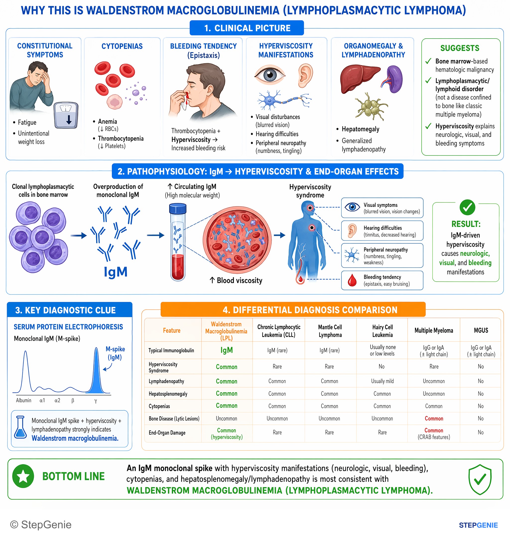
Waldenstrom macroglobulinemia: IgM monoclonal spike causing hyperviscosity (neurologic, visual, bleeding) with anemia, thrombocytopenia…
USMLE Step 1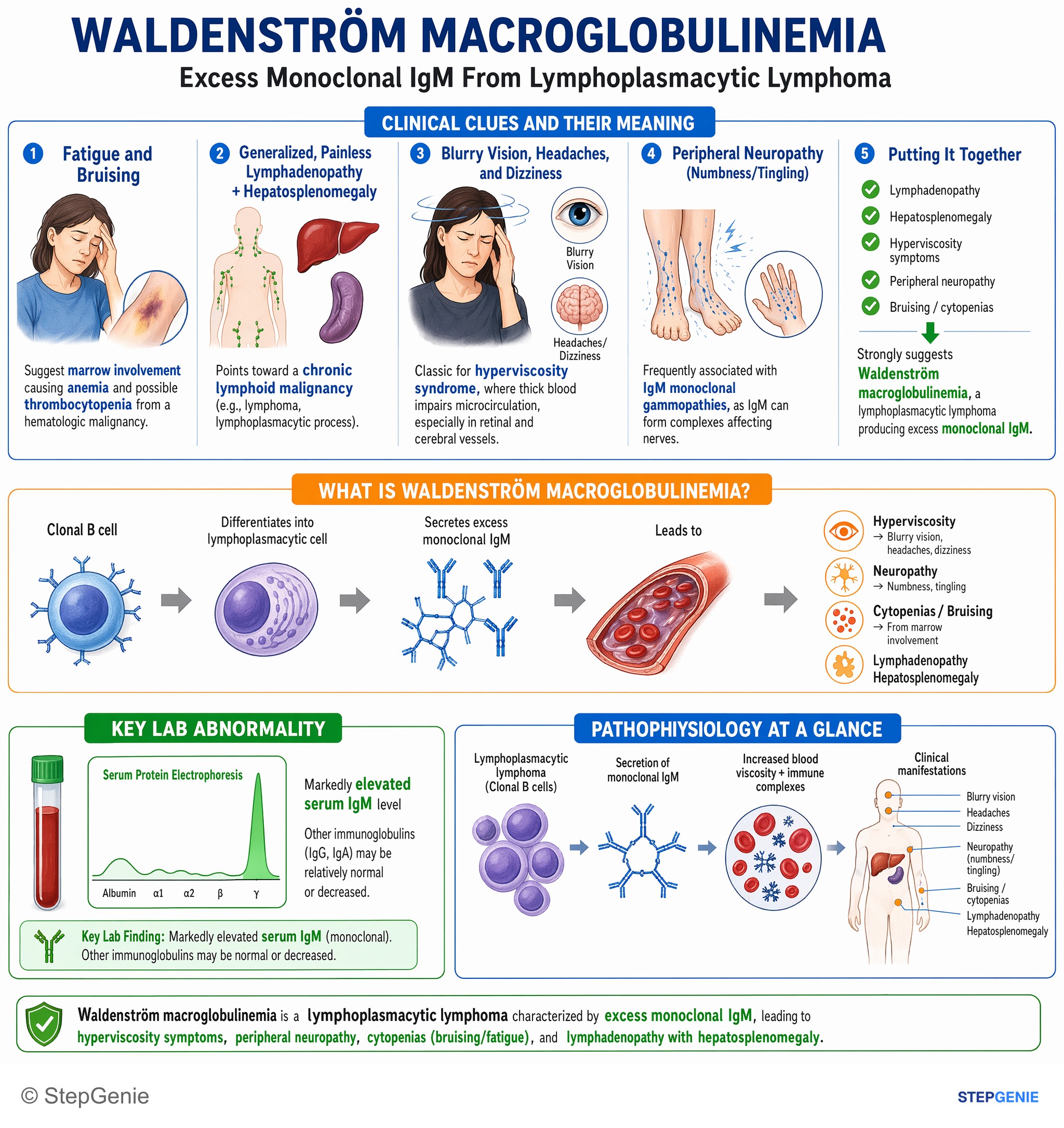
Waldenström macroglobulinemia from clonal B cells secreting excess IgM → hyperviscosity (blurry vision, headaches, dizziness, neuropathy)…
USMLE Step 1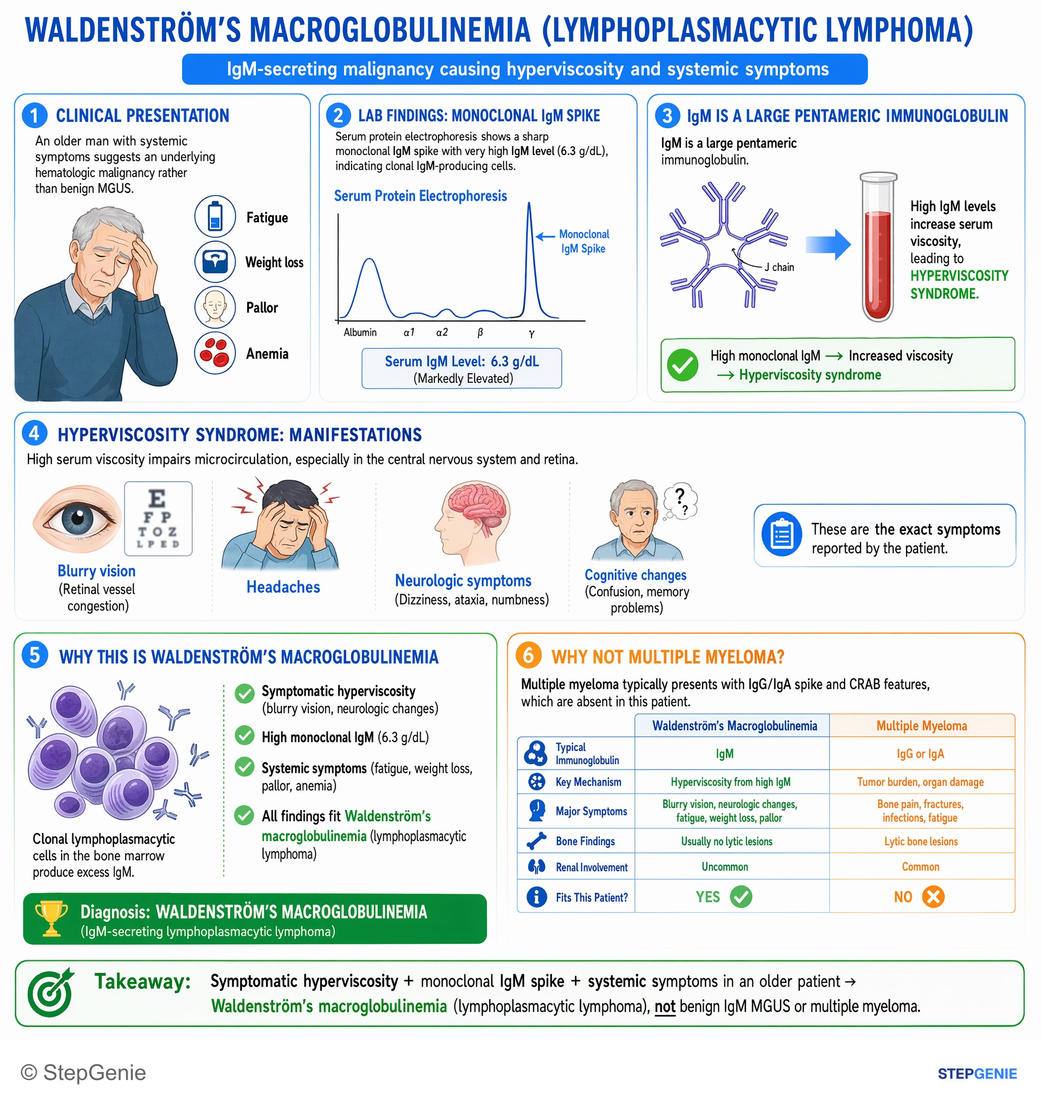
Waldenstrom’s macroglobulinemia: IgM-secreting lymphoplasmacytic lymphoma causing hyperviscosity (blurry vision, neurologic changes) with…
USMLE Step 2Frequently Asked Questions
Why does Waldenström macroglobulinemia cause headaches, blurry vision, and bleeding symptoms?
It causes these symptoms because the malignant cells secrete monoclonal IgM, a large pentamer that increases serum viscosity. This hyperviscosity syndrome leads to neurologic symptoms such as headache and dizziness, visual changes such as blurry vision, and bleeding like epistaxis or gingival bleeding due to impaired platelet function from sludging and coating.
How do you tell Waldenström macroglobulinemia from multiple myeloma on a vignette?
The key distinction is that Waldenström macroglobulinemia is an IgM-secreting lymphoplasmacytic lymphoma with hyperviscosity and often hepatosplenomegaly or lymphadenopathy, whereas multiple myeloma is usually an IgG or IgA plasma cell neoplasm with CRAB features. Myeloma classically has punched-out lytic bone lesions and renal injury, which are not classic findings in Waldenström macroglobulinemia.
What finding supports the diagnosis of Waldenström macroglobulinemia when there is an M spike?
A lymphoplasmacytic infiltrate in the bone marrow supports Waldenström macroglobulinemia, especially when paired with a monoclonal IgM paraprotein. Additional clues include rouleaux formation from increased serum proteins, hyperviscosity symptoms, and a MYD88 mutation, which is described as a very common association that is often tested as a detail supporting the diagnosis.
What is the first-line emergency treatment for symptomatic hyperviscosity in Waldenström macroglobulinemia?
The immediate treatment is plasmapheresis. It rapidly removes IgM from the plasma and is used when the patient has symptomatic hyperviscosity, such as vision changes, neurologic symptoms, or bleeding. Long-term control still requires treatment of the underlying malignant clone, such as rituximab-based therapy or BTK inhibitors.
Why is MGUS not the right answer if an older patient has an M spike plus vision changes and headaches?
MGUS is not the right diagnosis because it is asymptomatic by definition. An M protein can be present in MGUS, but there should be no end-organ damage and no hyperviscosity syndrome. If the vignette includes real symptoms like blurry vision, headaches, or bleeding, that points away from MGUS and toward a symptomatic disorder such as Waldenström macroglobulinemia.