Multiple myeloma questions are the definition of “the stem gives you everything you need, but only if you notice it.” A good Q-bank item will hand you 3–5 subtle clues (bone pain pattern, infections, labs, smear, kidney findings), then tempt you with distractors that are almost right. This post walks through a classic USMLE-style vignette, nails the correct answer, and then dissects every answer choice so you learn the pattern recognition and the why-not logic.
Tag: Heme/Onc > WBC Disorders & Lymphomas
The Clinical Vignette (USMLE-Style)
A 66-year-old man comes to clinic for progressive fatigue and low back pain for 4 months. He reports several recent “sinus infections” requiring antibiotics. Exam shows pallor and mild lumbar spine tenderness. Labs:
- Hemoglobin: 8.9 g/dL
- Creatinine: 2.3 mg/dL
- Calcium: 11.6 mg/dL
- Total protein: 9.8 g/dL (albumin 3.1 g/dL)
- Peripheral smear: rouleaux formation
- ESR: elevated
Urinalysis shows protein with few cells; dipstick is only mildly positive. Skeletal survey reveals multiple punched-out lytic lesions in the skull and vertebrae.
Question: Which of the following is the most likely underlying diagnosis?
Answer choices:
A. Chronic lymphocytic leukemia (CLL)
B. Multiple myeloma
C. Waldenström macroglobulinemia
D. Monoclonal gammopathy of undetermined significance (MGUS)
E. Primary amyloidosis (AL amyloid)
F. Metastatic prostate cancer to bone
The Correct Answer: B. Multiple myeloma
This stem basically screams CRAB + rouleaux + lytic lesions:
Why it’s multiple myeloma
Multiple myeloma is a malignant plasma cell disorder characterized by clonal proliferation of plasma cells in the bone marrow producing a monoclonal immunoglobulin (often IgG) and/or free light chains.
High-yield clinical/lab anchors:
- Calcium ↑: osteoclast activation (lytic bone destruction)
- Renal failure: light chains (Bence Jones) → cast nephropathy; also amyloid deposition
- Anemia: marrow crowding + inflammatory milieu
- Bone pain/lytic lesions: especially spine, ribs, skull
Plus: - Rouleaux formation: RBC stacking due to increased serum proteins
- Recurrent infections: impaired normal immunoglobulin production (functional hypogammaglobulinemia), classically encapsulated bacteria
The urinalysis clue everyone forgets
The UA shows protein, but the dipstick is only mildly positive. That points toward light chains, because standard urine dipsticks detect albumin better than free light chains.
Confirm with urine protein electrophoresis (UPEP) or serum free light chain assay.
What you’d expect on confirmatory testing
- SPEP/UPEP: M-spike (SPEP best for heavy chains; UPEP helps detect light chains)
- Bone marrow biopsy: clonal plasma cells (often )
- Serum free light chain ratio: abnormal
- β2-microglobulin: prognostic marker
Rapid Pattern Table: Multiple Myeloma vs Look-Alikes
| Condition | Key clue | Bone findings | Blood/viscosity | Typical immunoglobulin |
|---|---|---|---|---|
| Multiple myeloma | CRAB, rouleaux, recurrent infections, light chains | Lytic “punched-out” lesions | Not usually hyperviscosity (can happen, but less classic) | Usually IgG (also IgA); ± light chains |
| Waldenström macroglobulinemia | Hyperviscosity + neuropathy + mucosal bleeding | No lytic lesions | Hyperviscosity common | IgM |
| MGUS | M-protein but no end-organ damage | None | None | Any (small M-spike) |
| CLL | Lymphadenopathy + smudge cells | None | Autoimmune hemolysis possible | Not a plasma cell process |
| AL amyloidosis | Nephrotic syndrome, restrictive CM, macroglossia | Variable | Depends | Light chains (λ often) |
Now, Why Each Distractor Is Wrong (and When It Would Be Right)
A. Chronic lymphocytic leukemia (CLL)
Why it’s wrong here:
CLL is a malignancy of mature B lymphocytes, not plasma cells. The stem is dominated by CRAB, rouleaux, lytic lesions, and protein gap—all pointing to a monoclonal gammopathy with end-organ damage.
What would point to CLL instead:
- Older adult with painless lymphadenopathy
- Absolute lymphocytosis
- Smudge cells on smear
- Complications: autoimmune hemolytic anemia, hypogammaglobulinemia → infections
USMLE pearl: CLL cells commonly express CD5 and CD23 (a common testable immunophenotype).
C. Waldenström macroglobulinemia
Why it’s wrong here:
Waldenström is a lymphoplasmacytic lymphoma producing IgM, which is big and causes hyperviscosity—think:
- Headache, blurry vision, dizziness
- Mucosal bleeding
- Peripheral neuropathy
But it typically does not cause lytic bone lesions or the classic CRAB constellation.
What would point to Waldenström instead:
- Hyperviscosity syndrome
- No punched-out lesions
- Often hepatosplenomegaly/lymphadenopathy
- SPEP with IgM monoclonal spike
High-yield treatment tie-in: Hyperviscosity symptoms → plasmapheresis acutely (removes IgM).
D. Monoclonal gammopathy of undetermined significance (MGUS)
Why it’s wrong here:
MGUS is the “M-spike without the mayhem.” By definition, it’s monoclonal protein present but:
- No CRAB features (no end-organ damage)
- Bone marrow plasma cells <10%
- Lower M-protein burden (classically <3 g/dL)
This patient has anemia, renal failure, hypercalcemia, lytic lesions → that’s not MGUS.
When MGUS is correct:
- Incidental M-spike in an asymptomatic patient
- Normal calcium/creatinine/Hgb
- No bone lesions
USMLE pearl: MGUS can progress to myeloma at about 1% per year.
E. Primary amyloidosis (AL amyloid)
Why it’s wrong here (as the primary diagnosis):
AL amyloidosis is often driven by a plasma cell dyscrasia and can coexist with myeloma, but the stem is most classic for multiple myeloma with cast nephropathy + bone disease. AL amyloid would usually be signposted with “waxy” systemic features.
What would point to AL amyloidosis instead:
- Nephrotic syndrome (heavy proteinuria, edema; often albuminuria)
- Restrictive cardiomyopathy (HFpEF), arrhythmias
- Hepatosplenomegaly
- Macroglossia
- Periorbital purpura
- Biopsy with Congo red staining → apple-green birefringence
High-yield nuance: Amyloid kidney disease often presents with albuminuria (strong dipstick), whereas light chain cast nephropathy may have protein with a relatively weak dipstick.
F. Metastatic prostate cancer to bone
Why it’s wrong here:
Prostate cancer classically causes osteoblastic (sclerotic) bone metastases, not lytic “punched-out” lesions. It also doesn’t explain rouleaux formation or a protein gap.
What would point to prostate cancer instead:
- Older man with urinary symptoms, weight loss
- Elevated PSA
- Bone pain with imaging showing sclerotic lesions
- Possible increased alkaline phosphatase from osteoblastic activity
USMLE quick contrast:
- Myeloma → lytic lesions
- Prostate cancer → blastic lesions
- Renal cell carcinoma / thyroid / NSCLC → classically lytic metastases
High-Yield “What They’re Testing” in Multiple Myeloma
1) The “protein gap”
If total protein is high but albumin is relatively low, think excess globulins (immunoglobulins):
- This stem: total protein 9.8 with albumin 3.1 → large globulin gap
2) Rouleaux vs agglutination
- Rouleaux = stacked RBCs from high serum proteins (myeloma, inflammatory states)
- Agglutination = irregular clumping (often IgM cold agglutinins)
3) Bone pathophysiology
Myeloma cells stimulate osteoclasts via cytokines (notably IL-6 signaling is often emphasized), leading to:
- Bone pain
- Lytic lesions
- Hypercalcemia
- Pathologic fractures
4) Renal failure mechanisms (very testable)
- Light chain cast nephropathy (most classic): obstruction/toxicity in tubules
- AL amyloidosis: deposition in glomeruli → nephrotic syndrome
- Hypercalcemia-related nephropathy/dehydration can contribute
5) Infections
Even though myeloma makes lots of immunoglobulin, it’s monoclonal and dysfunctional, and normal antibody production drops → susceptibility to infections, especially encapsulated organisms.
Takeaway: How to Win These Q-Bank Stems Fast
When you see:
- Older patient
- Back/bone pain + lytic lesions
- Anemia + renal dysfunction
- Hypercalcemia
- High total protein / rouleaux
You should immediately think multiple myeloma and then use the distractors to prove to yourself:
- Waldenström = IgM + hyperviscosity, no lytic lesions
- MGUS = no end-organ damage
- CLL = lymphocytosis/smudge cells/lymphadenopathy
- AL amyloid = nephrotic/restrictive CM/macroglossia (often associated but not the primary pattern here)
- Prostate mets = osteoblastic lesions
Visual explanations from the StepGenie QBank
Infographics StepGenie drew to explain questions in its own QBank, matched to this article's topic. Each caption is the takeaway of the question it explains.
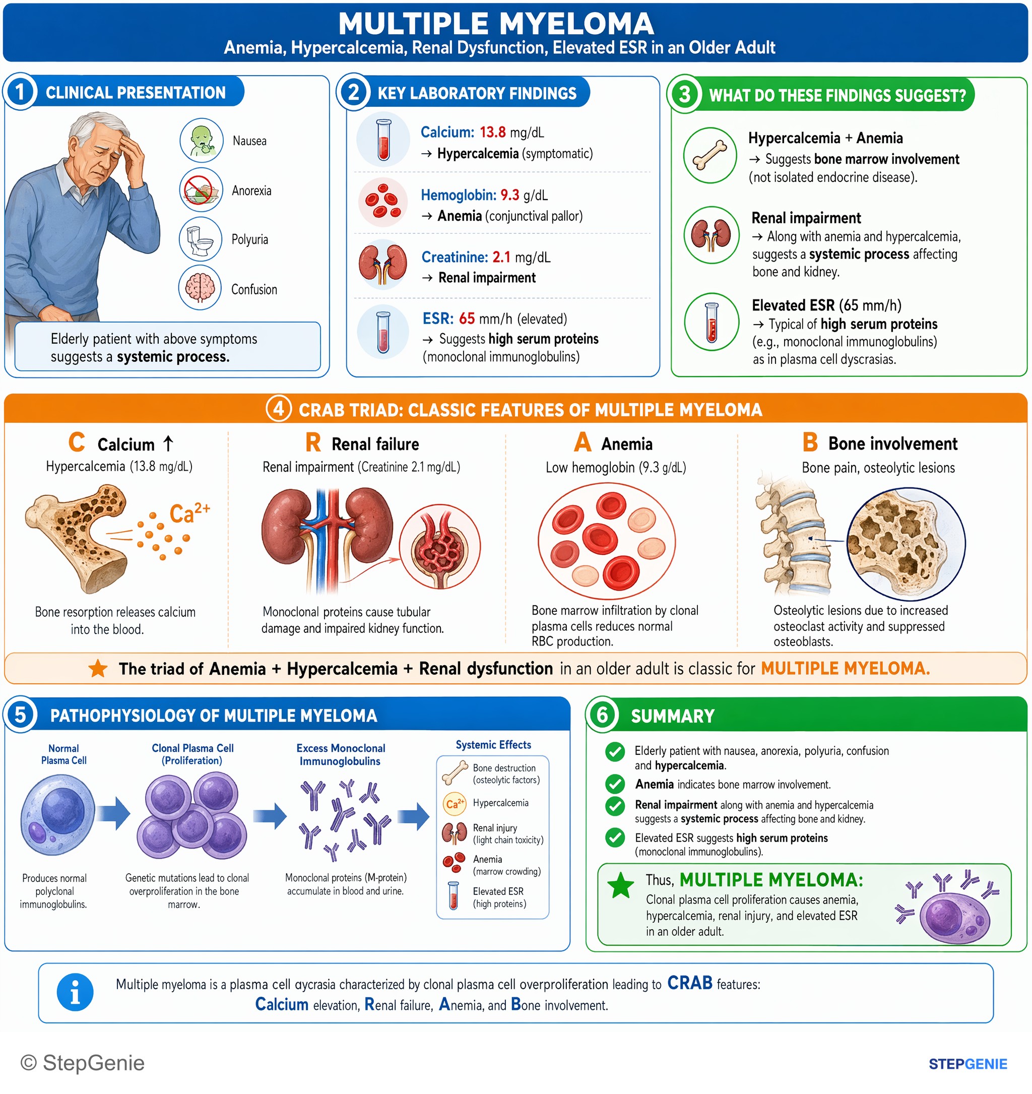
Multiple myeloma: clonal plasma cell proliferation causes anemia, hypercalcemia, renal injury, and elevated ESR in an older adult.
USMLE Step 1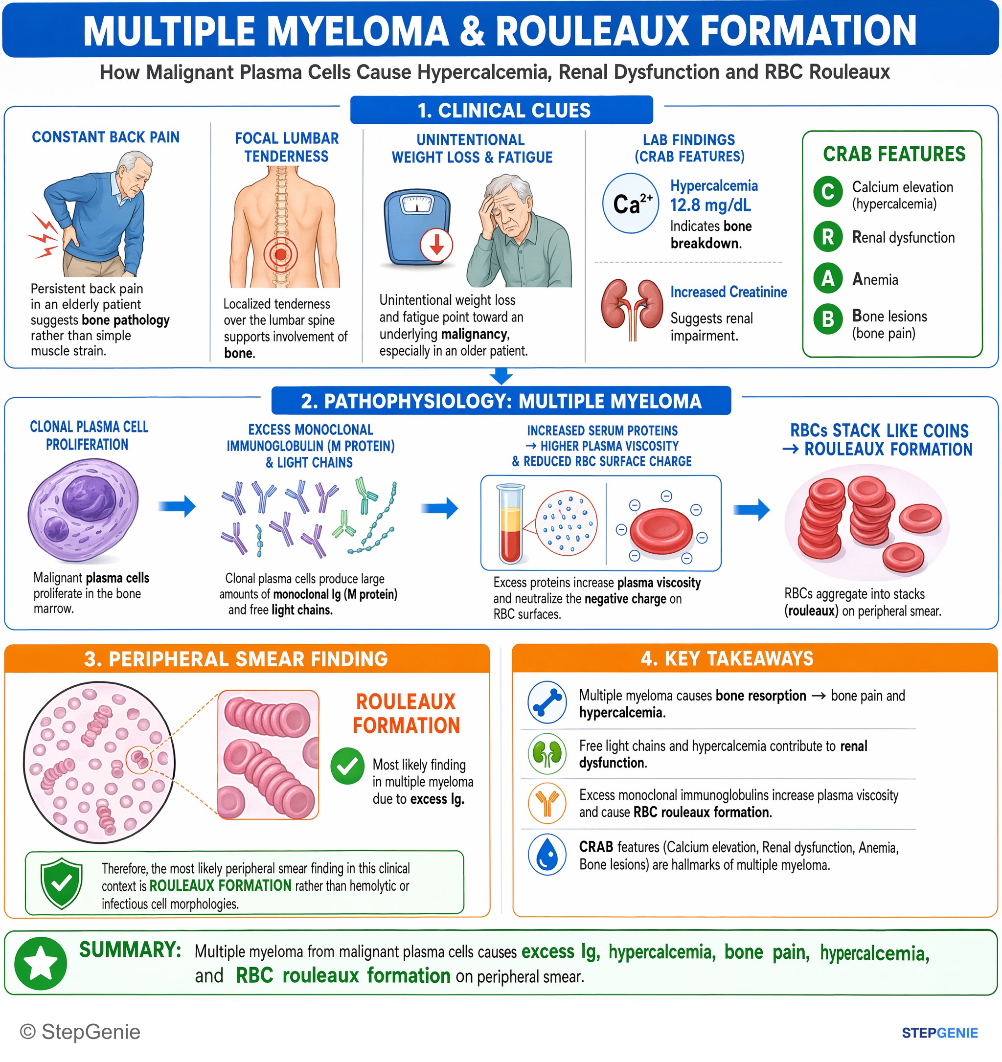
Multiple myeloma from malignant plasma cells causes excess Ig, hypercalcemia, bone pain, renal dysfunction, and RBC rouleaux formation on…
USMLE Step 1
Multiple myeloma suspected from bone pain, anemia, hypercalcemia, lytic skull lesions → confirm with serum protein electrophoresis for…
USMLE Step 2Frequently Asked Questions
What clues in a vignette point most strongly to multiple myeloma?
The strongest clues are the CRAB features plus rouleaux formation and lytic bone lesions. Multiple myeloma classically presents with hypercalcemia, renal failure, anemia, and bone pain from punched-out lytic lesions, especially in the spine, ribs, and skull. Rouleaux on smear reflects increased serum proteins, and recurrent infections fit impaired normal immunoglobulin production from a malignant plasma cell disorder.
Why can urinalysis show protein in multiple myeloma but the dipstick be only mildly positive?
This happens because the urine protein may be free light chains rather than albumin. Standard urine dipsticks detect albumin better than free light chains, so a patient with multiple myeloma can have protein in the urine with only mild dipstick positivity. That pattern should raise suspicion for Bence Jones proteins and be evaluated with urine protein electrophoresis or a serum free light chain assay.
How do you tell multiple myeloma from Waldenström macroglobulinemia on an exam question?
The key distinction is that multiple myeloma gives CRAB findings and punched-out lytic lesions, whereas Waldenström macroglobulinemia is more associated with hyperviscosity symptoms and usually no lytic lesions. Waldenström classically involves IgM and presents with headache, blurry vision, dizziness, mucosal bleeding, or neuropathy. If the stem emphasizes bone pain, hypercalcemia, renal dysfunction, and lytic lesions, multiple myeloma is more likely.
How do you distinguish multiple myeloma from MGUS in a question stem?
The main difference is end-organ damage. MGUS is a monoclonal protein without CRAB features, so patients do not have anemia, renal failure, hypercalcemia, or lytic bone lesions. Multiple myeloma does have those findings because it is a malignant plasma cell disorder causing clinically significant organ injury. An incidental M-spike in an otherwise asymptomatic patient favors MGUS rather than myeloma.
What confirmatory tests would you expect after a suspected diagnosis of multiple myeloma?
You would expect serum or urine studies showing a monoclonal protein and bone marrow evidence of clonal plasma cells. SPEP and UPEP can show an M-spike, with UPEP helping detect light chains, and the serum free light chain ratio is often abnormal. Bone marrow biopsy shows clonal plasma cells, and beta-2 microglobulin is used as a prognostic marker.