You’re going to miss Hodgkin lymphoma questions if you only memorize “Reed–Sternberg = CD15/CD30.” The USMLE loves to test why the other answers are wrong—because the distractors are often other lymphomas with overlapping B symptoms, lymph node findings, or immunophenotypes. Let’s walk through a classic vignette, then dismantle every answer choice like you would in a real Q-bank review.
Tag: Heme/Onc > WBC Disorders & Lymphomas
The Vignette (Classic Q-Bank Style)
A 24-year-old man presents with 2 months of intermittent fevers, drenching night sweats, and unintentional weight loss. He also reports pruritus and fatigue. Exam shows a nontender, rubbery left cervical lymph node. Chest imaging reveals a mediastinal mass. Excisional biopsy of the lymph node shows large binucleated cells with prominent nucleoli (“owl-eye” appearance) in a mixed inflammatory background. Immunohistochemistry is positive for CD15 and CD30.
Question: What is the most likely diagnosis?
The Correct Answer: Hodgkin Lymphoma (HL)
Why HL is the best fit
This vignette screams classic Hodgkin lymphoma:
- B symptoms: fever, night sweats, weight loss (high-yield: remember these are used in staging and prognosis)
- Painless lymphadenopathy
- Mediastinal mass (especially common in nodular sclerosis subtype)
- Reed–Sternberg (RS) cells: large atypical cells, often binucleate with “owl-eye” nucleoli
- Immunophenotype (classic HL): CD15+ and CD30+
High-yield pathology pearls
- RS cells are typically germinal center B-cell origin, but often have impaired B-cell gene expression.
- HL spreads contiguously (node-to-node), unlike many non-Hodgkin lymphomas.
- The lymph node background often contains reactive inflammatory cells (eosinophils, macrophages, T cells). Many symptoms are driven by cytokines.
Common classic HL subtypes you should know
| Subtype | Typical patient/profile | Key association |
|---|---|---|
| Nodular sclerosis | Young adults; mediastinal mass | Most common subtype |
| Mixed cellularity | Older patients; systemic symptoms | EBV association more common |
| Lymphocyte-rich | Better prognosis | More indolent |
| Lymphocyte-depleted | Worst prognosis; advanced disease | Seen in HIV, elderly |
Why Every Other Answer Choice Matters (Systematic Distractor Review)
Below are common “next-best” options test writers love. Learn the one feature that cleanly separates them from classic HL.
Distractor 1: Non-Hodgkin Lymphoma (NHL) — e.g., Diffuse Large B-Cell Lymphoma (DLBCL)
Why it’s tempting
- Can present with B symptoms
- Can present with rapidly enlarging lymph node mass
- Mediastinal involvement can occur (especially primary mediastinal large B-cell lymphoma)
Why it’s wrong here
- No Reed–Sternberg cells
- Immunophenotype would be B-cell markers:
- CD20+, PAX5+, often BCL6+
- Spread is typically noncontiguous and more likely to involve extranodal sites
High-yield DLBCL clue: aggressive course, “rapidly enlarging mass,” often CD20+, treat with R-CHOP.
Distractor 2: Follicular Lymphoma
Why it’s tempting
- Painless lymphadenopathy
- B symptoms can occur (though less classic early)
Why it’s wrong here
- Typically older adults and indolent
- Path: follicular/nodular pattern with centrocytes/centroblasts
- Genetics: → BCL2 overexpression (anti-apoptotic)
- Immunophenotype: CD20+, CD10+, BCL2+
- Not characterized by RS cells or CD15/CD30 positivity
High-yield pearl: Follicular lymphoma is often widespread at diagnosis and can transform to DLBCL.
Distractor 3: Burkitt Lymphoma
Why it’s tempting
- Lymphoma in a younger patient
- Aggressive and high-yield
Why it’s wrong here
- Burkitt is defined by:
- (MYC activation)
- “Starry-sky” appearance (macrophages amid rapidly proliferating tumor cells)
- Very high proliferation index (Ki-67 often near 100%)
- Typical presentations:
- Endemic (EBV-associated): jaw mass
- Sporadic: abdominal mass (ileocecal)
- Immunophenotype: CD20+, CD10+, BCL6+
- Not CD15/CD30, not RS cells
High-yield pitfall: If you see jaw mass + EBV → think endemic Burkitt, not HL.
Distractor 4: Chronic Lymphocytic Leukemia / Small Lymphocytic Lymphoma (CLL/SLL)
Why it’s tempting
- Can cause lymphadenopathy
- Lymphoid malignancy that’s frequently tested
Why it’s wrong here
- Usually older adults (median ~70)
- Often found incidentally with lymphocytosis
- Peripheral smear: smudge cells
- Immunophenotype: CD5+, CD23+, CD20 (dim) on B cells
- Can cause autoimmune hemolytic anemia (warm AIHA) and hypogammaglobulinemia
- Not associated with classic RS cells, CD15/CD30
High-yield pearl: CD5 is “T-cell marker,” but CLL/SLL is the classic CD5+ B-cell lymphoma (along with mantle cell).
Distractor 5: Mantle Cell Lymphoma
Why it’s tempting
- Lymphadenopathy and systemic symptoms can occur
- Another “CD5+ B-cell lymphoma” that confuses people with CLL/SLL
Why it’s wrong here
- Typically older men
- Genetics: → Cyclin D1 overexpression
- Immunophenotype: CD5+, Cyclin D1+, SOX11+, usually CD23−
- No RS cells; not CD15/CD30
High-yield rule:
- Mantle cell: CD5+, Cyclin D1+,
- CLL/SLL: CD5+, CD23+, smudge cells
Distractor 6: Infectious Mononucleosis (EBV)
Why it’s tempting
- Young patient
- Fever, fatigue, lymphadenopathy
- EBV is also linked to some lymphomas (including some HL subtypes)
Why it’s wrong here
- Classic mono findings:
- Sore throat, exudative pharyngitis
- Posterior cervical lymphadenopathy
- Atypical lymphocytes (reactive CD8+ T cells)
- Positive heterophile antibody (Monospot)
- Biopsy in mono does not show RS cells
- The vignette’s mediastinal mass + CD15/CD30 points to HL, not acute EBV infection
High-yield caution: EBV can be an association in mixed cellularity HL, but EBV infection alone doesn’t give you RS cells with that immunophenotype.
Distinguishers You Should Be Able to Rapid-Fire on Test Day
HL vs NHL (big picture)
| Feature | Hodgkin lymphoma | Non-Hodgkin lymphoma |
|---|---|---|
| Spread pattern | Contiguous | Noncontiguous, often extranodal |
| Key malignant cell | Reed–Sternberg cell | Malignant B/T/NK cell population |
| Typical markers | CD15+, CD30+ (classic) | Often CD20+ (B-cell NHL) |
| Background | Prominent inflammatory milieu | More “monomorphic” tumor cells |
Extra High-Yield HL Facts (Step 1 + Step 2 Favorites)
- Pruritus can be a symptom of HL (cytokine-related).
- Alcohol-induced pain at lymph nodes is a classic—but not common—association.
- Staging matters:
- Stage is based on Ann Arbor system (node regions, sides of diaphragm, extranodal spread).
- “B symptoms” add the B designation (e.g., IIIB).
- Mediastinal mass in young adult: nodular sclerosis HL is the board-style classic.
- Treatment overview (don’t overmemorize regimens, but know the vibe):
- Curable in many cases with combination chemo ± radiation.
- High cure rates, especially early-stage.
- Complications:
- Increased risk of secondary malignancies (especially after radiation/chemo).
- Therapy-related cardiopulmonary toxicity may be tested conceptually.
How to Approach These Questions Like a Q-Bank Assassin
When you see a lymphoma vignette, force yourself to answer in this order:
- Age + tempo (young vs old; indolent vs aggressive)
- Location (mediastinal? jaw? abdomen? extranodal?)
- Histology clue (RS cells? starry-sky? follicular pattern?)
- Immunophenotype (CD15/CD30 vs CD20 vs CD5/CD23 vs Cyclin D1)
- Genetic hallmark (, , )
That’s how you turn “I remember this vaguely” into “I can eliminate every distractor.”
Visual explanations from the StepGenie QBank
Infographics StepGenie drew to explain questions in its own QBank, matched to this article's topic. Each caption is the takeaway of the question it explains.
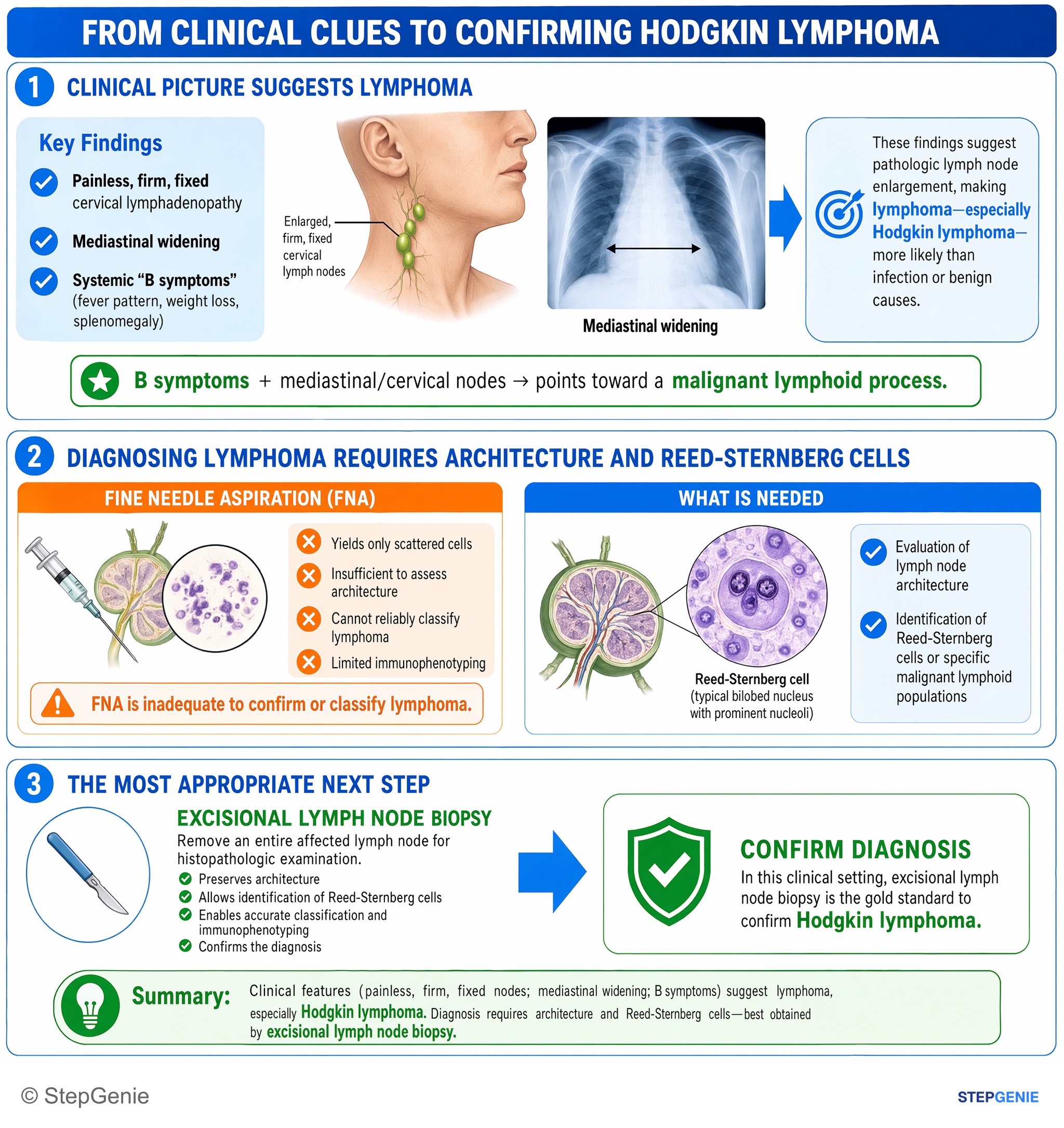
Hodgkin lymphoma suspected from B symptoms and mediastinal/cervical nodes; confirm with excisional lymph node biopsy for architecture and…
USMLE Step 1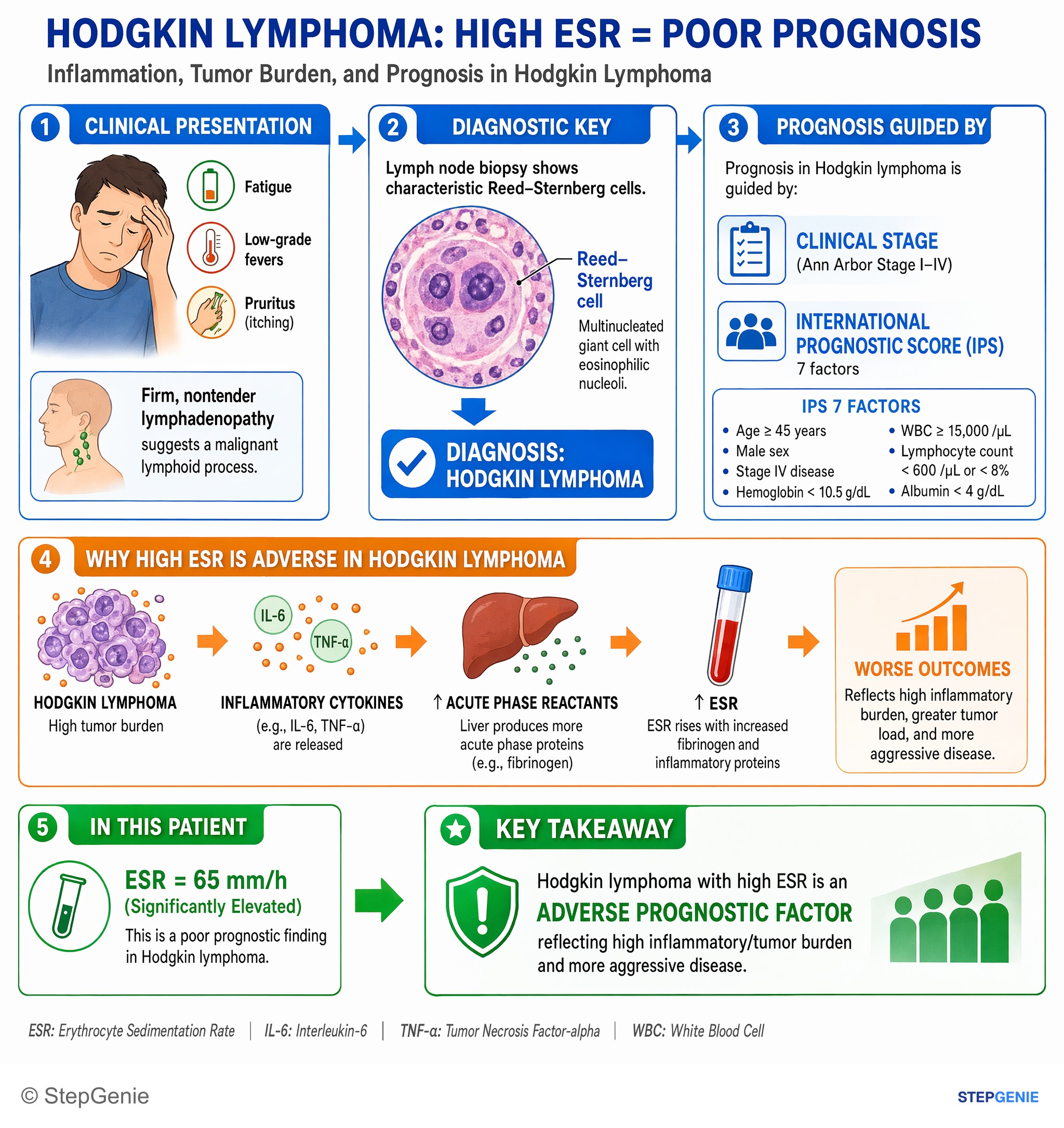
Hodgkin lymphoma with high ESR is an adverse prognostic factor reflecting high inflammatory/tumor burden and more aggressive disease.
USMLE Step 1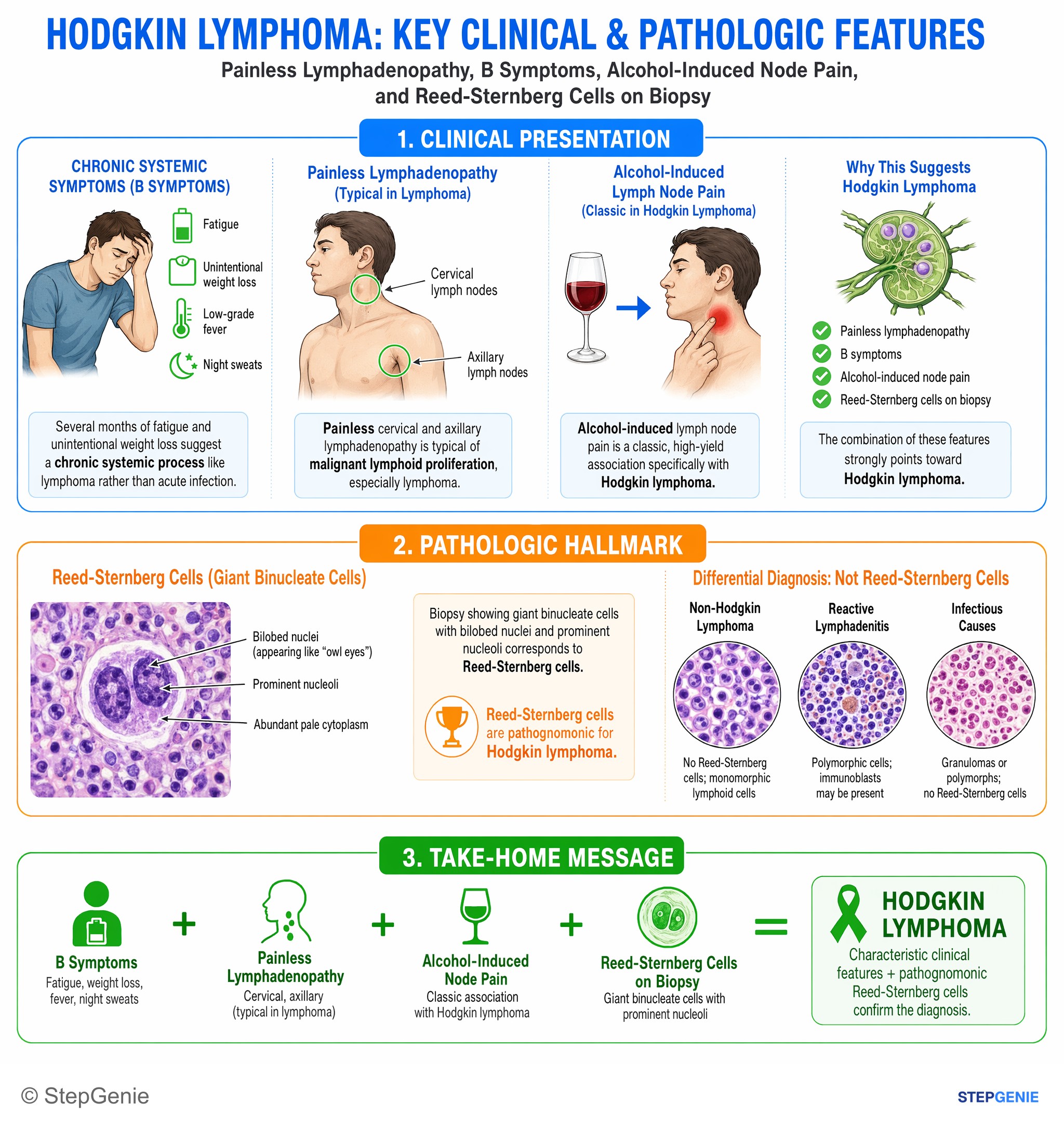
Hodgkin lymphoma presents with painless lymphadenopathy, B symptoms, alcohol-induced node pain, and Reed-Sternberg giant binucleate cells…
USMLE Step 1Frequently Asked Questions
How do you tell classic Hodgkin lymphoma from non-Hodgkin lymphoma in a vignette?
Classic Hodgkin lymphoma is suggested by Reed–Sternberg cells that are CD15 positive and CD30 positive, often with painless lymphadenopathy, B symptoms, and a mixed inflammatory background. Non-Hodgkin lymphoma lacks Reed–Sternberg cells and instead usually shows B-cell markers such as CD20 and PAX5, with spread that is more often noncontiguous and more likely to involve extranodal sites.
Why is a mediastinal mass a clue for Hodgkin lymphoma?
A mediastinal mass is a strong clue because it is especially common in the nodular sclerosis subtype of classic Hodgkin lymphoma. When that finding appears with a young adult, painless rubbery cervical lymphadenopathy, B symptoms, and biopsy showing binucleated owl-eye cells that are CD15 positive and CD30 positive, Hodgkin lymphoma becomes the best fit.
What immunophenotype points to classic Hodgkin lymphoma?
The key immunophenotype for classic Hodgkin lymphoma is CD15 positivity and CD30 positivity. In the right clinical setting, that pattern supports Reed–Sternberg cells as the malignant cells. This helps separate classic Hodgkin lymphoma from several B-cell lymphomas that instead express markers like CD20, CD10, BCL2, BCL6, CD5, CD23, or Cyclin D1.
Why can’t this vignette be infectious mononucleosis instead of Hodgkin lymphoma?
It is not infectious mononucleosis because the defining findings here are a mediastinal mass and biopsy showing Reed–Sternberg cells with CD15 and CD30 positivity. Infectious mononucleosis more classically causes sore throat, exudative pharyngitis, posterior cervical lymphadenopathy, atypical lymphocytes, and a positive heterophile antibody test, and its biopsy does not show Reed–Sternberg cells.
What are the highest-yield pathology clues for classic Hodgkin lymphoma on Step 1?
The highest-yield clues are B symptoms, painless lymphadenopathy, mediastinal mass, Reed–Sternberg cells, and CD15 and CD30 positivity. Reed–Sternberg cells are large atypical binucleated cells with owl-eye nucleoli in a reactive inflammatory background. Another major clue is that Hodgkin lymphoma spreads contiguously from node to node, unlike many non-Hodgkin lymphomas.