You’re cruising through a heme/onc Q-bank and suddenly get hit with an “easy” leukemia vignette—until the answer choices all look kind of right. Acute myeloid leukemia (AML) is a classic Step 1/2 trap because it shares real estate with ALL, CML, MDS, and APL, and the test writers love to make you prove you know the differences, not just the buzzword.
The Vignette (Q-bank style)
A 62-year-old man presents with 3 weeks of progressive fatigue, easy bruising, and intermittent fevers. He reports gum swelling and decreased appetite. Exam shows pallor, scattered petechiae, and gingival hypertrophy. Labs:
- Hemoglobin: 7.8 g/dL
- Platelets: 28,000/µL
- WBC: 38,000/µL
- Peripheral smear: large immature cells with Auer rods
- Coags: mildly prolonged PT and aPTT; fibrinogen is low
Bone marrow biopsy shows a hypercellular marrow with >20% myeloblasts.
Question: Which diagnosis best explains these findings?
A. Acute lymphoblastic leukemia (ALL)
B. Acute myeloid leukemia (AML)
C. Chronic myeloid leukemia (CML)
D. Myelodysplastic syndrome (MDS)
E. Hairy cell leukemia (HCL)
F. Acute promyelocytic leukemia (APL, M3 AML)
Step-by-Step: Why the Best Answer Is AML
✅ Correct answer: B. Acute myeloid leukemia (AML)
AML is a clonal proliferation of myeloid blasts that crowd out normal marrow, causing:
- Anemia → fatigue, pallor
- Thrombocytopenia → bruising, petechiae
- Neutropenia/dysfunctional WBCs → infections, fevers (even if WBC count is high)
Clues pointing to AML
- Older adult (median age ~65)
- Cytopenias + symptoms of marrow failure
- Myeloblasts with Auer rods (strongly supports myeloid lineage)
- Gingival hypertrophy (seen in monocytic variants like AML M5)
High-yield diagnostic cutoff
- myeloblasts in marrow = AML (general WHO rule)
What about the coag abnormalities?
- Low fibrinogen + prolonged PT/aPTT suggests DIC physiology, which should make you think of APL, but the question stem also shows Auer rods and classic AML presentation. If the smear had promyelocytes packed with Auer rods (“faggot cells”), APL would jump to the top.
Quick High-Yield AML Table
| Feature | AML (general) | APL (M3 subtype) |
|---|---|---|
| Typical age | Older adults | Any age (often middle-aged) |
| Smear | Myeloblasts, may have Auer rods | Promyelocytes, “faggot cells” with many Auer rods |
| Hallmark | Marrow failure (anemia, thrombocytopenia, infections) | DIC due to procoagulants from granules |
| Key genetics | Variable | t(15;17) → PML-RARA |
| Treatment highlight | Induction chemo (e.g., cytarabine + anthracycline) | ATRA ± arsenic trioxide (+ manage DIC) |
Why Each Distractor Is Wrong (and When It Would Be Right)
A. Acute lymphoblastic leukemia (ALL)
Why it’s wrong here
- ALL is more common in children
- Blasts are lymphoblasts, not myeloblasts
- Auer rods are myeloid, not lymphoid
When ALL is the right pick
- Child with bone pain, fever, infections
- Mediastinal mass (T-ALL)
- PAS-positive blasts, TdT positivity (lymphoblast marker)
- CNS/testicular involvement (classic Step association)
High-yield pearl: ALL can still present with cytopenias and blasts—so lineage markers matter (e.g., TdT for lymphoblasts; MPO for myeloblasts).
C. Chronic myeloid leukemia (CML)
Why it’s wrong here
- CML typically shows mature myeloid proliferation (not blasts) with:
- Very high WBC count with a left shift
- Basophilia
- Often splenomegaly
- Bone marrow: hypercellular with granulocytic hyperplasia, not blasts (unless blast crisis)
When CML is the right pick
- Middle-aged adult with fatigue, early satiety
- Smear: myelocytes, metamyelocytes, basophils
- Low leukocyte alkaline phosphatase (LAP) (vs leukemoid reaction = high LAP)
- BCR-ABL t(9;22) Philadelphia chromosome
- Treat: TKIs (imatinib, dasatinib)
Step trap: “Blast crisis” CML can mimic acute leukemia. The tell is a history of chronic phase signs + BCR-ABL.
D. Myelodysplastic syndrome (MDS)
Why it’s wrong here
- MDS is a clonal stem cell disorder with ineffective hematopoiesis
- Classically causes cytopenias but usually without big leukocytosis
- Blasts are <20% (once , it’s AML)
What you’d expect in MDS
- Older patient with chronic anemia (often macrocytic)
- Dysplastic cells:
- Hyposegmented neutrophils (pseudo–Pelger-Huët)
- Abnormal platelets
- Ring sideroblasts (in some subtypes)
- Risk of progression to AML
High-yield pearl: MDS = marrow is cellular but blood counts are low due to ineffective production.
E. Hairy cell leukemia (HCL)
Why it’s wrong here
- HCL is an indolent B-cell neoplasm with:
- Pancytopenia (not leukocytosis)
- Massive splenomegaly
- Dry tap on bone marrow due to fibrosis
- Peripheral smear: “hairy” cytoplasmic projections (not Auer rods)
When HCL is the right pick
- Middle-aged man with recurrent infections and splenomegaly
- TRAP positive (classic teaching)
- Treat: cladribine (purine analog)
F. Acute promyelocytic leukemia (APL, M3 AML)
Why it’s not the best answer here (but very tempting)
- The vignette includes DIC features (low fibrinogen, prolonged PT/aPTT), which is the big APL clue
- But the smear description emphasizes myeloblasts with Auer rods rather than promyelocytes or “faggot cells”
- If the question wanted APL, it would usually spotlight:
- t(15;17) PML-RARA
- Heavy granulation, promyelocytes, faggot cells
- Dramatic DIC/bleeding presentation
When APL is absolutely the right pick
- Any acute leukemia picture + DIC
- Smear shows promyelocytes with bundles of Auer rods
- Management includes:
- ATRA immediately (don’t wait—reduces DIC risk)
- Arsenic trioxide (commonly) and supportive care for coagulopathy
High-yield warning: APL is a “treat first, confirm second” scenario—ATRA can be life-saving.
The “Answer Choice Pattern” You Should Recognize on Test Day
If you see Auer rods
Think AML (myeloblasts) and keep APL on the short list.
If you see DIC
Think APL until proven otherwise.
If you see basophilia + splenomegaly + left shift
Think CML.
If you see pancytopenia + massive splenomegaly + dry tap
Think hairy cell leukemia.
If you see older patient + macrocytic anemia + dysplasia + blasts <20%
Think MDS.
Rapid-Fire AML High-Yield Facts (USMLE-friendly)
- Definition: myeloblasts in marrow (or certain defining genetic lesions)
- Smear: myeloblasts ± Auer rods; MPO positive
- Symptoms: fever/infections, fatigue, bleeding/bruising (marrow failure)
- Special association: gingival hypertrophy (monocytic differentiation)
- Emergencies: leukostasis (very high WBC), tumor lysis syndrome, severe infections, bleeding (especially in APL)
- APL genetics: t(15;17) → PML-RARA; treat with ATRA ± arsenic
Takeaway: How to Stop Missing “Easy” AML Questions
Don’t just hunt for a buzzword—force every option to earn its place. AML is about myeloblasts, marrow failure, and (often) Auer rods. APL is the AML subtype that adds a big red siren: DIC, t(15;17), and ATRA now.
Visual explanations from the StepGenie QBank
Infographics StepGenie drew to explain questions in its own QBank, matched to this article's topic. Each caption is the takeaway of the question it explains.
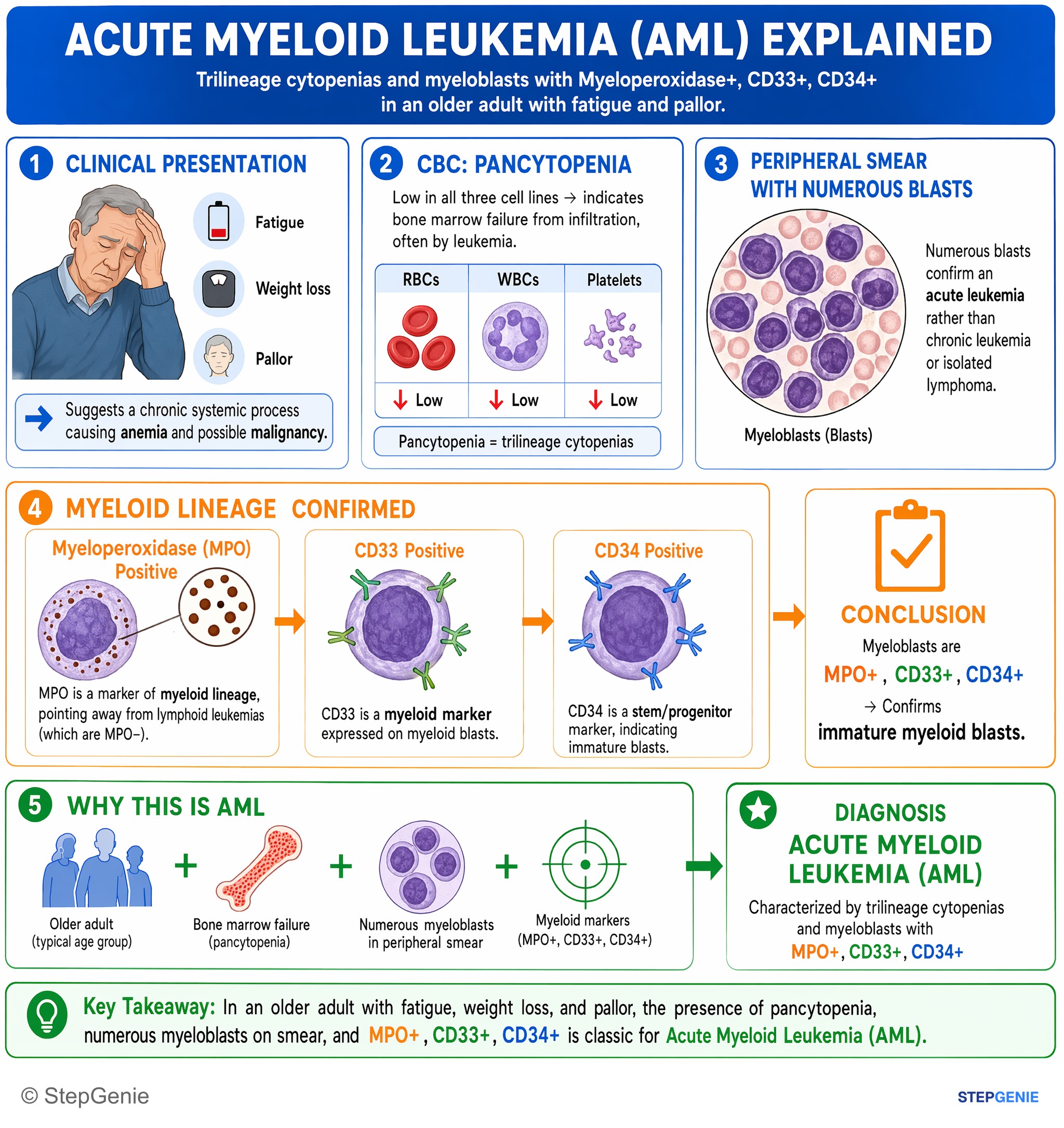
Acute myeloid leukemia (AML): trilineage cytopenias, myeloblasts with myeloperoxidase+, CD33+, CD34+ on smear in an older adult with…
USMLE Step 2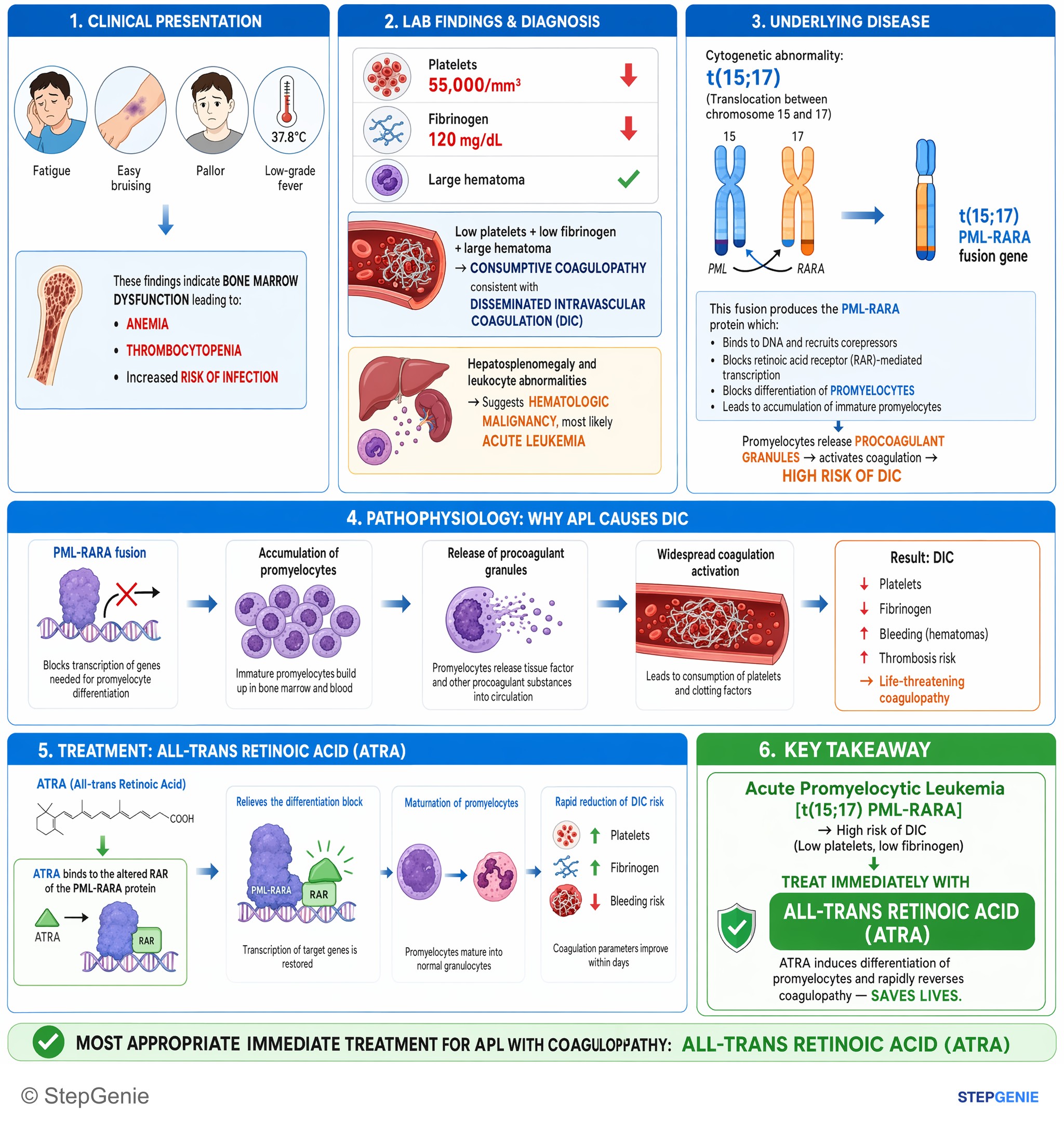
Acute promyelocytic leukemia (t(15;17)) → high DIC risk (low platelets, low fibrinogen) → treat immediately with all-trans retinoic acid…
USMLE Step 2Frequently Asked Questions
How do you tell AML from APL on a leukemia vignette?
AML is favored when the stem shows myeloblasts with Auer rods and marrow with at least 20% myeloblasts, while APL is suggested by promyelocytes packed with Auer rods, often called faggot cells, plus prominent DIC. Both can have Auer rods, but APL is the M3 subtype with t(15;17) PML-RARA and a stronger association with coagulopathy from procoagulants in granules.
Why can AML cause fatigue, bruising, petechiae, and fevers even when the white count is high?
AML causes these findings because clonal myeloid blasts crowd out normal bone marrow. That marrow failure leads to anemia causing fatigue and pallor, thrombocytopenia causing bruising and petechiae, and neutropenia or dysfunctional white cells causing infections and fevers. A high white count does not mean those cells work normally, so patients can still look infected despite leukocytosis.
What is the blast cutoff for diagnosing AML instead of MDS?
The key cutoff is 20% or more myeloblasts in the bone marrow, which supports AML. MDS is a clonal stem cell disorder with ineffective hematopoiesis and usually has less than 20% blasts. Once blasts reach or exceed that threshold, the diagnosis shifts from MDS to AML, even though both conditions can occur in older patients and cause cytopenias.
How do you tell AML from ALL on exam questions?
AML is suggested by myeloblasts with Auer rods in an older adult, while ALL is more common in children and involves lymphoblasts rather than myeloblasts. Lineage markers help separate them: Auer rods support myeloid lineage, and TdT supports lymphoblasts. ALL also classically has associations such as mediastinal mass in T-ALL and CNS or testicular involvement.
What is the first-line treatment for APL and why do you start it right away?
The treatment is ATRA immediately, often with arsenic trioxide, along with supportive care for coagulopathy. APL is treated urgently because it is strongly associated with DIC, and early ATRA reduces that risk. This is why APL is a treat-first, confirm-second scenario when an acute leukemia picture is paired with DIC and promyelocytes with bundles of Auer rods.