A good prion question feels unfair at first: the stem sounds like “dementia” (too broad), the MRI sounds like “tumor” (too scary), and the answer choices are packed with other neurodegenerative/infectious zebras. The trick—and what USMLE loves—is that Creutzfeldt-Jakob disease (CJD) is a pattern-recognition diagnosis where small details (time course, myoclonus, MRI/EEG/CSF clues) make every distractor clearly wrong.
Tag: Neurology > CNS Tumors & Infections
The Clinical Vignette (Q-bank style)
A 66-year-old man is brought by family for rapidly progressive confusion and memory loss over 6 weeks. He has startle-induced myoclonus and difficulty walking. Exam shows ataxia and hyperreflexia. He is afebrile. MRI brain (DWI) shows cortical ribboning with hyperintensity in the caudate and putamen. EEG shows periodic sharp wave complexes. CSF has elevated 14-3-3 protein.
Most likely diagnosis? → Sporadic Creutzfeldt-Jakob disease (sCJD).
Why the Correct Answer Is CJD
The “USMLE fingerprint” for sporadic CJD
CJD = rapidly progressive dementia + myoclonus + characteristic MRI/EEG/CSF.
High-yield features:
- Time course: weeks to months (often < 1 year), not years
- Symptoms:
- Rapid dementia
- Myoclonus (often stimulus-sensitive)
- Ataxia
- Can progress to akinetic mutism
- MRI (best early test): DWI/FLAIR hyperintensity in:
- Cortex (“cortical ribboning”)
- Basal ganglia (caudate/putamen)
- EEG: periodic sharp wave complexes
- CSF: supportive markers:
- 14-3-3, tau (markers of neuronal injury; not perfectly specific)
- RT-QuIC is highly specific where available (prion “seeding” assay)
- Pathology: spongiform change, neuronal loss, gliosis; no inflammation
- Agent: misfolded prion protein PrP inducing conformational change of normal PrP
- Prognosis: uniformly fatal (often within months)
Classic prion path buzzwords
- Spongiform encephalopathy
- No nucleic acid (protein-only infectious particle)
- Transmissible (iatrogenic risk: neurosurgical instruments, corneal transplant, dura mater graft)
The Distractors: Why Every Answer Choice Matters
Below is a “Q-bank autopsy” of common wrong answers—and the exact stem details that rule them out.
Quick comparison table (high yield)
| Condition (Common distractor) | Time course | Key clinical clues | Imaging/Tests | What rules it out vs CJD |
|---|---|---|---|---|
| Alzheimer disease | Years | Early memory impairment, later neuro deficits | MRI: hippocampal atrophy | Too slow, no myoclonus, no DWI ribboning |
| Dementia with Lewy bodies (DLB) | Months–years | Visual hallucinations, fluctuating cognition, parkinsonism | Often normal/atrophy; sensitivity to antipsychotics | Hallucinations + parkinsonism dominate; not “weeks” |
| Frontotemporal dementia (Pick disease) | Years | Early personality/language changes | Frontal/temporal atrophy | Early behavior/language > memory; slow |
| HSV-1 encephalitis | Days | Fever, headache, seizures, AMS | MRI: temporal lobe; CSF lymphocytes, ↑RBC; PCR+ | Febrile, inflammatory CSF; temporal lobe predilection |
| Subacute sclerosing panencephalitis (SSPE) | Years after measles | Child/teen, cognitive decline + myoclonus | High measles Ab; EEG periodic complexes | Age/history wrong; measles link |
| Normal pressure hydrocephalus | Months–years | “Wet, wobbly, wacky” | Enlarged ventricles | Gait/urinary dominate; not DWI ribboning |
| Primary CNS lymphoma | Weeks–months | Immunosuppressed, focal deficits | Enhancing lesion; EBV association | Mass lesion/contrast enhancement pattern differs |
| Glioblastoma | Weeks–months | Focal deficits, headache, seizures | Ring-enhancing mass crossing corpus callosum | Mass effect/enhancing tumor; not cortical ribboning |
| Progressive multifocal leukoencephalopathy (PML) | Weeks–months | Immunosuppressed; focal deficits | Nonenhancing demyelinating WM lesions | White matter demyelination pattern; JC virus |
Distractor Deep Dive (how USMLE traps you)
1) Alzheimer disease
Why it tempts you: “dementia in an older adult.”
- Rule-out clue: Alzheimer is insidious (years), not a 6-week collapse.
- No classic myoclonus, and MRI won’t show DWI cortical ribboning.
- Path: Aβ plaques + tau tangles, not spongiform change.
Test-day pearl: If the stem screams weeks to months, stop thinking Alzheimer.
2) Dementia with Lewy bodies (DLB)
Why it tempts you: Dementia + movement issues.
- DLB core features:
- Fluctuating cognition
- Well-formed visual hallucinations
- Parkinsonism
- Rule-out clue: time course is typically more gradual than CJD, and MRI/EEG/CSF prion markers aren’t the story.
Pearl: DLB is the dementia that “acts psychotic” early (visual hallucinations) and hates antipsychotics.
3) Frontotemporal dementia (FTD)
Why it tempts you: Dementia + weird neuro signs.
- Rule-out clue: FTD is a behavior/language-first dementia (disinhibition, apathy, primary progressive aphasia).
- Still typically slow, not “rapidly progressive dementia with myoclonus.”
4) HSV-1 encephalitis
Why it tempts you: Altered mental status can progress quickly.
- HSV-1 clues:
- Fever, headache, seizures
- MRI: temporal lobe involvement
- CSF: lymphocytic pleocytosis, ↑protein, RBCs (hemorrhagic)
- Confirm: CSF PCR
- Rule-out clue in this vignette: afebrile + supportive prion findings (DWI ribboning, periodic sharp waves, 14-3-3).
Pearl: When HSV is in the differential, the test wants you to say acyclovir now—but you need the stem’s inflammatory clues.
5) Subacute sclerosing panencephalitis (SSPE)
Why it tempts you: Myoclonus + periodic EEG sounds prion-like.
- SSPE is due to persistent measles infection.
- Rule-out clue: occurs in children/teens, years after measles infection; progressive cognitive decline + myoclonus.
Pearl: Periodic EEG is not exclusive to CJD—age and history matter.
6) Normal pressure hydrocephalus (NPH)
Why it tempts you: Gait problems + cognitive issues.
- Triad: gait disturbance (earliest/most prominent) + urinary incontinence + dementia
- Imaging: ventriculomegaly
- Rule-out clue: NPH dementia is not usually lightning-fast, and myoclonus/MRI DWI changes aren’t part of it.
7) Primary CNS lymphoma
Why it tempts you: The tag includes “tumors & infections,” and CNS lymphoma can evolve over weeks.
- Often in immunocompromised (HIV/AIDS, transplant); linked to EBV.
- Imaging: enhancing lesion, often periventricular; may have mass effect; steroid responsiveness can obscure biopsy.
- Rule-out clue: CJD is diffuse neurodegeneration with DWI cortical/basal ganglia changes, not a focal enhancing mass.
8) Glioblastoma
Why it tempts you: Older patient + rapid decline + dramatic MRI = tumor?
- Imaging: heterogeneous ring-enhancing necrotic mass; may cross corpus callosum (“butterfly”).
- Rule-out clue: Tumor gives focal deficits, seizures, headaches, and mass effect—not myoclonus + classic prion DWI findings.
9) Progressive multifocal leukoencephalopathy (PML)
Why it tempts you: Rapid neuro decline over weeks-months.
- Caused by JC virus reactivation in immunosuppressed (HIV with low CD4, natalizumab).
- Imaging: nonenhancing demyelinating lesions in white matter (often parietal/occipital).
- Rule-out clue: PML is white matter disease with immunosuppression; CJD is gray matter signal changes + prion markers.
High-Yield CJD Subtypes (Step-friendly)
Sporadic CJD (most common)
- No clear exposure; older adults
- Classic vignette: rapid dementia + myoclonus
Variant CJD (vCJD)
- Associated with bovine spongiform encephalopathy (“mad cow”)
- Younger patients
- Prominent psychiatric symptoms early
- “Pulvinar sign” on MRI sometimes described (thalamic involvement)
Iatrogenic CJD
- Prior neurosurgery, corneal transplant, dura graft
- Think instrument sterilization issues (prions resist standard methods)
Familial prion disease
- PRNP gene mutation; family history
Board-Style Takeaways (what to memorize)
- Rapidly progressive dementia (weeks–months) + myoclonus → think CJD first.
- MRI DWI/FLAIR: cortical ribboning + caudate/putamen hyperintensity.
- EEG: periodic sharp wave complexes.
- CSF: 14-3-3/tau supportive; RT-QuIC highly specific.
- Path: spongiform changes, neuronal loss, gliosis, no inflammation.
- Not treatable; fatal. Management is supportive and involves infection-control precautions for high-risk tissues/instruments.
Mini Self-Check (1-minute)
If the stem says:
- Fever + temporal lobe + RBCs in CSF → HSV encephalitis
- Immunosuppressed + nonenhancing white matter lesions → PML
- Psych symptoms in a younger patient + prion exposure → variant CJD
- Weeks + myoclonus + cortical ribboning → sporadic CJD
Visual explanations from the StepGenie QBank
Infographics StepGenie drew to explain questions in its own QBank, matched to this article's topic. Each caption is the takeaway of the question it explains.
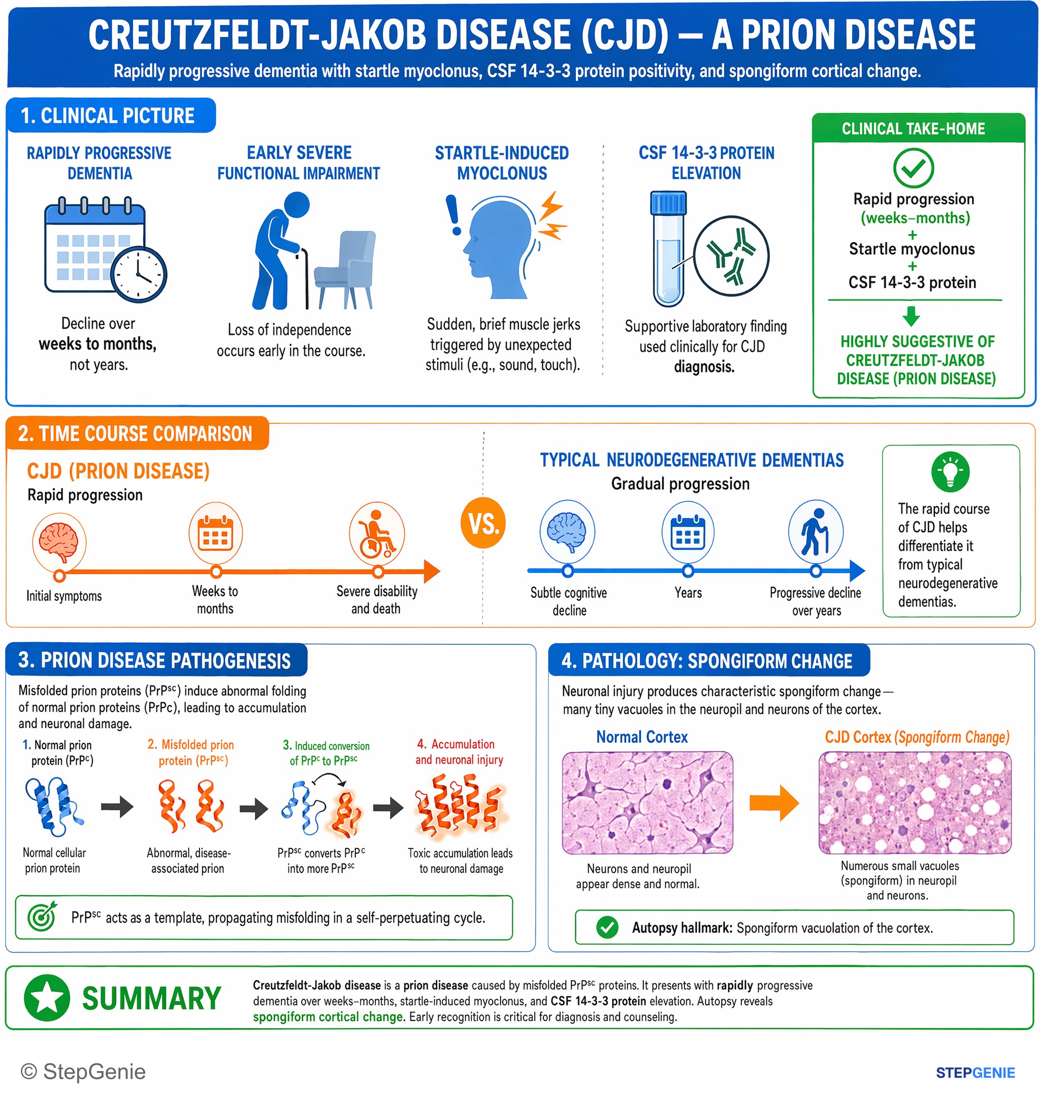
Creutzfeldt-Jakob disease (prion disease) causes rapidly progressive dementia with startle myoclonus and CSF 14-3-3 protein; autopsy shows…
USMLE Step 1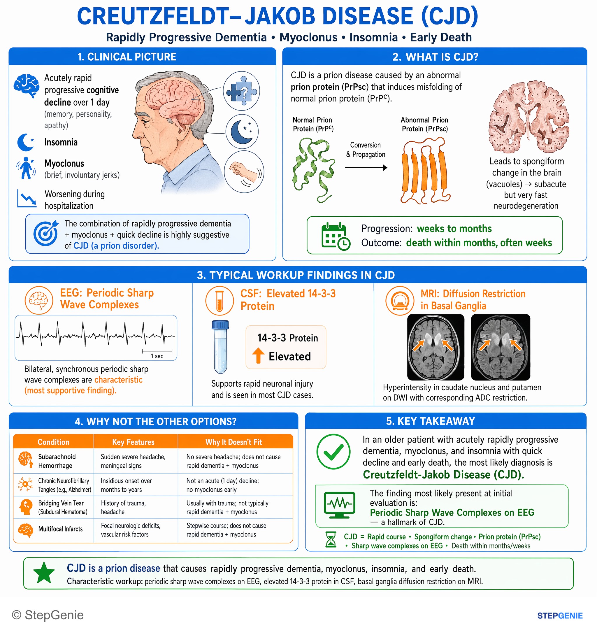
Creutzfeldt-Jakob disease (prion disease) causes rapidly progressive dementia, myoclonus, insomnia, and early death, with characteristic…
USMLE Step 1Frequently Asked Questions
What is the classic USMLE pattern for sporadic Creutzfeldt-Jakob disease?
The classic pattern is rapidly progressive dementia over weeks to months plus myoclonus and characteristic MRI, EEG, and CSF findings. Typical clues include stimulus-sensitive myoclonus, ataxia, and a course often shorter than a year. MRI shows cortical ribboning and basal ganglia hyperintensity, EEG shows periodic sharp wave complexes, and CSF may have supportive markers such as 14-3-3 protein.
How do you tell CJD from Alzheimer disease on a question stem?
You separate CJD from Alzheimer disease mainly by the time course and associated neurologic clues. CJD causes a rapid decline over weeks to months, often with myoclonus and ataxia, whereas Alzheimer disease is insidious over years and does not classically cause myoclonus. MRI in CJD shows DWI cortical ribboning, while Alzheimer disease is associated instead with hippocampal atrophy.
Why is HSV-1 encephalitis less likely than CJD in an afebrile patient with myoclonus and cortical ribboning?
HSV-1 encephalitis is less likely because it usually presents with inflammatory and temporal lobe clues rather than the prion pattern. Typical HSV-1 findings include fever, headache, seizures, temporal lobe involvement on MRI, and CSF lymphocytic pleocytosis with increased protein and red blood cells. An afebrile patient with cortical ribboning, periodic sharp waves, and CSF 14-3-3 protein fits CJD much better.
What MRI and EEG findings point toward CJD?
The MRI finding that strongly points toward CJD is DWI or FLAIR hyperintensity in the cortex and basal ganglia, especially cortical ribboning with caudate and putamen involvement. The EEG clue is periodic sharp wave complexes. When these findings appear in a patient with rapidly progressive dementia and myoclonus, they form a high-yield pattern that strongly supports sporadic CJD.
What causes CJD, and what would pathology show?
CJD is caused by a misfolded prion protein, PrPSc, which induces conformational change of the normal prion protein PrPC. It is a protein-only infectious particle with no nucleic acid. Pathology shows spongiform change, neuronal loss, and gliosis without inflammation. This explains why CJD is classified as a transmissible spongiform encephalopathy rather than a typical inflammatory CNS infection.