Progressive focal neuro deficits in an immunocompromised patient are a classic “don’t-miss” Step presentation—and PML is one of the highest-yield diagnoses in that lane. The trick is that many answer choices will sound plausible (toxoplasmosis! lymphoma! MS!), but the stem details and imaging pattern are what let you lock in the right call.
Clinical Vignette (Q-bank style)
A 36-year-old man with HIV (CD4 count 48/mm³) presents with 3 weeks of progressive difficulty speaking and clumsiness of his right hand. He denies fever or headache. Exam shows expressive aphasia and right upper extremity weakness. MRI brain shows multiple asymmetric, non-enhancing lesions in the subcortical white matter that are hyperintense on T2/FLAIR. Lumbar puncture shows normal opening pressure and mild lymphocytic pleocytosis.
Which pathogen is most likely responsible?
A. Toxoplasma gondii
B. JC virus
C. Epstein–Barr virus
D. Cryptococcus neoformans
E. HSV-1
Correct Answer: B. JC virus (Progressive multifocal leukoencephalopathy, PML)
Why it’s JC virus
PML is a demyelinating disease caused by reactivation of JC virus (a polyomavirus) in the setting of impaired cell-mediated immunity.
Key stem clues
- Subacute, progressive focal neurologic deficits (weeks)
- No fever, minimal systemic symptoms
- MRI: multifocal asymmetric white matter lesions
- Classically non-enhancing (or minimal enhancement) and no mass effect
- Setting: advanced HIV (also occurs with natalizumab, rituximab, transplant)
Pathophysiology (high-yield)
- JC virus infects oligodendrocytes → demyelination
- Leads to multifocal deficits depending on lesion locations (motor, speech, vision, ataxia)
Diagnosis
- CSF JC virus PCR is commonly tested and very high-yield
- Brain biopsy: enlarged oligodendrocyte nuclei with viral inclusions (rarely needed in Step-style questions)
Treatment
- Immune reconstitution is the main therapy
- HIV: start/optimize ART
- Medication-associated (e.g., natalizumab): stop the agent
- Clinical caveat: immune reconstitution can trigger IRIS (transient worsening due to inflammation)
The Imaging Pattern You Must Recognize
| Feature | PML (JC virus) | Toxo | Primary CNS lymphoma |
|---|---|---|---|
| Lesion location | White matter, subcortical | Basal ganglia/corticomedullary junction | Periventricular/deep |
| Enhancement | None/minimal | Ring-enhancing | Homogeneous (often), can ring in HIV |
| Mass effect/edema | Minimal | Prominent | Variable, often present |
| Symptoms | Progressive focal deficits | Headache, fever, seizures common | Focal deficits + constitutional sx possible |
| Best test | CSF JC PCR | Serology + response to therapy | EBV+ in CSF, biopsy |
Why Each Distractor Is Wrong (and when it would be right)
A. Toxoplasma gondii
Why it’s tempting: AIDS + brain lesions is a classic toxo setup.
Why it’s wrong here:
- Toxo typically presents with headache, fever, seizures
- MRI usually shows multiple ring-enhancing lesions with significant edema and mass effect
- Often located in basal ganglia
When to pick it on Step:
- HIV patient with CD4 <100, ring-enhancing lesions, mass effect
- Risk factor: exposure to cat feces/undercooked meat
- Empiric therapy often started; improvement supports diagnosis
- Prophylaxis: TMP-SMX (also covers PCP)
C. Epstein–Barr virus (EBV)
Most associated diagnosis: Primary CNS lymphoma in AIDS/transplant.
Why it’s wrong here:
- Primary CNS lymphoma tends to produce enhancing lesions (often homogeneous), frequently periventricular
- More likely to have mass effect than PML
- PML is demyelination—not a tumor mass
When to pick EBV:
- HIV (often CD4 <50) with enhancing periventricular lesion(s)
- CSF: EBV DNA positive
- Often responds to high-dose methotrexate; ART is still important
D. Cryptococcus neoformans
Why it’s wrong here:
- Cryptococcus usually causes meningitis/meningoencephalitis → headache, fever, neck stiffness
- Increased intracranial pressure is common
- Not a classic “multifocal white matter demyelination” picture
When to pick cryptococcus:
- AIDS patient with subacute meningitis, elevated opening pressure
- Diagnosis: cryptococcal antigen in CSF (or India ink historically)
- Treatment: amphotericin B + flucytosine, then fluconazole; manage ICP with serial LPs
E. HSV-1
Why it’s wrong here:
- HSV-1 encephalitis is typically acute, with fever, altered mental status, seizures
- Predilection for the temporal lobes
- Imaging: hemorrhagic necrosis; CSF: elevated RBCs can be seen
When to pick HSV-1:
- Rapid onset encephalitis + temporal lobe signs (aphasia, personality changes)
- Diagnosis: CSF HSV PCR
- Treatment: IV acyclovir
PML High-Yield “One-Liners” for USMLE
- JC virus reactivation in immunosuppression → PML
- Infects oligodendrocytes → demyelination
- Presents with progressive focal neurologic deficits (no fever is common)
- MRI: multifocal asymmetric non-enhancing white matter lesions, minimal mass effect
- Diagnosis: CSF JC virus PCR
- Management: restore immune function (ART, stop offending immunosuppressant); watch for IRIS
Rapid Differentiation: PML vs MS vs ADEM (common confusion)
| Condition | Typical patient | Time course | Imaging | Key clue |
|---|---|---|---|---|
| PML (JC) | Immunocompromised | Subacute progressive | Non-enhancing WM lesions, minimal mass effect | HIV/natalizumab |
| MS | Young adult, often female | Relapsing-remitting | Periventricular plaques, may enhance if active | Optic neuritis, internuclear ophthalmoplegia |
| ADEM | Child after viral infection/vaccine | Acute, monophasic | Large, bilateral WM lesions | Encephalopathy prominent |
Test-Day Strategy: How to “Earn” PML in One Pass
If you see:
- Immunocompromised +
- Progressive focal deficits (weeks) +
- MRI: multifocal asymmetric white matter lesions with no enhancement/mass effect
→ pick JC virus (PML), then confirm with CSF PCR in your head.
Visual explanations from the StepGenie QBank
Infographics StepGenie drew to explain questions in its own QBank, matched to this article's topic. Each caption is the takeaway of the question it explains.
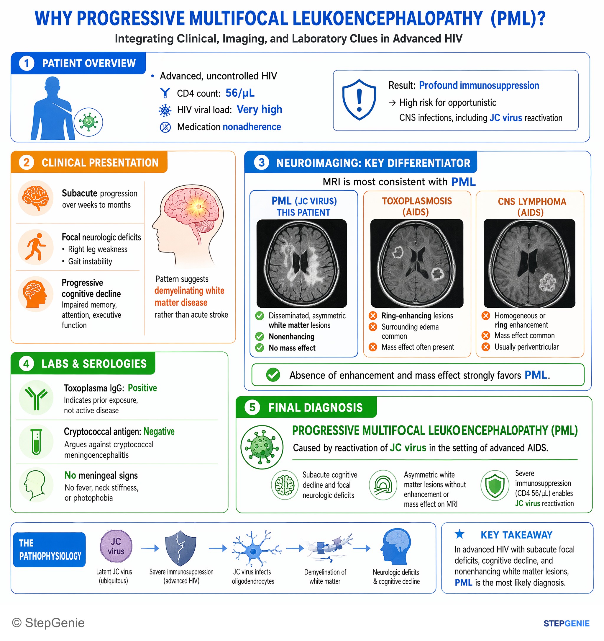
Progressive multifocal leukoencephalopathy (PML) from JC virus reactivation in advanced AIDS causes subacute cognitive decline and…
USMLE Step 2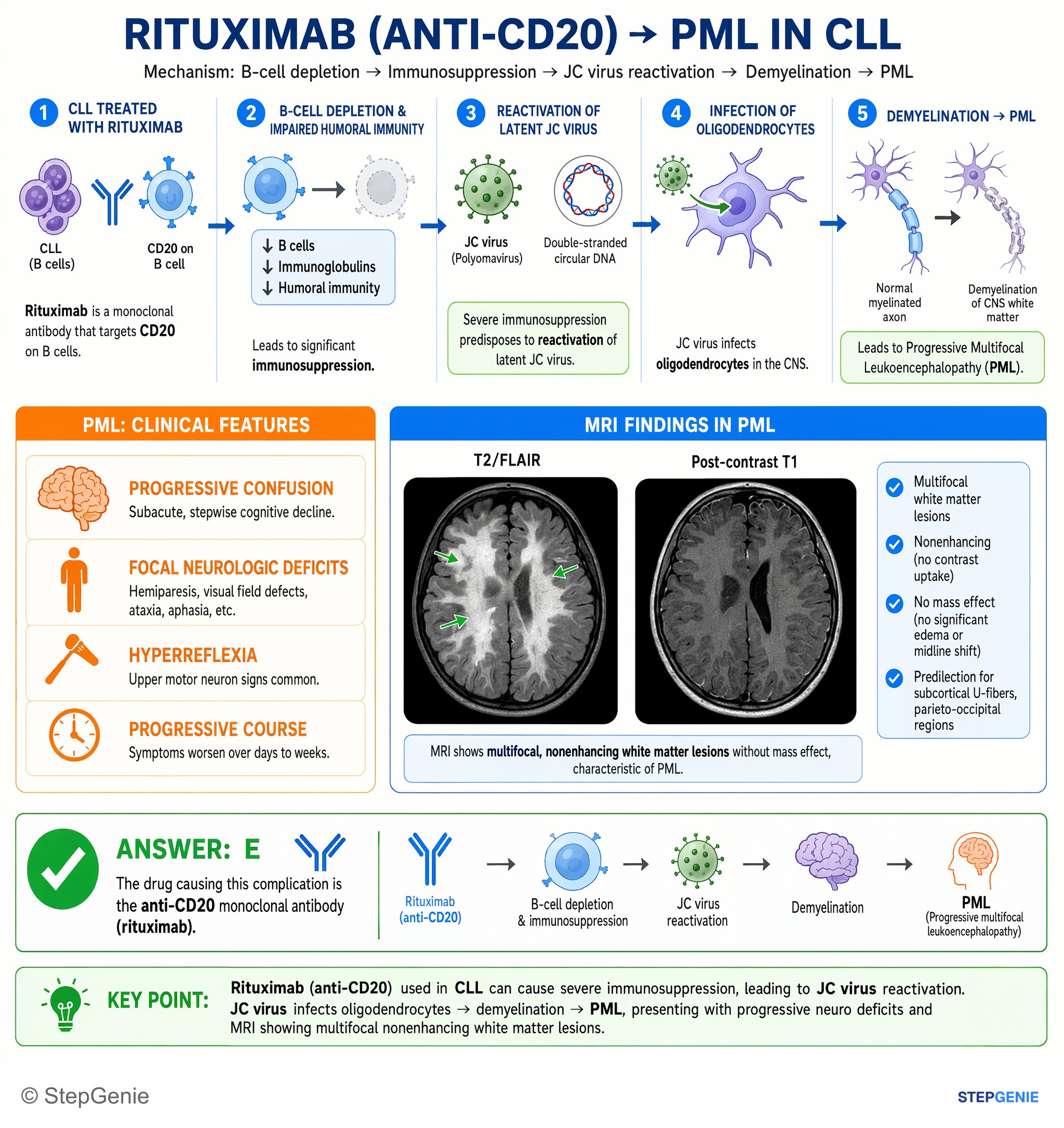
Progressive multifocal leukoencephalopathy (JC virus reactivation) from rituximab (anti-CD20) used for CLL causes nonenhancing…
USMLE Step 1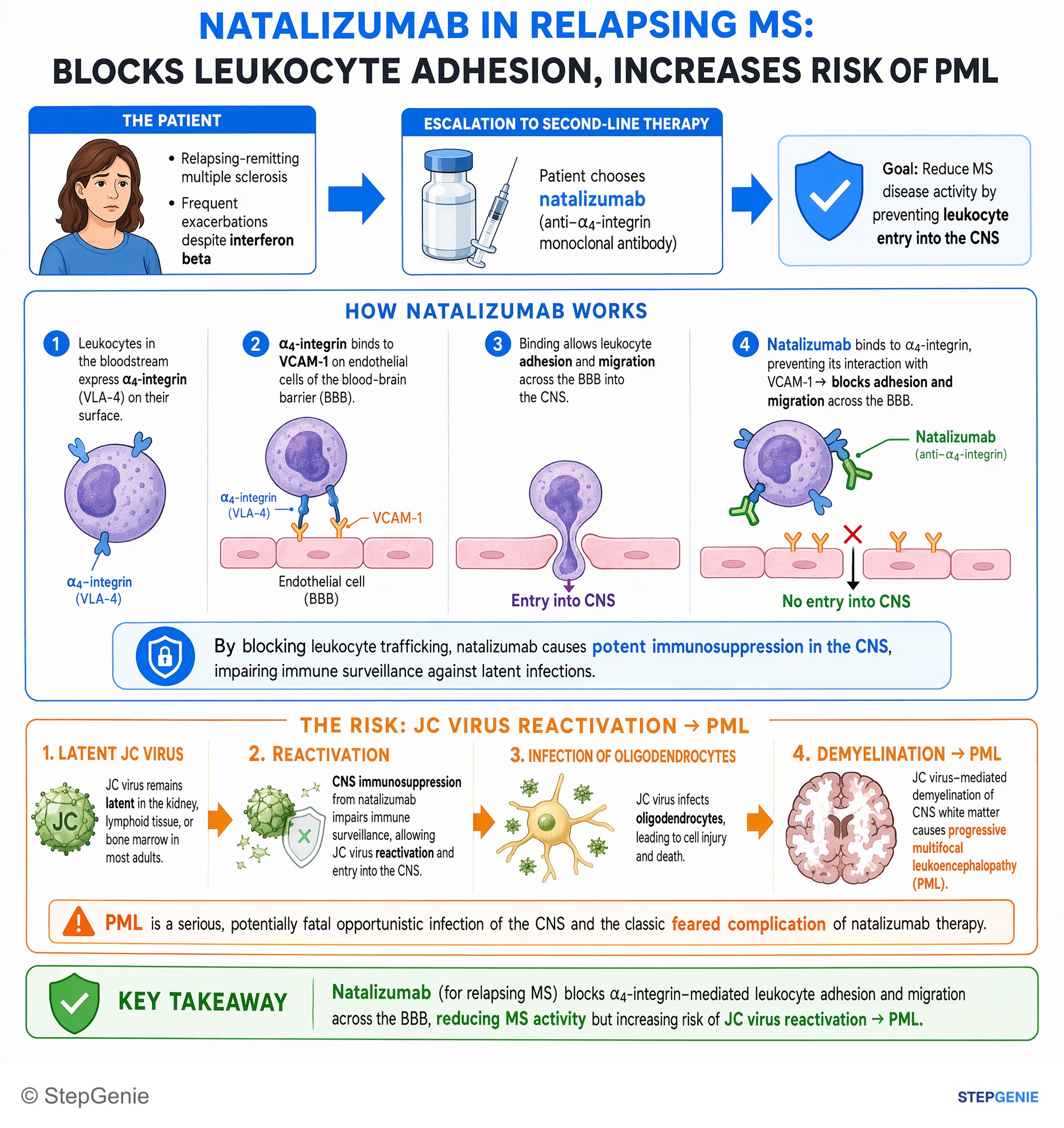
Natalizumab (for relapsing MS) blocks leukocyte adhesion and increases risk of JC virus reactivation → progressive multifocal…
USMLE Step 1Frequently Asked Questions
How do you recognize PML on MRI in an immunocompromised patient?
PML is suggested by multifocal asymmetric subcortical white matter lesions that are non-enhancing or only minimally enhancing, with little to no mass effect. In an immunocompromised patient, that imaging pattern plus subacute progressive focal neurologic deficits and minimal systemic symptoms strongly points to JC virus reactivation causing progressive multifocal leukoencephalopathy.
Why does JC virus cause progressive focal neurologic deficits?
JC virus causes progressive focal deficits because it infects oligodendrocytes and produces demyelination. As different white matter regions are affected, patients develop deficits based on lesion location, such as motor weakness, speech problems, visual symptoms, or ataxia. The presentation is typically subacute over weeks rather than a sudden or febrile encephalitic illness.
How do you tell PML from toxoplasmosis in a patient with AIDS and brain lesions?
PML is more likely when an AIDS patient has progressive focal deficits without fever and MRI shows non-enhancing white matter lesions with minimal mass effect. Toxoplasmosis usually causes headache, fever, or seizures, and MRI classically shows multiple ring-enhancing lesions with significant edema and mass effect, often in the basal ganglia or corticomedullary junction.
What is the best test for suspected PML?
The best test for suspected PML is CSF JC virus PCR. This is a commonly tested, high-yield way to confirm the diagnosis in a patient with the right clinical setting and MRI pattern. Brain biopsy can show enlarged oligodendrocyte nuclei with viral inclusions, but it is rarely needed in Step-style questions.
What is the main treatment for PML?
The main treatment for PML is immune reconstitution. In HIV-associated disease, that means starting or optimizing antiretroviral therapy, and in medication-associated cases it means stopping the offending immunosuppressive agent such as natalizumab. A key clinical caveat is that immune recovery can trigger IRIS, causing transient worsening due to inflammation.