TIA is one of those Step 1 topics that seems “small” until you realize it’s the testable bridge between vascular risk factors, neuroanatomy localization, and real-world stroke prevention. The key is to think of TIA as a warning shot: the brain was ischemic long enough to cause symptoms, but not long enough to infarct (at least not on imaging). On exams, that single concept drives the definition, the workup, and the urgency.
What Exactly Is a TIA?
Transient ischemic attack (TIA) = transient focal neurologic dysfunction caused by ischemia without evidence of acute infarction.
The modern (testable) definition
- Clinical: sudden-onset focal neuro deficits (not generalized)
- Imaging-based: no acute infarct on diffusion-weighted MRI (DWI)
- Time-based (older): symptoms resolve within <24 hours
- Still shows up in question stems, but the current concept is tissue-based (no infarct).
High-yield wording:
- TIA = “stroke-like symptoms that resolve” and “no infarct on imaging.”
- Stroke = deficits with infarction (even if symptoms resolve quickly).
Pathophysiology: Why TIAs Happen
Think of TIA mechanisms as temporary reductions in cerebral blood flow.
1) Large artery atherosclerosis (most classic)
- Carotid atherosclerosis → plaque rupture → platelet thrombus → artery-to-artery embolus to MCA territory
- Classic board setup: older patient + vascular risk factors + “amaurosis fugax” or unilateral weakness.
2) Cardioembolism
- Atrial fibrillation (left atrial appendage thrombus)
- Recent MI → LV mural thrombus
- Valvular disease (mechanical valves, endocarditis)
- Paradoxical embolus (PFO) in younger patients (less Step 1, more Step 2)
3) Small vessel (lacunar) disease
- Lipohyalinosis from HTN or diabetes
- More commonly causes lacunar infarcts, but can present as transient deficits.
4) Hypoperfusion (watershed ischemia)
- Severe carotid stenosis + hypotension
- Symptoms can be transient but risk is high.
What’s happening at the cellular level? (Step 1 tie-in)
Ischemia → ↓ATP → Na⁺/K⁺ pump failure → depolarization → glutamate release → Ca²⁺ influx → cellular injury.
In TIA, perfusion is restored before irreversible infarction occurs.
Clinical Presentation: Recognize the Pattern
Key features
- Sudden onset
- Focal neuro deficits (localize to vascular territory)
- Symptoms typically last minutes to <1 hour, but can last longer
- Complete resolution of deficits (by definition, clinically)
Common symptoms (anterior circulation: carotids/MCA/ACA)
- Unilateral weakness (face/arm > leg often suggests MCA)
- Unilateral sensory loss
- Aphasia (dominant hemisphere)
- Transient monocular vision loss (amaurosis fugax)
- Think retinal ischemia from ipsilateral carotid embolus.
Posterior circulation (vertebrobasilar) symptoms
- Diplopia
- Dysarthria
- Dysphagia
- Ataxia
- Vertigo (when paired with other focal brainstem/cerebellar signs)
Classic “TIA vs not TIA” exam traps
More consistent with TIA/stroke
- Unilateral weakness or numbness
- Aphasia, dysarthria
- Visual field cuts, amaurosis fugax
- Ataxia with other brainstem signs
Less consistent with TIA (consider mimics)
- Isolated confusion (without focal deficits)
- Syncope
- Generalized weakness
- Isolated headache (think SAH, migraine)
- Positive spreading symptoms (migraine aura)
Rapid table: TIA vs common mimics
| Condition | Typical onset | Key clues | Why it’s not TIA |
|---|---|---|---|
| TIA | Sudden | Negative symptoms (loss of function), focal deficits | Vascular territory pattern |
| Seizure + Todd paralysis | Sudden | Postictal confusion, tongue biting, witnessed convulsion | Neuro deficit follows seizure |
| Migraine aura | Gradual spread | Positive symptoms (zig-zags, tingling marching) | Slow progression, headache history |
| Hypoglycemia | Variable | Diaphoresis, tremor, altered mental status | Metabolic, not focal vascular |
| Bell palsy | Hours–days | Forehead involvement | Peripheral CN VII lesion |
Diagnosis: What You Do (and Why It’s Urgent)
TIA is a medical emergency because early stroke risk is high (especially within the first 48 hours). The workup is designed to:
- Exclude hemorrhage and stroke
- Identify embolism sources
- Find treatable vascular lesions (like carotid stenosis)
Imaging: what Step questions want you to pick
- MRI brain with DWI: best for ruling out acute infarct
- TIA: typically no DWI lesion
- Noncontrast CT head: often first in the ED
- Good for hemorrhage, may be normal in ischemia early
- Vascular imaging (to find the culprit vessel):
- Carotid duplex ultrasound
- CTA or MRA head/neck
Cardiac evaluation (embolism hunt)
- ECG: atrial fibrillation, recent MI clues
- Echocardiography if cardioembolic source suspected
- Consider prolonged rhythm monitoring if occult afib suspected (more Step 2 vibe, but fair game)
Labs (often in vignettes)
- Glucose (rule out hypoglycemia mimic)
- Lipids, A1c (risk stratification)
- CBC/coags if indicated
Risk stratification concept (Step 2-leaning but useful)
ABCD2 score exists, but for Step 1: remember TIA = high near-term stroke risk → urgent workup.
Treatment: Prevention Mode Activated
TIA management is essentially secondary stroke prevention—you’re treating the cause and reducing recurrence risk.
Immediate priorities (exam logic)
- Start antiplatelet therapy unless anticoagulation is indicated
- Address vascular risk factors aggressively
- Evaluate for carotid stenosis and intervene when appropriate
- Anticoagulate if atrial fibrillation or other high-risk cardioembolic source
Antiplatelet therapy (non-cardioembolic TIA)
- Aspirin is the classic answer.
- Clopidogrel is an alternative.
- Dual antiplatelet therapy may be used short-term in select high-risk cases (more Step 2/clinical guidelines), but Step 1 usually wants: aspirin.
Anticoagulation (cardioembolic TIA)
- If atrial fibrillation → anticoagulation (e.g., DOACs or warfarin depending on scenario)
- Key exam distinction:
- Non-cardioembolic → antiplatelet
- Cardioembolic (afib) → anticoagulation
Carotid endarterectomy (CEA): the board-relevant trigger
Indicated for symptomatic carotid stenosis, classically:
- 70–99% stenosis → CEA recommended if perioperative risk acceptable
- 50–69% may be considered in select patients (often beyond Step 1 level)
Phrase to recognize: “TIA symptoms + carotid bruit + ultrasound shows high-grade ipsilateral carotid stenosis.”
Statins + BP + diabetes + lifestyle (don’t neglect these)
- High-intensity statin for atherosclerotic disease
- Tight blood pressure control (long-term)
- Control diabetes
- Smoking cessation, diet/exercise
High-Yield Associations & Classic Vignettes
Amaurosis fugax
- Transient monocular blindness (“curtain coming down”)
- Often from ipsilateral carotid atherosclerosis → retinal ischemia
- Strongly suggests a TIA-equivalent needing urgent evaluation.
Carotid bruit
- Suggests turbulent flow from carotid stenosis (not perfectly sensitive/specific)
- Bruit + TIA symptoms should prompt carotid imaging.
Watershed ischemia
- Hypotension + carotid stenosis → border-zone symptoms (can be transient)
- Step 1 loves the mechanism: hypoperfusion, not embolus.
Lacunar vs TIA
- Lacunar infarcts: small penetrating arteries (HTN/DM), can cause pure motor/pure sensory syndromes.
- TIA can mimic these deficits but has no infarct.
First Aid Cross-References (Where This Lives in Your Memory Palace)
Use these to anchor TIA into your existing FA framework:
- Cerebrovascular disease / Stroke syndromes: vascular territories (MCA, ACA, PCA) and classic deficits
- Atherosclerosis & HTN: risk factors and complications
- Atrial fibrillation: cardioembolism, anticoagulation concept
- Hypercoagulability (less common for TIA, but can appear in young patients)
- Neuroanatomy localization: motor/sensory cortex, language areas, visual pathways
(Edition/page numbers vary, so treat these as topic cross-links rather than exact citations.)
How It’s Tested: Rapid-Fire High-Yield Points
- TIA = transient focal neuro deficits due to ischemia without infarction (often normal DWI).
- “Resolved symptoms” does not mean “not dangerous.” Early stroke risk is high.
- Amaurosis fugax → think carotid stenosis (retinal ischemia).
- Noncontrast CT may be normal; MRI DWI is more sensitive for acute infarct.
- Antiplatelet for non-cardioembolic TIA; anticoagulation for afib/cardioembolic sources.
- Carotid endarterectomy for symptomatic high-grade carotid stenosis (classically 70–99%).
- Always consider mimics: seizure/Todd paralysis, migraine aura, hypoglycemia.
Mini Self-Check (USMLE Style)
- A 68-year-old with HTN, DM has 10 minutes of right arm weakness and aphasia that fully resolves. CT is normal. Next best step?
- Think: TIA workup + secondary prevention → start antiplatelet and evaluate carotids/heart. MRI DWI helps exclude infarct.
- A 72-year-old has transient left monocular vision loss. Carotid ultrasound shows 85% left ICA stenosis. Best definitive prevention?
- Carotid endarterectomy (symptomatic high-grade stenosis).
- A 70-year-old with irregularly irregular rhythm has transient unilateral weakness. What long-term therapy reduces recurrence?
- Anticoagulation (cardioembolic source likely afib).
Visual explanations from the StepGenie QBank
Infographics StepGenie drew to explain questions in its own QBank, matched to this article's topic. Each caption is the takeaway of the question it explains.
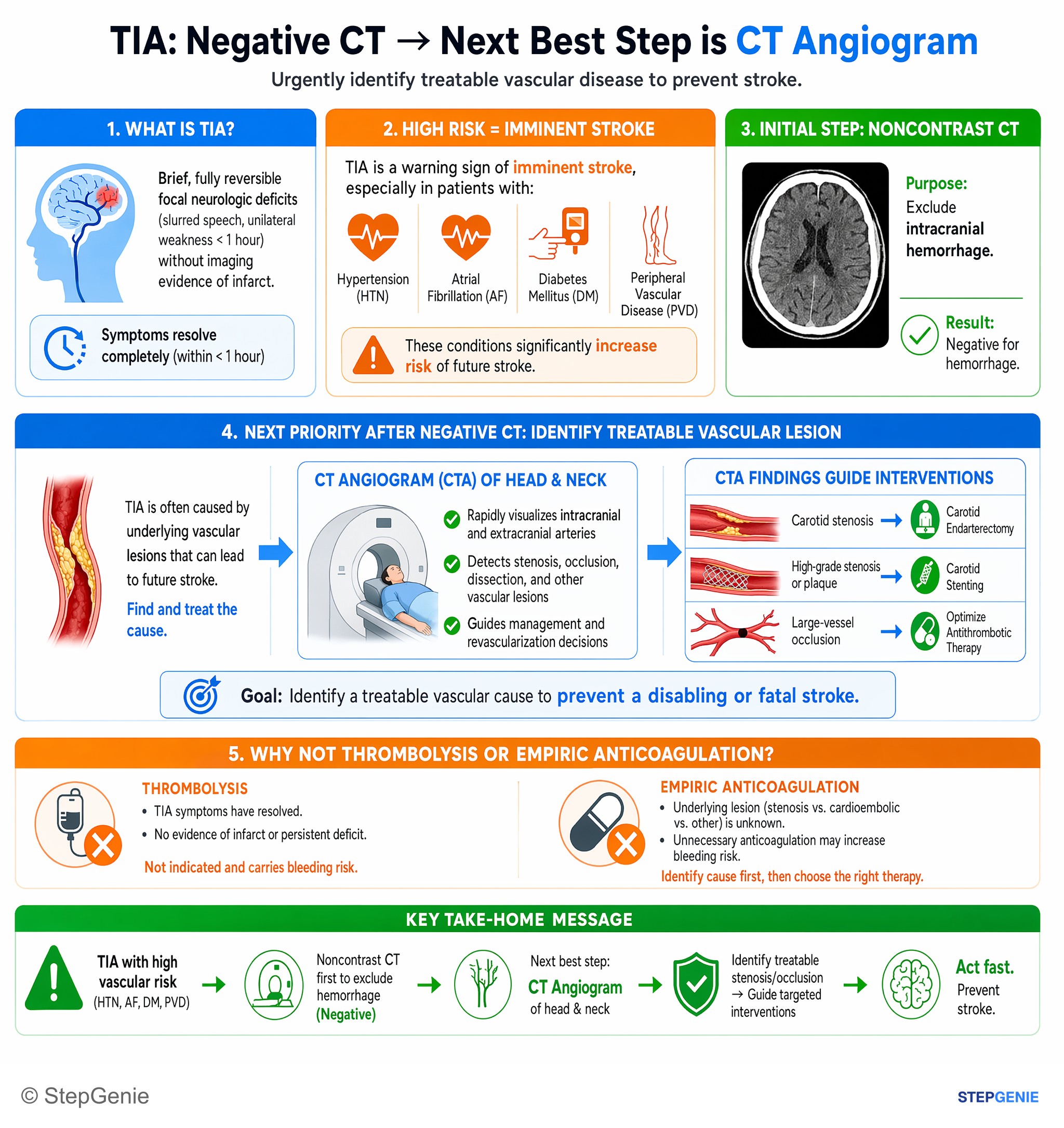
Transient ischemic attack (TIA) with high vascular risk → urgently image cervicocephalic vessels (e.g., CT angiogram) to identify…
USMLE Step 2Frequently Asked Questions
What is the modern definition of a TIA on Step 1?
A transient ischemic attack is transient focal neurologic dysfunction caused by ischemia without evidence of acute infarction. The modern testable definition is tissue-based, so the key feature is no acute infarct on diffusion-weighted MRI. Older question stems may still mention symptoms resolving within less than 24 hours, but the current concept is ischemic symptoms without infarction.
How do you tell a TIA from a stroke when symptoms resolve quickly?
You distinguish them by whether infarction is present, not just by symptom duration. A TIA causes stroke-like focal deficits that resolve and has no infarct on imaging, especially diffusion-weighted MRI. A stroke has deficits with infarction, even if the symptoms improve or resolve quickly. That is why imaging is central when a vignette makes the diagnosis feel ambiguous.
What symptoms are most consistent with a TIA instead of a mimic?
TIA is most consistent with sudden-onset focal negative neurologic deficits that localize to a vascular territory. Common examples include unilateral weakness or numbness, aphasia, dysarthria, visual field loss, amaurosis fugax, and ataxia when paired with other brainstem signs. Isolated confusion, syncope, generalized weakness, isolated headache, or gradually spreading positive symptoms point more toward mimics than TIA.
Why does carotid atherosclerosis cause amaurosis fugax in TIA?
Carotid atherosclerosis can cause amaurosis fugax because plaque rupture and platelet thrombus can send an embolus from the carotid circulation to the retina, producing transient monocular vision loss. This is a classic TIA clue, especially in an older patient with vascular risk factors. It reflects transient ischemia in tissue supplied by the ipsilateral carotid artery rather than permanent infarction.
What is the first-line treatment approach for TIA?
The first-line approach is urgent secondary stroke prevention, starting with antiplatelet therapy unless anticoagulation is indicated. Aspirin is the classic answer for non-cardioembolic TIA, with clopidogrel as an alternative. Management also includes aggressive control of vascular risk factors, evaluation for carotid stenosis, and anticoagulation when atrial fibrillation or another high-risk cardioembolic source is present.