Multiple sclerosis (MS) questions are where USMLE writers love to test your clinical pattern recognition and your ability to eliminate tempting distractors. The key isn’t just spotting the right diagnosis—it’s knowing why every other option is wrong, and what diagnosis it would fit instead.
Clinical Vignette (Q-Bank Style)
A 29-year-old woman comes to clinic for 2 weeks of blurry vision and pain with eye movement in her right eye. She also reports intermittent tingling in her left leg and urinary urgency over the past year, with symptoms that “come and go.” She had an episode of right arm weakness 8 months ago that resolved over several weeks without treatment. Neurologic exam shows decreased visual acuity in the right eye and a right afferent pupillary defect. MRI brain and spine reveals multiple periventricular T2 hyperintense lesions, some enhancing with gadolinium and others nonenhancing.
Which of the following is the most likely underlying diagnosis?
A. Neuromyelitis optica spectrum disorder (aquaporin-4 antibody mediated)
B. Multiple sclerosis
C. Myasthenia gravis
D. Amyotrophic lateral sclerosis
E. Progressive multifocal leukoencephalopathy
Step 1/2: Identify the “MS Pattern” First
This stem is screaming MS:
- Young woman (typical demographic)
- Neurologic deficits separated in time and space
- Time: different episodes months apart
- Space: optic neuritis (CN II), sensory symptoms (spinal cord/brain), urinary urgency (spinal cord)
- Optic neuritis: painful eye movements + afferent pupillary defect (RAPD)
- MRI: periventricular lesions + both enhancing and nonenhancing lesions
- Enhancement = active inflammation (new lesion)
- Nonenhancing = older lesions
→ classic imaging proof of dissemination in time
Correct Answer: B. Multiple Sclerosis
What MS is (high yield)
An autoimmune demyelinating disease of the CNS characterized by inflammatory plaques with subsequent gliosis.
Core buzzwords & “must-know” associations
- Symptoms: optic neuritis, internuclear ophthalmoplegia (INO), sensory symptoms, weakness, spasticity, Lhermitte sign, bladder dysfunction
- MRI: periventricular plaques (often described as Dawson fingers)
- CSF:
- Oligoclonal IgG bands
- ↑ IgG index
- Uhthoff phenomenon: neurologic symptoms worsen with heat/exercise
- Most common subtype: relapsing-remitting MS
Treatment (board-relevant)
- Acute relapse: high-dose IV corticosteroids
- Disease-modifying therapy (reduce relapses/new lesions):
- interferon-β, glatiramer
- fingolimod, dimethyl fumarate, teriflunomide
- monoclonals (e.g., natalizumab, ocrelizumab)
- Symptomatic: baclofen/tizanidine for spasticity, dalfampridine for gait, etc.
Why Each Distractor Is Wrong (and What It Would Fit)
A. Neuromyelitis optica spectrum disorder (NMOSD)
Why it’s tempting: optic neuritis + spinal symptoms can look MS-ish.
Why it’s wrong here:
- NMOSD classically causes:
- Severe optic neuritis (often bilateral) and
- Longitudinally extensive transverse myelitis (spinal cord lesion spanning ≥3 vertebral segments)
- Brain MRI in NMOSD is often less “classic MS”; MS is the one with periventricular lesions and dissemination in time/space patterns like this.
High-yield differentiators
- NMOSD: aquaporin-4 IgG (AQP4) positive (often)
- Treatment differs: immunosuppression (e.g., rituximab, eculizumab) rather than typical MS DMT approach
- Important trap: giving interferon-β (MS therapy) can worsen NMOSD in some cases.
Board clue for NMOSD: profound myelitis + long cord lesion + AQP4 Ab.
C. Myasthenia gravis (MG)
Why it’s tempting: young woman + neuro symptoms.
Why it’s wrong here:
- MG is a neuromuscular junction disorder, not CNS demyelination.
- MG causes fatigable weakness (ptosis, diplopia, bulbar symptoms) that worsens with use and improves with rest.
- MG does not cause:
- sensory loss/tingling
- bladder dysfunction
- CNS MRI plaques
- optic neuritis with RAPD (MG eye symptoms are from extraocular muscle weakness, not optic nerve inflammation)
High-yield MG facts
- Autoantibodies: ACh receptor (most) or MuSK
- Associations: thymoma, thymic hyperplasia
- Diagnosis: AChR Ab, MuSK Ab, EMG; edrophonium is historical
- Treatment: pyridostigmine, immunosuppression; thymectomy for thymoma (and many generalized cases)
D. Amyotrophic lateral sclerosis (ALS)
Why it’s tempting: progressive neuro disease.
Why it’s wrong here:
- ALS is UMN + LMN degeneration:
- UMN: hyperreflexia, spasticity, Babinski
- LMN: weakness, atrophy, fasciculations
- No sensory symptoms (tingling is a big red flag against ALS)
- No optic neuritis and no characteristic periventricular MRI plaques
- Typically presents later than classic MS (often 50s–60s), though age alone isn’t enough.
High-yield ALS facts
- Cognition usually intact, but can overlap with frontotemporal dementia
- Death often due to respiratory failure
- Treatments: riluzole, edaravone (modest benefit)
E. Progressive multifocal leukoencephalopathy (PML)
Why it’s tempting: demyelinating disease with MRI lesions.
Why it’s wrong here:
- PML = JC virus infection causing demyelination in immunosuppressed patients (HIV/AIDS, transplant, or natalizumab use).
- Presentation is typically subacute progressive focal deficits (weeks to months), not relapsing-remitting episodes separated in time.
- MRI classically shows nonenhancing multifocal lesions in white matter (often parieto-occipital), and CSF JC virus PCR can help.
High-yield tie-in to MS
- Natalizumab (MS therapy) increases risk of PML
→ “MS patient on natalizumab develops progressive neuro deficits” = think PML.
Rapid-Fire High-Yield MS Pearls (Exam Day Stuff)
Classic symptom clusters
- Optic neuritis: painful eye movement, decreased visual acuity, RAPD
- INO: lesion of MLF → impaired adduction of one eye + abducting nystagmus of the other
- Spinal cord involvement: sensory changes, weakness, bladder dysfunction
- Heat sensitivity (Uhthoff): worse symptoms after hot shower/exercise
MS diagnosis: what the question writers want you to say
- Dissemination in time: enhancing + nonenhancing lesions; new lesions on follow-up; distinct clinical attacks
- Dissemination in space: lesions in different CNS regions (periventricular, juxtacortical, infratentorial, spinal cord)
Quick Comparison Table: MS vs Common Look-Alikes
| Condition | Key Pathology | Classic Clues | Sensory Symptoms? | MRI Pattern | “Giveaway” Test/Association |
|---|---|---|---|---|---|
| Multiple sclerosis | Autoimmune CNS demyelination | Optic neuritis, INO, neuro deficits separated in time/space | Yes | Periventricular plaques, enhancing + nonenhancing | CSF oligoclonal bands |
| NMOSD | AQP4-IgG astrocytopathy | Severe optic neuritis + longitudinally extensive myelitis | Yes | Long spinal cord lesions (≥3 segments) | AQP4 antibody |
| Myasthenia gravis | NMJ (AChR/MuSK) | Fatigable weakness, ptosis/diplopia | No | Normal CNS MRI | Thymoma, AChR Ab |
| ALS | UMN + LMN degeneration | Fasciculations + spasticity, no sensory loss | No | Not MS-like | Riluzole modest benefit |
| PML | JC virus demyelination | Immunosuppressed, progressive deficits | Variable | Multifocal white matter lesions (often nonenhancing) | JC virus PCR; natalizumab risk |
Takeaway: Why Every Answer Choice Matters
In MS questions, the test makers don’t just want “young woman + optic neuritis = MS.” They want you to notice relapsing symptoms, CNS localization, and the MRI time-stamp of inflammation (enhancing vs nonenhancing). Then they want you to prove you can distinguish MS from:
- NMOSD (AQP4, long spinal lesions),
- MG (fatigable weakness, no sensory/CNS lesions),
- ALS (UMN+LMN, no sensory),
- PML (JC virus in immunosuppressed, progressive course).
If you can articulate why each distractor doesn’t fit the stem, you’ll stop missing the “easy” MS questions that are designed to be tricky.
Visual explanations from the StepGenie QBank
Infographics StepGenie drew to explain questions in its own QBank, matched to this article's topic. Each caption is the takeaway of the question it explains.
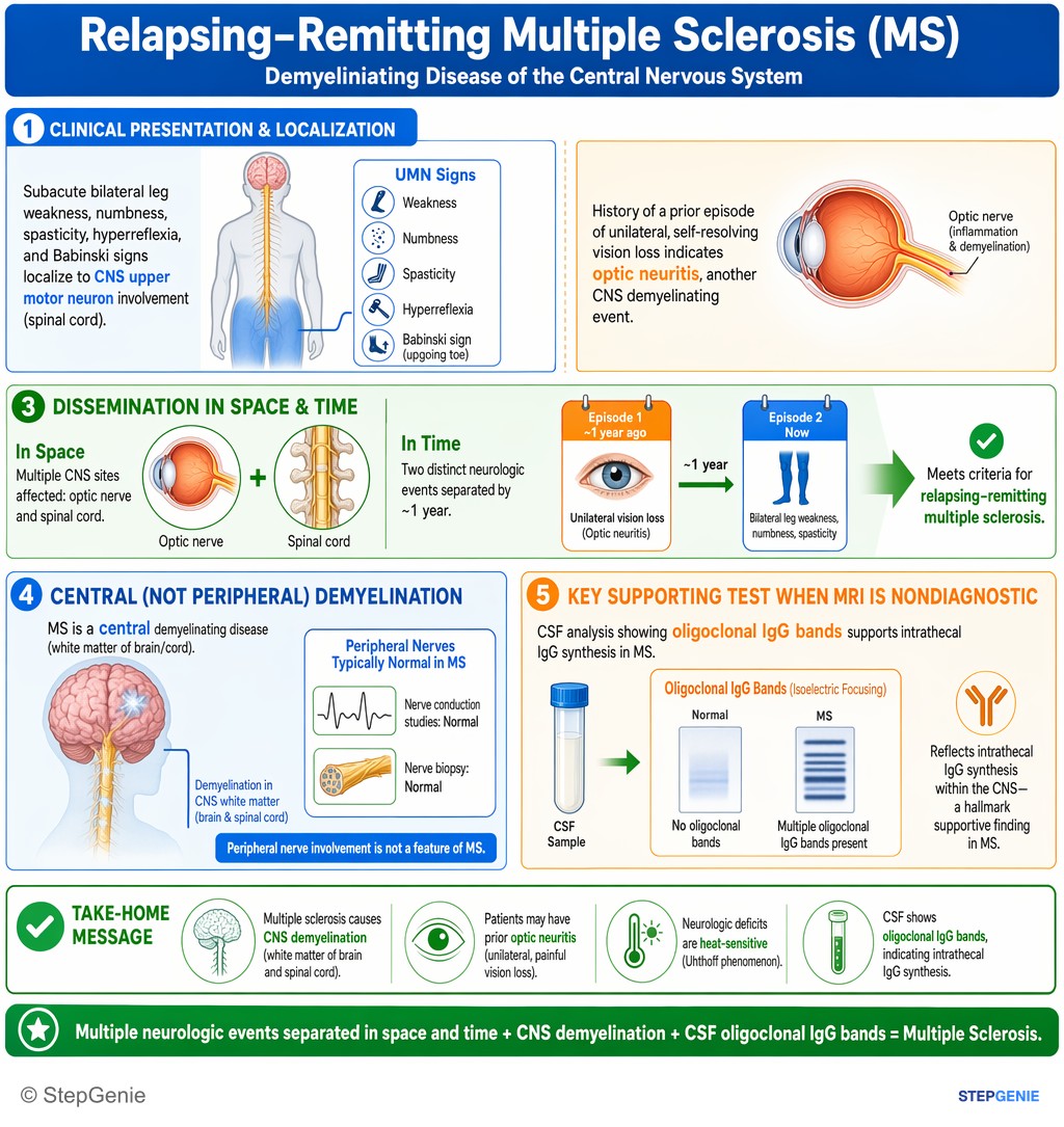
Multiple sclerosis causes CNS demyelination with heat-sensitive neurologic deficits and prior optic neuritis; CSF shows oligoclonal IgG…
USMLE Step 1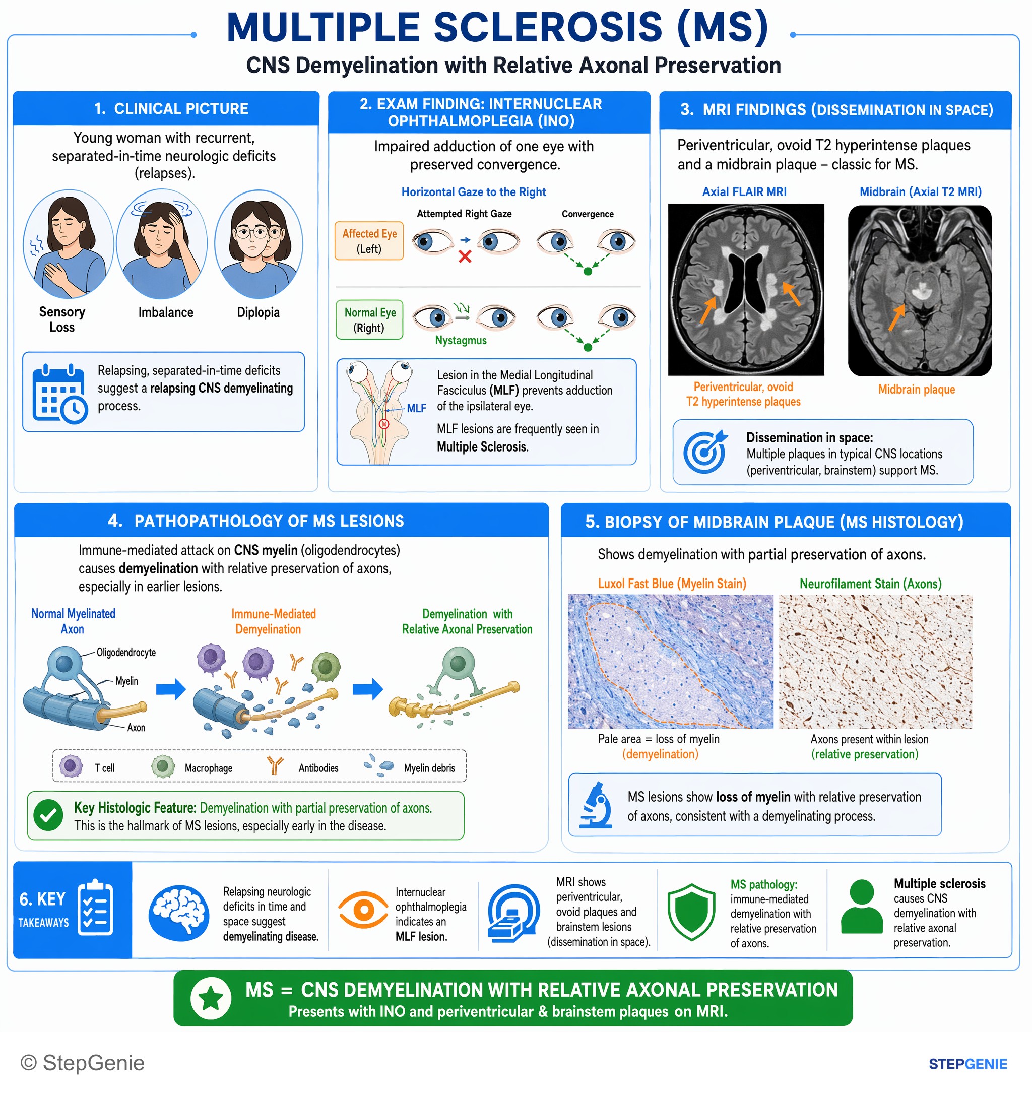
Multiple sclerosis causes CNS demyelination with relative axonal preservation, presenting with internuclear ophthalmoplegia and…
USMLE Step 1Frequently Asked Questions
How do you recognize the classic multiple sclerosis pattern in a vignette?
The classic pattern is a young woman with neurologic deficits separated in time and space. A stem pointing to multiple sclerosis often includes episodes months apart that affect different CNS locations, such as optic neuritis, sensory symptoms, weakness, or bladder dysfunction. MRI support comes from periventricular T2 lesions with both gadolinium-enhancing and nonenhancing plaques, showing active and older lesions.
Why do both enhancing and nonenhancing MRI lesions point to multiple sclerosis?
They point to dissemination in time, which is a classic clue for multiple sclerosis. Gadolinium-enhancing lesions represent active inflammation and therefore newer plaques, while nonenhancing lesions represent older lesions. When both are present along with typical periventricular lesions, the MRI shows lesions of different ages, supporting relapsing disease in the CNS rather than a single event.
How do you tell multiple sclerosis from neuromyelitis optica spectrum disorder?
Multiple sclerosis is favored when there are classic periventricular brain lesions and a dissemination-in-time-and-space pattern, whereas NMOSD is suggested by severe optic neuritis, often bilateral, plus longitudinally extensive transverse myelitis spanning at least 3 vertebral segments. NMOSD is also often associated with aquaporin-4 IgG. This distinction matters because treatment differs, and interferon-β can worsen NMOSD in some cases.
Why isn’t myasthenia gravis the cause of optic neuritis, tingling, and bladder symptoms?
Myasthenia gravis is not the cause because it is a neuromuscular junction disorder that produces fatigable weakness, not CNS demyelination. MG commonly causes ptosis, diplopia, and bulbar weakness that worsen with use and improve with rest. It does not cause sensory loss, tingling, bladder dysfunction, CNS MRI plaques, or optic neuritis with a relative afferent pupillary defect.
What is the board-relevant treatment for multiple sclerosis relapses and long-term control?
Acute multiple sclerosis relapses are treated with high-dose IV corticosteroids, while long-term control uses disease-modifying therapy to reduce relapses and new lesions. Board-relevant options listed include interferon-β, glatiramer, fingolimod, dimethyl fumarate, teriflunomide, and monoclonal antibodies such as natalizumab and ocrelizumab. Symptomatic therapy can also be added for issues like spasticity or gait difficulty.