Diverticulosis is simply having colonic diverticula, false pouches of mucosa and submucosa pushed through the muscle wall, usually in the sigmoid colon; it is mostly silent, and its classic complication is sudden, painless hematochezia from a ruptured vasa recta. Diverticulitis is inflammation of a diverticulum: left lower quadrant (LLQ) pain and tenderness, fever and leukocytosis, confirmed by CT of the abdomen and pelvis. Mild uncomplicated diverticulitis is treated at home, with antibiotics used selectively rather than routinely (AGA 2020, ACP 2022, ACG 2026); an abscess, fistula, obstruction or perforation makes it complicated. Colonoscopy waits until the attack has settled (usually 6-8 weeks) and is advised mainly after complicated disease.
The acronym trick: -OSIS oozes, -ITIS hurts
Let the suffix answer the stem. DiverticulOSIS Oozes blood without pain; diverticulITIS is Inflamed, and It hurts.
| DiverticulOSIS: "Oozes" | DiverticulITIS: "It hurts" | |
|---|---|---|
| What it is | Pouches are present, no inflammation | Inflammation, with or without infection, of a diverticulum |
| Pain | None | LLQ pain and tenderness (right-sided disease is more common in people of Asian or African descent) |
| Blood | Sudden, painless bright red or maroon hematochezia | Bleeding is uncommon |
| Fever and WBC | Normal | Fever, leukocytosis, raised CRP |
| First test | Colonoscopy after rapid bowel prep; CT angiography first if bleeding is ongoing and hemodynamically significant | CT of the abdomen and pelvis with IV contrast |
| Colonoscopy | Finds and treats the bleeding vessel (clips, epinephrine, coagulation) | Not during the attack; later, to exclude a cancer, when indicated |
| Treatment | Most bleeds stop on their own; the rest get endoscopic therapy, embolization or, rarely, surgery | Outpatient care with selective antibiotics; drainage or surgery for complications |
SIGMOID: the diverticulitis checklist
Diverticulitis lives in the sigmoid, so let the organ carry the whole workup:
| Letter | Stands for | What to remember |
|---|---|---|
| S | Sigmoid, LLQ | "Left-sided appendicitis": LLQ pain and tenderness, often a palpable tender sigmoid, sometimes suprapubic pain |
| I | Inflammation | Fever, nausea and vomiting, leukocytosis, raised CRP; urinary symptoms if the bladder is irritated |
| G | Get a CT | Abdomen and pelvis: bowel wall thickening and pericolic fat stranding, plus any abscess or free air. Ultrasound is an alternative; MRI suits pregnant and young patients |
| M | Mild = manage at home | Uncomplicated and well: clear liquids, advancing as the pain settles; antibiotics only for patients with risk features |
| O | Other organs (fistula) | Colovesical is the most common: pneumaturia, fecaluria, UTIs. Colovaginal: feculent vaginal discharge |
| I | Intervene if complicated | IV antibiotics; percutaneous drainage for abscesses over about 3-4 cm; emergency resection for free perforation or feculent peritonitis |
| D | Delay the scope | No colonoscopy during the attack (an inflamed colon can be damaged or ruptured); scope after recovery, usually 6-8 weeks, mainly after complicated disease, to exclude a cancer mimic |
Diverticulosis: why the pouches form and why they bleed
- False diverticula. A colonic diverticulum contains only mucosa and submucosa, so it is a pseudodiverticulum. The classic true diverticulum (all layers of the wall) is Meckel diverticulum.
- Where. At weak points in the muscle next to the penetrating intramural vessels (vasa recta). Mostly the sigmoid in Western patients, the right colon in Asian countries, and rarely the rectum.
- Why. Raised intraluminal pressure (probably from spasm of the muscle layer) pushes mucosa out through those weak points. Associations: age, a diet low in fiber or high in red meat, obesity, inactivity, smoking, NSAIDs and aspirin, glucocorticoids, opioids and genetics. ADPKD also brings more colonic diverticula (see the ADPKD vs ARPKD mnemonic).
- Myth. Nuts, seeds, corn and popcorn are not linked to diverticulosis, diverticulitis or bleeding; patients no longer need to avoid them.
- Numbers (Merck). Half of people aged 60 or older and about 70% of those 80 or older have diverticulosis. About 75% have no symptoms (or only intermittent constipation); diverticulitis develops in only about 1-5%, and bleeding in 10-15%.
- The bleed. The torn vessel is an arteriole, so blood loss is usually moderate to severe: bright red or maroon, painless and usually without diverticulitis. Most diverticula in Western patients are left-sided, yet the right colon is the source in more than half of diverticular bleeds. Most bleeds stop spontaneously, but rebleeding is common.
- Rank. Diverticular bleeding is the most common cause of acute lower GI bleeding in adults (30-65% in Merck's figures).
Painless rectal bleeding: sort the mimics
| Cause | Classic stem | Discriminator |
|---|---|---|
| Diverticular bleeding | Older adult, sudden large painless bright red or maroon bleed | Vasa recta rupture; stops on its own in most |
| Angiodysplasia (vascular ectasia) | Older adult; associations include renal failure and aortic stenosis | Flat, bright red ectatic vessels, typically in the cecum and ascending colon |
| Meckel diverticulum | Young child with painless bright red bleeding | True diverticulum; acid from ectopic gastric mucosa ulcerates the ileum; Tc-99m pertechnetate (Meckel) scan |
Diverticulitis: diagnosis and who needs antibiotics
- CT is the test. Merck gives CT a sensitivity of 95-99% and a specificity of 95-100%. ACG (2026) advises CT at the first presentation, to confirm the diagnosis, exclude alternatives, grade severity and localize disease; ACP (2022) advises CT when the diagnosis is uncertain.
- Why image at all: appendicitis, colon or ovarian cancer and IBD can look the same, and in about 10% of patients CT can't separate diverticulitis from colon cancer.
- Pathogenesis is shifting. The classic model is micro-perforation of a diverticulum with bacterial spillover, but in some patients acute diverticulitis appears to be more inflammatory than infectious, which is why antibiotics are now selective.
| Uncomplicated | Complicated | |
|---|---|---|
| Definition | Localized inflammation only | Abscess, fistula, obstruction (stricture) or free perforation |
| How common | Most patients | About 12% present this way (Merck) |
| Setting | Outpatient for most (ACP) | Hospital: IV fluids, bowel rest |
| Antibiotics | Selective (below) | Yes: IV, covering gram-negative rods and anaerobes |
| Procedures | None | Drain abscesses over about 3-4 cm (CT-guided); resect for free perforation, feculent peritonitis or no response in 3-5 days |
| Colonoscopy afterward | Only with alarm features or if not up to date on screening (ACG 2026) | Yes, once recovered (AGA, ACP, ACG) |
Antibiotics can often be skipped (ACG 2026) in patients who are immunocompetent, hemodynamically stable, managed as outpatients, tolerating oral intake without SIRS or complicated disease, not frail, and able to follow up reliably. AGA calls for antibiotics "selectively, rather than routinely" in immunocompetent patients with mild uncomplicated disease, and ACP suggests managing select patients without them.
Give antibiotics to patients who are immunocompromised, frail or medically complex, vomiting or unable to eat, worsening, or who have markedly raised inflammatory markers (AGA: CRP above 140 mg/L or WBC above 15 × 10⁹/L), a fluid collection or long inflamed segment on CT, or no reliable follow-up. Oral options: amoxicillin-clavulanate, or metronidazole plus ciprofloxacin (or levofloxacin) or TMP-SMX; moxifloxacin alone suits patients who can't take penicillins or metronidazole.
After the attack: colonoscopy, prevention and surgery
- Colonoscopy timing. AGA: delay it 6-8 weeks or until symptoms fully resolve, whichever is longer, and sooner if alarm symptoms appear. It is advised after complicated diverticulitis (ACP: if no recent colonoscopy).
- After uncomplicated disease the guidance has narrowed: ACG (2026) limits colonoscopy to patients with alarm symptoms (weight loss, change in bowel habits, iron-deficiency anemia, bloody stools, persistent pain) or who are not up to date with screening, whereas AGA (2020) advised it after a first episode unless a high-quality colonoscopy was done within a year. Routine intervals are in the colorectal cancer screening flowchart.
- Prevention. High-fiber, plant-forward diet, normal weight, regular activity, no smoking, and no regular NSAIDs (AGA exempts aspirin for secondary cardiovascular prevention). ACP recommends against mesalamine; AGA also advises against rifaximin and probiotics for prevention.
- Elective colectomy is individualized, not counted. AGA advises against recommending resection based on the number of episodes. Complicated disease is most often the first presentation, and the risk of complicated disease decreases with recurrences. ACG quotes recurrence risks of 22% after a first episode and 55% after a second; surgery lowers, but does not eliminate, that risk.
- Don't confuse it with sigmoid volvulus, the other classic sigmoid emergency of older adults: massive distension and obstipation with a coffee-bean loop, not fever with focal LLQ tenderness.
Practice question
A 64-year-old man has 4 days of worsening left lower quadrant pain and fevers. Temperature is 38.8°C (101.8°F), pulse 104/min and blood pressure 128/78 mm Hg. He has LLQ tenderness with a vague mass but no rebound or rigidity. WBC count is 17,000/mm³. CT of the abdomen and pelvis with IV contrast shows sigmoid wall thickening, pericolic fat stranding and a 5-cm pericolic fluid collection, with no free intraperitoneal air. IV fluids and IV antibiotics are started. Which is the most appropriate next step?
A. CT-guided percutaneous drainage of the collection B. Colonoscopy now to exclude colon cancer C. Oral amoxicillin-clavulanate and outpatient follow-up D. Emergency sigmoid resection with colostomy E. Clear liquids at home without antibiotics
Answer: A. This is complicated diverticulitis with a 5-cm abscess. Abscesses larger than about 3-4 cm are drained percutaneously alongside IV antibiotics; surgery is kept for abscesses that are multiloculated, inaccessible or not improving.
- B: colonoscopy during the acute attack can damage or rupture the inflamed colon. It comes later, after complicated disease: once the attack has resolved, usually 6-8 weeks on.
- C: an abscess, fever and leukocytosis rule out outpatient oral therapy; this patient needs admission.
- D: emergency resection is for free perforation, feculent peritonitis or failure of nonoperative management, and none is present.
- E: skipping antibiotics is only for select patients with mild, uncomplicated disease, not an abscess.
Key takeaways
- -OSIS oozes, -ITIS hurts: painless hematochezia = diverticulosis; LLQ pain, fever and leukocytosis = diverticulitis.
- Colonic diverticula are false (mucosa and submucosa only), form where the vasa recta pierce the muscle, and cluster in the sigmoid; yet more than half of diverticular bleeds come from the right colon.
- CT of the abdomen and pelvis confirms diverticulitis; no colonoscopy during the attack.
- Mild uncomplicated disease: outpatient care, clear liquids and selective antibiotics (AGA, ACP, ACG).
- Complicated disease: IV antibiotics, percutaneous drainage for abscesses over about 3-4 cm, surgery for free perforation or peritonitis.
- Colovesical fistula = pneumaturia, fecaluria and UTIs; most fistulas run from the sigmoid to the bladder.
- Colonoscopy 6-8 weeks after complicated diverticulitis; elective colectomy is individualized, not decided by the number of attacks.
Sources
- Colonic Diverticulosis — Merck Manual Professional Edition (2026)
- Colonic Diverticulitis — Merck Manual Professional Edition (2026)
- Diverticulitis — Merck Manual Consumer Version
- Medical Management of Colonic Diverticulitis: AGA Clinical Practice Update — American Gastroenterological Association (2020)
- Diagnosis and Management of Acute Left-Sided Colonic Diverticulitis: ACP Clinical Guideline — Annals of Internal Medicine (2022)
- Colonoscopy for Diagnostic Evaluation and Interventions to Prevent Recurrence After Acute Left-Sided Colonic Diverticulitis: ACP Clinical Guideline — Annals of Internal Medicine (2022)
- ACG Guideline Highlights: Diagnosis and Management of Diverticulitis — American College of Gastroenterology (2026)
- Vascular Gastrointestinal Lesions — Merck Manual Professional Edition
- Meckel Diverticulum — Merck Manual Professional Edition
- Autosomal Dominant Polycystic Kidney Disease (ADPKD) — Merck Manual Professional Edition
Visual explanations from the StepGenie QBank
Infographics StepGenie drew to explain questions in its own QBank, matched to this article's topic. Each caption is the takeaway of the question it explains.
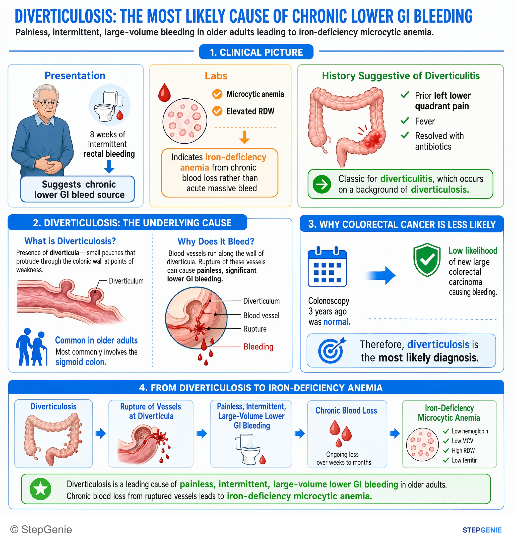
Diverticulosis causes painless, intermittent, large-volume lower GI bleeding in older adults; chronic blood loss leads to iron-deficiency…
USMLE Step 2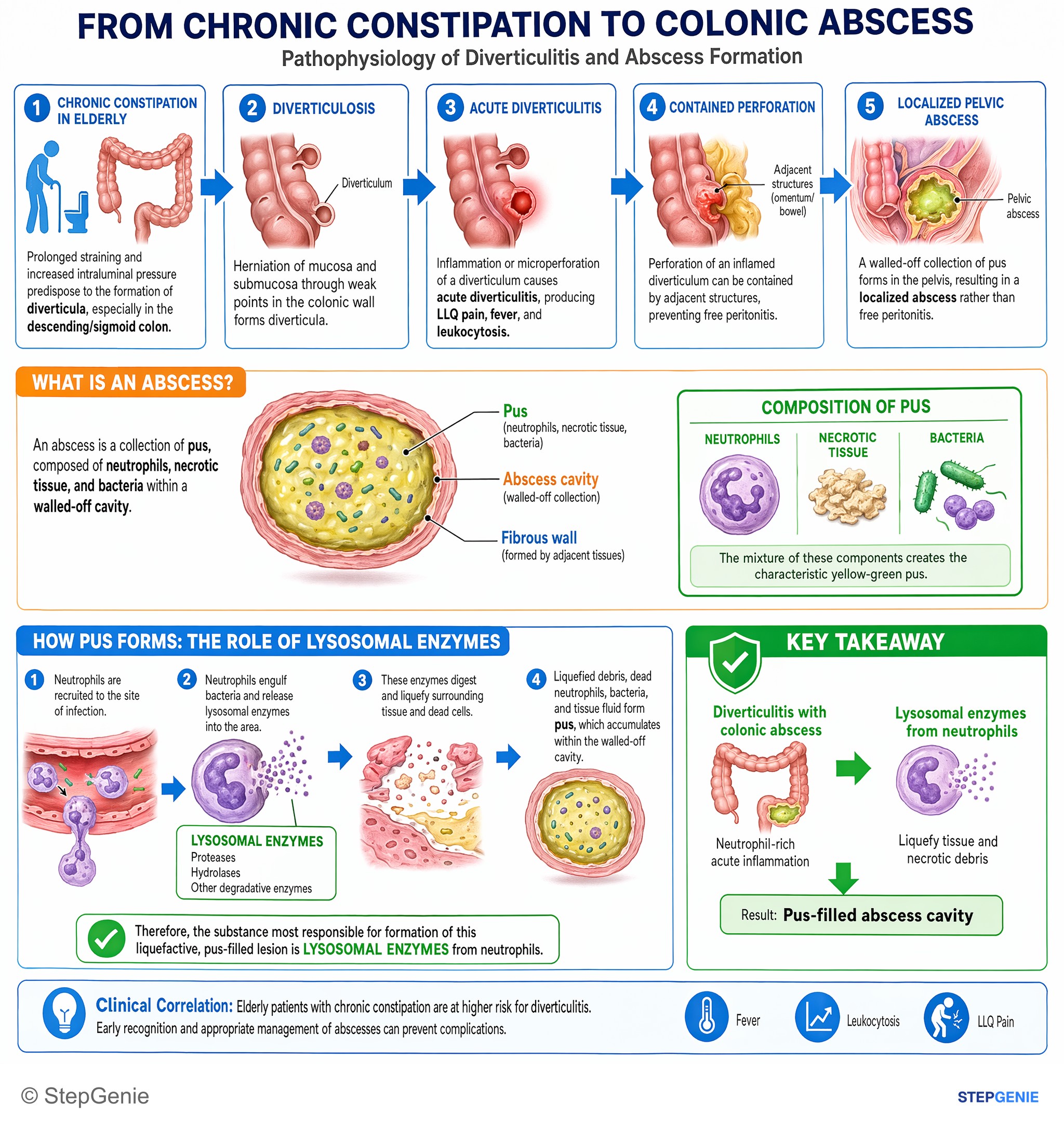
Diverticulitis with colonic abscess: neutrophil-rich acute inflammation; lysosomal enzymes from neutrophils liquefy tissue, forming a…
USMLE Step 1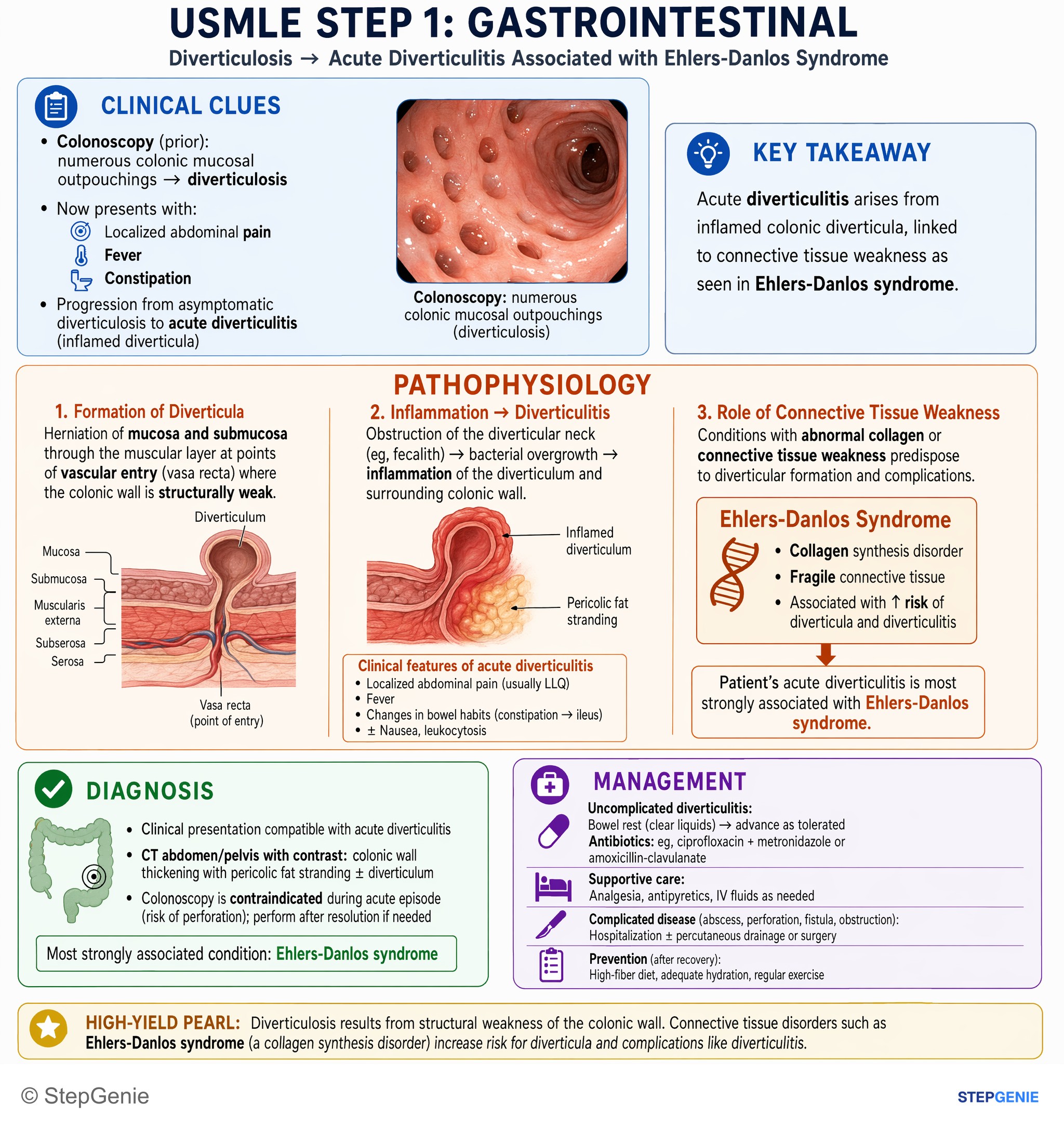
Acute diverticulitis arises from inflamed colonic diverticula, linked to connective tissue weakness as seen in Ehlers-Danlos syndrome.
USMLE Step 1Frequently Asked Questions
What is the difference between diverticulosis and diverticulitis?
Diverticulosis means pouches (diverticula) are present in the colon wall, most often in the sigmoid colon; most people have no symptoms, and its classic complication is sudden, painless rectal bleeding. Diverticulitis is inflammation of one of those pouches, which causes left lower quadrant pain and tenderness, fever and a raised white cell count. A simple way to remember it: diverticulOSIS oozes, diverticulITIS hurts.
Why does diverticulosis cause painless bleeding?
Colonic diverticula are false diverticula that push out at weak points where small arteries (the vasa recta) run through the muscle wall, so the vessel ends up stretched over the pouch. When that arteriole ruptures, the bleeding is sudden, painless and often large, with bright red or maroon blood. Most episodes stop on their own, and colonoscopy can find and treat the bleeding point.
What is the best test to diagnose diverticulitis?
CT of the abdomen and pelvis, usually with IV contrast, is the preferred test because it confirms the diagnosis and shows complications such as an abscess or perforation. Ultrasound is an alternative in some settings, and MRI can be used in pregnant and young patients. Colonoscopy is not used during the acute attack.
Does diverticulitis always need antibiotics?
No. Current US guidance from the AGA (2020), ACP (2022) and ACG (2026) supports managing select patients with mild, uncomplicated diverticulitis without antibiotics if they are immunocompetent, stable, able to eat and drink, and able to follow up reliably. Antibiotics are still given for complicated disease and for patients who are immunocompromised, frail, vomiting, worsening or who have markedly raised inflammatory markers.
When should you have a colonoscopy after diverticulitis?
Not during the attack, because an inflamed colon can be damaged. After complicated diverticulitis, colonoscopy is advised once the episode has resolved, usually 6 to 8 weeks later, to rule out a colon cancer that can look like diverticulitis on CT. After uncomplicated diverticulitis, the 2026 ACG guidance limits it to people with alarm symptoms or who are not up to date with colorectal cancer screening.
Should people with diverticulosis avoid nuts, seeds and popcorn?
No. Nuts, seeds, corn and popcorn have not been linked to diverticulosis, diverticulitis or diverticular bleeding, and avoiding them is no longer recommended. The advised prevention measures are a high-fiber diet, a normal body weight, regular physical activity, not smoking and avoiding regular NSAID use.