Restrictive cardiomyopathy (RCM) is a stiff, non-dilated ventricle that fills poorly: a small increase in filling volume causes a disproportionate rise in diastolic pressure, so patients develop heart failure with a preserved ejection fraction, biatrial enlargement and prominent right-sided congestion (JVD, edema, ascites, hepatomegaly). Cardiac amyloidosis is the most common cause in developed countries; sarcoidosis, storage diseases such as hemochromatosis and Fabry disease, hypereosinophilic (Löffler) endocarditis, endomyocardial fibrosis and radiation round out the list. The classic exam clue is low QRS voltage despite thick ventricular walls (amyloid), and the key look-alike is constrictive pericarditis, which pericardiectomy can cure.
Where RCM fits among the cardiomyopathies
| Restrictive (RCM) | Dilated (DCM) | Hypertrophic (HCM) | |
|---|---|---|---|
| Core problem | Filling (stiff myocardium) | Pumping (weak myocardium) | Thickened myocardium |
| Ventricle on echo | Not dilated; walls normal or thick; both atria enlarged | Dilated | Thick walls, small cavity |
| Ejection fraction | Preserved early, can fall later | Reduced | Preserved |
RCM is therefore a cause of heart failure with preserved EF; the HFrEF vs HFpEF guide covers the broader picture, and dilated cardiomyopathy and HOCM have their own pages.
Pathophysiology: why a stiff ventricle fails
- Diastolic dysfunction dominates. A stiff, noncompliant ventricle fills poorly and needs high filling pressures; systolic function is usually preserved early and may decline as disease advances.
- Fixed stroke volume. The small, stiff ventricle can't increase its stroke volume much, so cardiac output depends on heart rate.
- Biatrial enlargement from chronically high filling pressures predisposes to atrial fibrillation, which is poorly tolerated because the atrial "kick" is lost.
- Back-pressure: pulmonary venous hypertension, then right ventricular dysfunction and systemic venous congestion.
- Other consequences: functional mitral or tricuspid regurgitation, and intracardiac thrombi (especially in endomyocardial fibrosis, hypereosinophilic syndrome and cardiac amyloidosis).
Causes: the RCM list
A common mnemonic is "Puppy LEASH": Postradiation fibrosis, Löffler endocarditis, Endocardial fibroelastosis, Amyloidosis, Sarcoidosis, Hemochromatosis. Merck groups causes into four families:
| Category | Causes | Exam clue |
|---|---|---|
| Infiltrative | Amyloidosis (AL and ATTR), sarcoidosis, primary hyperoxaluria | Amyloid: low voltage with thick walls, carpal tunnel, neuropathy, nephrotic syndrome (see AL vs AA vs ATTR amyloidosis). Sarcoid: noncaseating granulomas, heart block |
| Storage | Hemochromatosis, Fabry disease, Gaucher, glycogen storage diseases, mucopolysaccharidoses (Hurler, Hunter), Niemann-Pick | Hemochromatosis: cardiomyopathy with heart failure is its second most common fatal complication |
| Endomyocardial | Hypereosinophilic syndrome (Löffler endocarditis), endomyocardial fibrosis, endocardial fibroelastosis, carcinoid, metastatic cancer, radiation, drugs (hydroxychloroquine, ergotamine, methysergide, busulfan) | Eosinophilia, mural thrombi with emboli, mitral or tricuspid regurgitation |
| Non-infiltrative | Idiopathic RCM, systemic sclerosis, pseudoxanthoma elasticum | Idiopathic RCM only after excluding other causes |
Hypereosinophilic syndrome (eosinophils above 1,500/mcL with organ damage; traditionally persisting 6 months or more, though an alternative definition accepts 2 or more counts at least 2 weeks apart) causes endomyocardial disease with embolizing mural thrombi; it is treated with glucocorticoids, or imatinib for FIP1L1::PDGFRA-associated disease.
Presentation
- Symptoms: exertional dyspnea, orthopnea, fatigue and exercise intolerance, palpitations, abdominal distension, leg edema.
- Signs: marked JVD with a rapid y descent, a low-volume rapid carotid pulse, crackles, S3 and/or S4, functional mitral or tricuspid regurgitation murmurs, and, in advanced disease, ascites and edema. Pulsus paradoxus is typically absent.
- Kussmaul sign (JVP rising on inspiration) is the classic sign of constrictive pericarditis. Sources disagree on RCM: StatPearls says the JVP may rise with inspiration in RCM, while Merck's 2026 chapter lists the absence of a Kussmaul sign as typical. Don't use it alone to separate the two.
Diagnosis
- ECG: nonspecific (prominent P waves from atrial enlargement, ST-T changes, conduction disease). Conduction disturbances raise suspicion for infiltration. Voltage can be low or high depending on the cause; low voltage despite thick walls points to amyloid.
- Echocardiogram (key initial test, with ECG and chest x-ray): non-dilated LV with preserved EF, biatrial enlargement, normal or increased wall thickness, and a restrictive filling pattern: E/A above 1.5, short mitral deceleration time, low mitral annular velocities (e′) and a high E/e′.
- Cardiac MRI: best tissue characterization and especially useful for separating RCM from constrictive pericarditis. Amyloid typically shows subendocardial or transmural late gadolinium enhancement with abnormal gadolinium kinetics, a markedly raised native T1 and a high extracellular volume.
- Targeted tests: iron studies, serum and urine protein electrophoresis with immunofixation, serum free light chains, CBC with eosinophil count, genetic testing; FDG-PET for cardiac sarcoidosis; technetium-99m pyrophosphate scintigraphy for ATTR amyloid.
- BNP: elevated BNP is far more common in RCM; a BNP under 100 pg/mL favors constriction (StatPearls).
- Catheterization: high diastolic pressures with rapid early filling and equalized diastolic pressures (the "square root" sign), but minimal respiratory variation and ventricular interdependence, unlike constriction.
- Endomyocardial biopsy is the gold standard in selected cases: it can show amyloid, granulomas, eosinophilic infiltration, fibrosis or Fabry inclusions.
Cardiac amyloidosis: AL vs ATTR
| AL (light chain) | ATTR wild-type | ATTR variant (hereditary) | |
|---|---|---|---|
| Protein | Clonal immunoglobulin light chains | Normal transthyretin (TTR) | Mutant TTR |
| Typical patient | Plasma cell disorder | Older men (over 90% male, usually over 70) | Autosomal dominant; the V122I variant is carried by about 3-4% of Black Americans |
| Other clues | Proteinuria or nephrotic syndrome, neuropathy, macroglossia, periorbital bruising | Carpal tunnel syndrome, spinal stenosis, biceps tendon rupture, which may precede the heart disease | Neuropathy and/or cardiomyopathy |
| Diagnosis | Monoclonal protein plus tissue (Congo red, apple-green birefringence) | Grade 2-3 cardiac uptake on technetium scintigraphy (SPECT) with no monoclonal protein (free light chains, serum and urine immunofixation) = no biopsy needed | Same, plus TTR genetic testing |
| Treatment | Daratumumab plus cyclophosphamide, bortezomib and dexamethasone (Dara-CyBorD); selected patients get high-dose melphalan with stem cell transplant | Tafamidis, acoramidis or vutrisiran | Same drugs; liver transplant can help some variants if done early, but not wild-type disease |
FDA-labeled drugs for ATTR cardiomyopathy (checked Oct 2026): the TTR stabilizers tafamidis (Vyndaqel, Vyndamax; approved 2019) and acoramidis (Attruby; approved November 2024) bind TTR at its thyroxine-binding sites and slow the tetramer breakup that drives amyloid formation. The silencer vutrisiran (Amvuttra), a GalNAc-conjugated siRNA that degrades mutant and wild-type TTR mRNA, gained its cardiomyopathy indication in March 2025. The other gene silencers, patisiran and eplontersen, are used for ATTR polyneuropathy; eplontersen's label (Wainua, updated April 2026) covers hereditary ATTR polyneuropathy only.
RCM vs constrictive pericarditis
| Feature | Restrictive cardiomyopathy | Constrictive pericarditis |
|---|---|---|
| What is stiff | Myocardium | Pericardium |
| Common causes | Amyloid, sarcoid, storage disease, radiation | Idiopathic or viral, prior cardiac surgery, radiation; tuberculosis worldwide |
| Extra sound | S3 and/or S4 | Pericardial knock (early diastolic) |
| Pericardium on imaging | Normal | Thickened (over 4 mm) on CT or MRI; calcification may show on a lateral chest x-ray |
| Mitral annular e′ | Low | Normal or high; septal e′ greater than lateral ("annulus reversus") |
| Inspiratory fall in mitral inflow | Under 15% | Over 25% |
| Respiratory variation, interdependence | Minimal | Marked (septal bounce, discordant ventricular pressures) |
| RV systolic pressure | Often over 50 mm Hg | Less often |
| BNP | High | Lower; under 100 pg/mL favors constriction |
| Definitive therapy | Treat the cause; transplant in selected patients | Pericardiectomy (stable, newly diagnosed cases without chronic constriction may get a 3-month anti-inflammatory trial instead) |
Radiation can cause both, so look for pericardial thickening versus infiltrative myocardial findings.
Treatment
- Treat the cause: immunosuppression for sarcoidosis, iron removal for hemochromatosis (phlebotomy is simplest), enzyme replacement for Fabry disease, and amyloid-specific therapy (above).
- Diuretics are the cornerstone for congestion, but use them cautiously: these patients are highly preload dependent, and over-diuresis drops cardiac output.
- Heart failure drugs: Merck's 2026 chapter advises an SGLT2 inhibitor for all patients, with mineralocorticoid receptor antagonists and renin-angiotensin inhibitors for selected patients; afterload reducers can cause hypotension.
- Atrial fibrillation: rhythm control (antiarrhythmic drugs or ablation) is generally preferred; low-dose beta-blockers or nondihydropyridine calcium channel blockers only with caution. Anticoagulate with atrial fibrillation or flutter, or a documented intracardiac thrombus.
- Amyloid caution: Merck's amyloidosis chapter calls digoxin, ACE inhibitors or ARBs, calcium channel blockers and beta-blockers poorly tolerated and contraindicated in amyloid heart failure.
- Advanced disease: pacemakers for bradyarrhythmias and conduction disease; heart transplant in carefully selected patients. A small LV cavity and RV dysfunction may preclude a durable LV assist device.
How USMLE tests it
- HFpEF + big atria + right-sided congestion + normal-sized ventricles = think RCM.
- Low voltage despite thick walls = amyloid; add carpal tunnel or spinal stenosis in an older man for wild-type ATTR.
- Biopsy stain: Congo red with apple-green birefringence under polarized light = amyloid; noncaseating granulomas = sarcoid; eosinophils = Löffler.
- Pericardial knock, calcified pericardium, prior cardiac surgery or TB, low BNP = constrictive pericarditis, treated definitively with pericardiectomy.
- Drug-mechanism pairs: tafamidis and acoramidis stabilize TTR; vutrisiran silences TTR production; bortezomib and daratumumab treat AL.
Practice question
A 78-year-old man has 6 months of exertional dyspnea and leg swelling. He had carpal tunnel release on both wrists 8 years ago. He has JVD, hepatomegaly and pitting edema. ECG shows low QRS voltage in the limb leads. Echocardiography shows a 17-mm LV wall, normal ejection fraction, biatrial enlargement and reduced mitral annular e′ velocities. Serum free light chain ratio is normal, and serum and urine immunofixation detect no monoclonal protein. Which is the most appropriate next step to establish the diagnosis?
A. Technetium-99m pyrophosphate scintigraphy with SPECT B. Endomyocardial biopsy C. Abdominal fat pad biopsy D. CT of the chest to measure pericardial thickness E. FDG-PET of the heart
Answer: A. Low voltage despite thick walls, biatrial enlargement and a history of bilateral carpal tunnel syndrome in an older man point to wild-type ATTR cardiac amyloidosis. With AL excluded (normal free light chains and negative immunofixation), grade 2-3 myocardial uptake on technetium scintigraphy confirms ATTR-CM without a biopsy; TTR genetic testing then separates wild-type from hereditary disease. Treatment would be tafamidis, acoramidis or vutrisiran.
- B: endomyocardial biopsy is the gold standard for selected cases, but it is invasive and unnecessary when noninvasive criteria are met. If a monoclonal protein had been found, scintigraphy alone could not make the diagnosis and tissue would be needed.
- C: fat pad biopsy detects amyloid in about 80% of patients with AL but fewer than 25% with wild-type ATTR, so a negative result would not rule it out.
- D: pericardial thickening over 4 mm suggests constrictive pericarditis, but reduced e′ and thick, low-voltage myocardium point to an infiltrative cardiomyopathy.
- E: FDG-PET evaluates active cardiac sarcoidosis, a cause of RCM and heart block; it is not the test for ATTR amyloid.
Key takeaways
- RCM = stiff, non-dilated ventricles, impaired filling, biatrial enlargement, preserved EF early, right-sided congestion.
- Amyloidosis is the most common cause in developed countries; also sarcoid, storage diseases (hemochromatosis, Fabry), Löffler endocarditis, endomyocardial fibrosis and radiation.
- Low QRS voltage with thick walls = amyloid; apple-green birefringence on Congo red.
- ATTR-CM: technetium scintigraphy grade 2-3 plus no monoclonal protein = diagnosis without biopsy; treat with tafamidis, acoramidis or vutrisiran.
- Vs constriction: RCM has low e′, high BNP and minimal respiratory variation; constriction has a knock, thick pericardium and responds to pericardiectomy.
- Diurese gently, anticoagulate atrial fibrillation, and avoid digoxin, beta-blockers and calcium channel blockers in amyloid heart disease.
Sources
- Restrictive Cardiomyopathy — Merck Manual Professional Edition (2026)
- Restrictive Cardiomyopathy — StatPearls, NCBI Bookshelf (2023)
- Pericarditis (constrictive pericarditis) — Merck Manual Professional Edition (2026)
- Amyloidosis — Merck Manual Professional Edition (2026)
- Transthyretin Amyloid Cardiomyopathy—2025 Update: Current Diagnostic Approaches and Emerging Therapeutic Options — Journal of Clinical Medicine (2025)
- Vyndaqel and Vyndamax (tafamidis) prescribing information — DailyMed, NLM (2023)
- Attruby (acoramidis) prescribing information — DailyMed, NLM (2026)
- Amvuttra (vutrisiran) prescribing information — DailyMed, NLM (2025)
- Wainua (eplontersen) prescribing information — DailyMed, NLM (2026)
- Transthyretin Amyloidosis — MedlinePlus Genetics (2023)
- Hypereosinophilic Syndrome — Merck Manual Professional Edition (2026)
- Hereditary Hemochromatosis — Merck Manual Professional Edition (2025)
Visual explanations from the StepGenie QBank
Infographics StepGenie drew to explain questions in its own QBank, matched to this article's topic. Each caption is the takeaway of the question it explains.
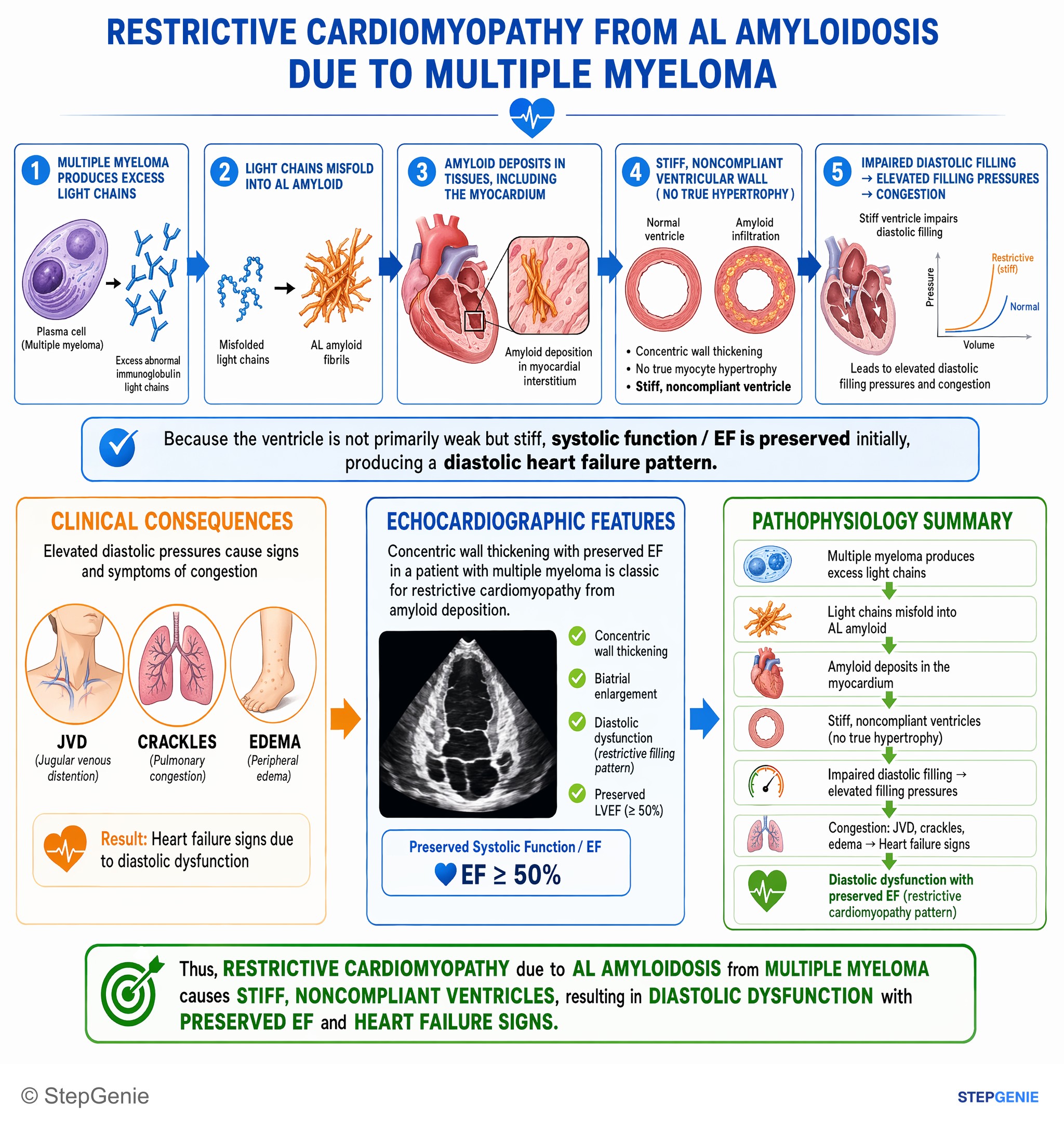
Restrictive cardiomyopathy due to AL amyloidosis from multiple myeloma causes stiff, noncompliant ventricles → diastolic dysfunction with…
USMLE Step 1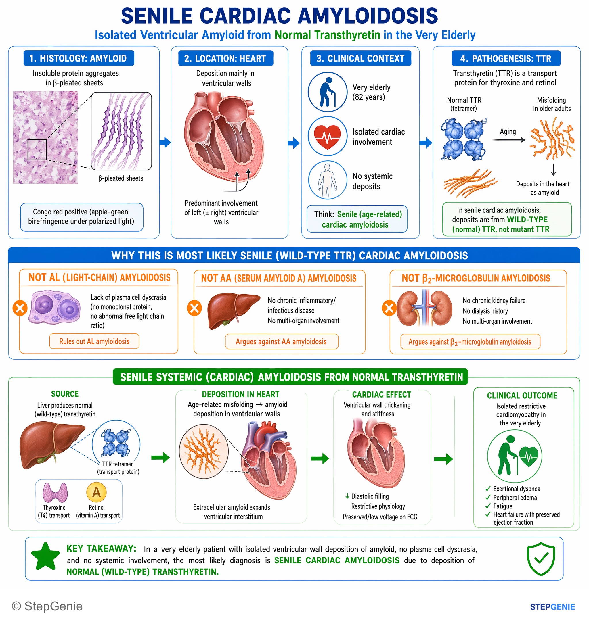
Senile systemic (cardiac) amyloidosis from deposition of normal transthyretin causes isolated ventricular wall thickening/restrictive…
USMLE Step 1Frequently Asked Questions
What is restrictive cardiomyopathy?
Restrictive cardiomyopathy is a heart muscle disease in which stiff, non-dilated ventricles fill poorly, so a small increase in volume causes a large rise in diastolic pressure. Systolic function and ejection fraction are usually preserved early, both atria enlarge from the high filling pressures, and patients present with heart failure with preserved ejection fraction, often with prominent right-sided signs such as jugular venous distention, edema, ascites and hepatomegaly. Atrial fibrillation is common and poorly tolerated.
What are the most common causes of restrictive cardiomyopathy?
Cardiac amyloidosis, either light-chain (AL) or transthyretin (ATTR), is the most common cause in developed countries. Other causes include sarcoidosis, storage diseases such as hemochromatosis, Fabry disease and glycogen storage diseases, hypereosinophilic syndrome (Löffler endocarditis), endomyocardial fibrosis, endocardial fibroelastosis, carcinoid, radiation, systemic sclerosis and some drugs. The mnemonic Puppy LEASH covers the classic exam list: postradiation fibrosis, Löffler endocarditis, endocardial fibroelastosis, amyloidosis, sarcoidosis and hemochromatosis.
How do you tell restrictive cardiomyopathy from constrictive pericarditis?
Both cause impaired filling, high equalized diastolic pressures and right-sided congestion. Constrictive pericarditis has a thickened pericardium on CT or MRI, a pericardial knock, marked respiratory variation with ventricular interdependence, normal or high mitral annular e′ velocities and a lower BNP (under 100 pg/mL favors constriction), and pericardiectomy can cure it. Restrictive cardiomyopathy has low e′ velocities, minimal respiratory variation, a normal pericardium and a high BNP, and cardiac MRI often shows the infiltrating disease.
What are the echo findings in restrictive cardiomyopathy?
Echocardiography shows a non-dilated left ventricle with a preserved ejection fraction, enlargement of both atria, normal or increased wall thickness and a restrictive filling pattern: an E/A ratio above 1.5, a short mitral deceleration time, low mitral annular e′ velocities and a high E/e′ ratio, all reflecting high filling pressures. In cardiac amyloidosis the walls look thick, and longitudinal strain is typically abnormal with relative sparing of the apex.
How is ATTR cardiac amyloidosis diagnosed and treated?
When serum free light chains and serum and urine immunofixation show no monoclonal protein, grade 2 or 3 myocardial uptake on technetium scintigraphy with SPECT confirms ATTR cardiac amyloidosis without a biopsy, and TTR genetic testing separates hereditary from wild-type disease. FDA-labeled treatments for ATTR cardiomyopathy include the stabilizers tafamidis and acoramidis and the gene silencer vutrisiran. Liver transplant can help only some hereditary forms, and digoxin, beta-blockers and calcium channel blockers are poorly tolerated.
How is restrictive cardiomyopathy treated?
Treatment targets congestion and the underlying cause. Loop diuretics relieve congestion but must be used cautiously because stiff ventricles are preload dependent. Merck's 2026 chapter recommends an SGLT2 inhibitor for all patients as part of HFpEF therapy, generally prefers rhythm control for atrial fibrillation, and calls for anticoagulation with atrial fibrillation, flutter or intracardiac thrombus. Cause-specific options include amyloid therapies, immunosuppression for sarcoidosis, iron removal for hemochromatosis and enzyme replacement for Fabry disease; selected patients need a pacemaker or heart transplant.